Iranian Journal of Basic Medical Sciences
ijbms.mums.ac.ir
Melatonin treatment reduces astrogliosis and apoptosis in
rats with traumatic brain injury
Abdolreza Babaee , Seyed (assan Eftekhar‐Vaghefi , Majid Asadi‐shekaari , Nader
Shahrokhi , Samereh Dehghani Soltani , Reza Malekpour‐Afshar , Mohsen Basiri *
Department of Anatomical Sciences, Afzalipour School of Medicine, Kerman University of Medical Sciences, Kerman, )ran Neuroscience Research Center, )nstitute of Neuropharmacology, Kerman University of Medical Sciences, Kerman, )ran Department of Physiology, Afzalipour School of Medicine, Kerman University of Medical Sciences, Kerman, )ran Department of Pathology, Afzalipour School of Medicine, Kerman University of Medical Sciences, Kerman, )ran A R T ) C L E ) N F O A B S T R A C T
Article type:
Original article Objective(s):neuroprotectionMelatonin is known as an anti‐inflammatory agent, and it has been proven to exert through inhibition of cell death apoptosis in several models of brain injury. Secondary injury following the primary traumatic brain injury TB) results in glial cells activation, especially astrocytes. )n fact, astrocyte activation causes the production of pro‐inflammatory cytokines that may lead to secondary injury. Since most TB) research studies have focused on injured neurons and paid little attention to glial cells, the aim of current study was to investigate the effects of melatonin against astrocytes activation astrogliosis , as well as inhibition of apoptosis in brain tissue of male rats after TB).
Materials and Methods: The animals were randomly allocated into five groups: sham group, TB)+
vehicle group % ethanol in saline and TB)+ melatonin groups mg/kg, mg/kg and mg/kg . All rats were intubated and then exposed to diffuse TB), except for the sham group. )mmunohistochemical methods were conducted using glial fibrillary acidic protein GFAP marker and TUNEL assay to evaluate astrocyte reactivity and cell death, respectively.
Results: The results showed that based on the number of GFAP positive astrocytes in brain cortex,
astrogliosis was reduced significantly P< . in melatonin‐ treated groups no dose dependent compared to the vehicle group. Furthermore, based on TUNEL results, melatonin treatment considerably reduced the number of apoptotic cells P< . .
Conclusion:)n total, the present findings suggest that melatonin treatment following TB) diminishes
astrocyte reactivity and neuronal cells apoptosis in brain cortex in the rat model.
Article history:
Received: Jan , Accepted: May ,
Keywords:
Apoptosis Astrogliosis GFAP Melatonin
Traumatic brain injury
►Please cite this article as:
Babaee A, Eftekhar‐Vaghefi S(, Asadi‐shekaari M, Shahrokhi N, Dehghani Soltani S, Malekpour‐Afshar R, Basiri M. Melatonin treatment reduces astrogliosis and apoptosis in rats with traumatic brain injury. )ran J Basic Med Sci ; : ‐ .
Introduction
Traumatic brain injury TB) is the leading cause of death and disability among the youth in developed countries. Annually, . million people lose their
lives due to TB) worldwide . Based on an
epidemiological study, accidents are the second
leading cause of mortality and morbidity in )ran .
TB) following motorcycle accidents is the most common cause of permanent disability of individuals in )ran . TB) is known to cause primary mechanical injury of neurons; however, the secondary damage following injury is complex with a broad spectrum of symptoms. The impact can be devastating on person’s life . Neural inflammation and oxidative stress are the main pathological mechanisms of neuronal cell apoptosis after TB) , . The role of inflammatory
cascade activated after TB) is very important in brain tissue homeostasis, mediated by the release of pro‐ and
anti‐inflammatory cytokines and chemokine
)nterleukin, TNF‐ , Fas Ligand , which are normally scarcely detectable in healthy brain tissue, but quickly upregulated in response to stressful or pathological conditions .
Different strategies such as melatonin treatment can be adopted to reduce the complications of TB), using varieties of antioxidant compounds . Melatonin
N‐acetyl ‐methoxytryptamine , originally identified
by Lerner in , is a tryptophan derivatives that is
produced mainly in the pineal gland. Control of circadian rhythm and sleep induction are the main physiological functions of melatonin , . There is some evidence showing the reduction of melatonin
*Corresponding author: Mohsen Basiri. Neuroscience Research Center, )nstitute of Neuropharmacology, Kerman University of Medical Sciences, Kerman,
secretion after TB) ; hence, some scientists tried to use the exogenous treatment of melatonin to evaluate its neuroprotective effects on different neural cell injuries. For instance, administration of melatonin after TB) in an animal contusion model facilitates neurobehavioral recovery , . Besides, experimental
and clinical data confirm that melatonin has an important role in the reduction of adhesion
molecules and pro‐inflammatory cytokines. Moreover, experimental evidence supports its function as a direct and indirect antioxidant, free radicals scavenger and
antioxidant enzymes stimulating agent .
Most TB) research studies have focused on injured neurons, while little attention has been paid to glial cells. Glial cells, particularly astrocytes become activated after TB) and have an important
role in additional injury ‐ . Activated astrocytes
are one of the main sources of pro‐inflammatory cytokines, which are the main cause of prominent
histological brain injury . Nevertheless, not
enough attention has been paid to the effects of melatonin on astrocytes function and activation. Since brain tissue is susceptible to inflammation and
free radicals , we conducted a study to examine
the neuroprotective effects of melatonin on the prevention of neuronal cells apoptosis and astrocytes activation in male rats after TB).
Materials
and
Methods
Animalsandexperimentalprotocols
All experiments of this study were performed in accordance with the animal experimental protocols approved by the Ethics Committee of Kerman University of Medical Sciences EC/KNRC/ ‐ .
Animals NMR) adult male rats weighing – g
maintained in a climate controlled room – °C ,
with a ‐hr light/dark cycle and free access to
laboratory chow and water.
A total of adult male rats were randomly
divided into five groups before TB) induction; sham intact group ; a vehicle‐treated TB) group that
received an injection of vehicle % ethanol in
saline after TB) at the corresponding points in time; and melatonin‐treated TB) groups TB)+M , TB)
M , TB)+M that were exposed to brain trauma
and received mg/kg, mg/kg, and mg/kg
melatonin )P, respectively Sigma, St. Louis, MO at ,
, and hr post‐TB) . Eventually, the brain
tissues of animals from each group were processed for histological examinations.
InductionofTBI
All rats were intubated and then exposed to diffuse TB) moderate type . As previously described
by
marmarou
, in this method a g weight isdropped onto the head of the anesthetized male rat from free‐falling tube with a height of m , while a mm thick steel’s disc with a diameter of mm
was attached to the animal’s skull. After induction of brain trauma, the animals were connected to a respiratory pump Germany, TSA animal respiratory compact , and following spontaneous breathing and recovery, they were returned to the individual cages
.
Preparingthebraintissue
For the histological assessment of brain tissue,
animals were deeply anesthetized with chloral
hydrate mg/kg, )P, Merck hr after TB), and
sacrificed by cardiac perfusion with – ml of
. % heparinized normal saline followed by to
ml of cold % paraformaldehyde in phosphate buffered saline PBS, p( of . . The perfusion lasted for to min until the lungs and liver were clear of blood . Then, the brains were carefully removed and immediately immersed in an aldehyde fixative % formaldehyde in . M sodium phosphate‐
buffered and kept overnight at °C. They were then
dehydrated in alcohol solution and eventually
embedded in paraffin , .
Immunohistochemistry
Coronal serial sections μm of the brain tissue including the cortex and underlying white matter were cut on a microtome using stereotaxic
coordinates of the Bregma AP + . to ─+ . mm,
with ‐mm interval , mounted on poly‐l‐lysine‐ coated slides St Louis, MO, Sigma Chemical Co , and
dried at room temperature overnight . Three
sets of sections were obtained from each brain. One set of the sections was stained with (emotoxylin and Eosin for morphologic analysis, and the other two sets were subjected to immunohistochemistry )(C by using glial fibrillary acidic protein GFAP antibody and TUNEL staining.
Mouse monoclonal anti‐GFAP antibody : dilutions, Dako A/S Denmark was used to assess astrocytic reactivity. For this purpose, sections were
deparaffinized in a microwave oven °C for
min and then in xylene min . After rinsing in PBS, the sections were put in the citrate buffer mM, p( of and temperature of °C for hr and then
kept in hydrogen peroxide . % ( O in %
methanol/PBS for min to inhibit endogenous
peroxidase activity. The sections were incubated
with primary antibody and stored overnight at °C
having been rinsed three times in PBS, and
were incubated for . hr at °C with a :
diluted secondary antibody rabbit anti‐mouse antibody . To detect antibody‐antigen complex, . %
diaminobenzidine was used in the presence of
. % ( O .
The TUNEL assay was performed on the cerebral sections to detect the cell death using the Kit POD;
)ndianapolis, Roche, )N . After incubation with
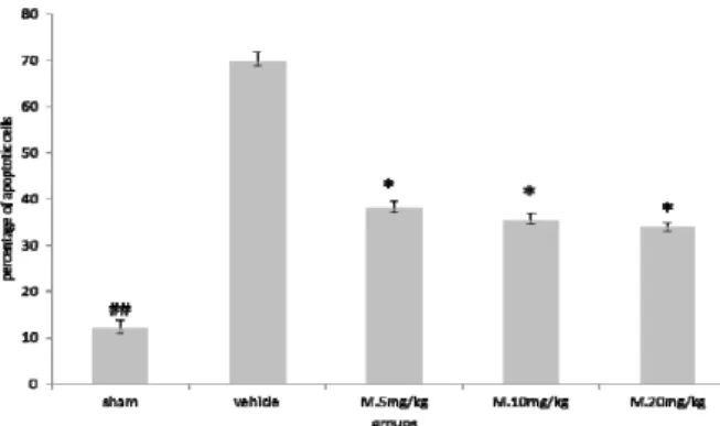
Figure 1. Melatonin treatment reduced apoptosis of neurons
induced by TB). Results are expressed as mean±SEM and data were analyzed by one‐way ANOVA followed by Tukey Kramer multiple comparisons test
* Significantly different from the vehicle group P< .
## Significantly different from the vehicle group P< .
dark brown color revealed damage to neuronal
perikarya .
Cellcounting
Five coronal sections were chosen for cell counting between the Bregma AP ─ . and ─ . mm . The number of surviving and degenerating neurons was counted in five random and non‐overlapping regions
at a magnification of X . Percent of neuronal
damage was calculated based on the ratio of the number of degenerated neurons to that of both surviving and degenerated neurons. )n addition, the number of astrocytes were counted in five random and separated regions of the cortex at the level between
Bregma AP‐ . and + . .
Statisticalanalyses
The data were expressed as mean±SEM. One way
ANOVA and Tukey‐Kramer multiple posthoc test were
used to evaluate the differences between the groups, and P< . was designated for statistical significance.
Results
Apoptoticcellsinbraincortex
(istological studies in the vehicle group animals showed severe morphological changes including extensively dark pyknotic nuclei and shrunken cytoplasm in the brain cortex following TB). (owever, in melatonin treatment groups, the severity of
Table 1. The effect of melatonin on cortical neuronal death
following TB)
DC/CC ₓ oo *** DC** CC* Groups
. ## Sham
. Vehicle
. # M. mg/kg
. # M. mg/kg
. # M. mg/kg
*counted cells
**Average number of the degenerated cells DC *** Average percentage of dead cells
# P< . for melatonin , and mg/kg ## P< . sham vs Vehicle
Figure2. )mmunohistochemical analysis of TUNEL in brain cortex
of rat. Arrows show the apoptotic cells. Bar= µm
degenerative changes in the nucleus and cytoplasm was lower than those in the vehicle and sham groups
P< . Table . )n addition, there were few TUNEL
positive cells of brain cortex in the sham group, while many TUNEL positive cells were detected in the vehicle group Figure l .
The results of neuronal cell counting showed significant difference between TB)+ mg/kg melatonin
P< . , TB)+ mg/kg melatonin P< . , and
TB)+ mg/kg melatonin P< . groups, compared
to the vehicle group Figure .
Activatedastrocytesnumbers
Our findings showed that TB) causes a dramatic increase in the number of activated astrocytes in brain cortex. But, melatonin treatment significantly decreased the number of GFAP positive astrocytes
P< . that were not dose dependent Figure
and .
Figure 3. Melatonin treatment reduced the number of glial
fibrillary acidic protein GFAP positive astrocytes. Results are expressed as mean±SEM, and data were analyzed by One‐way ANOVA followed by Tukey‐Kramer multiple comparisons test *Significantly different from the vehicle group P< .
Figure4. )mmunohistochemical analysis of glial fibrillary acidic protein GFAP positive cells in brain cortex of rat. Arrows show the GFAP
positive cells. Bar= µm
Discussion
The results of this study have shown the neuroprotective effect of melatonin after TB) in male rat. Our findings indicate that TB) causes a dramatic increase in neuronal cell death in brain tissue, and based on TUNEL assay, the percentage of apoptotic cells significantly decreased in the melatonin treatment groups. )t seems that melatonin plays a great role in antiapoptotic activities via the inhibition of intrinsic apoptotic pathways, as well as, the activation of several associated signal molecules in
various brain regions in TB) model . )n an
experimental study, it was evaluated that treatment
with melatonin and mg/kg, )P improved the
survival rate in a stroke model of mice .
Melatonin has been evaluated as effective in TB), through increasing superoxide dismutase SOD and
glutathione peroxidase GPx activities .
The result of the present study does not show any significant difference between three different doses of melatonin in our animal groups. A previous study has also shown that melatonin significantly attenuated neuronal cell death in hippocampal CA and CA regions and dentate gyrus of immature rats after head trauma, which was equally effective at doses of and
mg/kg . )n another experimental animal model,
melatonin treatment led to glial cell death reduction in white matter in mid‐gestation fetal sheep following
umbilical cord occlusion .
The anti‐inflammatory effect of melatonin has been introduced as a chief protective mechanism
against brain injury , . (owever, the
modulation behaviors of melatonin to suppress activated astrocytes have not been particularly investigated in a research with respect to TB). Astrocytes activation occurs in response to many CNS pathologies, such as trauma, tumor formation, and neurodegenerative disease. The process of astrocyte activation results in so‐called "reactive astrogliosis", which is a reaction with specific structural and functional characteristics including
hypertrophy of cellular process of astrocytes .
Up‐regulation of intermediate filament proteins, particularly GFAP by reactive astrocytes may be applied to distinguish reactive astrogliosis. (ence, the present study has focused on reactive
astrogliosis based on GFAP immunoreactivity .
)n fact, astrocytes, as the largest and the most abundant glial cells in CNS, may be the target of melatonin. We have demonstrated that the number of GFAP positive cells is reduced in the melatonin treatment groups, which indicates the alleviation of astrogliosis process induced by TB). Melatonin has been shown to modify immunity and the stress response, and has an antioxidant activity scavenge
free radicals . Melatonin has a greater
antioxidant effect than vitamin E, and can indirectly increase the expression of other endogenous
antioxidant enzymes . Ananth and colleagues
demonstrated that domoic acid‐induced astrocyte activation is attenuated significantly by the intravenous administration of melatonin in the
hippocampus of adult rats . Also, studies have
in the acute post‐traumatic period, impairment of
the blood–brain barrier BBB has a major role and allows the entry of circulating lymphocytes, monocytes, and neutrophils into the injured site, which directly affects inflammation and glial cell reactivities that may lead to neural cell death , .
)n addition, it was shown that melatonin administration significantly up regulated STAT DNA‐ binding activity, and led to the reduction of pro‐ inflammatory cytokines such as )L‐ , and Nitric Oxide
Synthase NOS . (owever, the modulation behaviors
of melatonin to astrocyte,an important source of pro‐
inflammatory cytokines, have not yet been extensively explored. Proliferation of astrocytes and their migration toward the lesion site can be recorded by GFAP
immunoreactivity .
Also, we showed the importance of the therapeutic time‐window after the initial insult of TB). )t is critical
to start interventions as soon as possible upon TB) .
)n our experiment, the treatment started one hr after TB) and continued until hr, which is a critical time period for histological examination.
)t would be necessary to mention that there are several limitations to our study. Firstly, the mechanisms by which melatonin reduces GFAP positive astrocytes are not explained. )n addition, this study only reveals the neuroprotective effects of melatonin after a short time period following TB). Therefore, further investigations need to be performed to clarify the molecular mechanisem of melatonin in suppression of astrocyte reactivity.
Conclusion
Findings support that melatonin is highly protective of both neurons and glial cells following TB) in our animal model. )n general, the present findings suggest that melatonin treatment of TB) markedly diminish astrocyte reactivity astrogliosis , as well as the number of apoptotic neurons in brain cortex of traumatic brain injury in rat model.
Acknowledgment
The present study was financially supported by Neuroscience Research Center, )nstitute of Neuro‐ pharmacology, Kerman University of Medical Sciences, Kerman, )ran. This work was part of MSc thesis of Mr Babaee at the Department of Anatomical Sciences. The authors have no conflicts of interest to declare.
References
.Chua KSG NY, Yap SGM, Bok C. A brief review of traumatic brain injury rehabilitation. Ann Acad Med
Singapore ; : ‐ .
. Khorasani‐Zavareh D, Mohammadi R, Khankeh (R, Laflamme L, Bikmoradi A, (aglund BJ. The requirements and challenges in preventing of road traffic injury in )ran. A qualitative study. BMC Public (ealth ; : .
. Aghakhani N, Azami M, Jasemi M, Khoshsima M, Eghtedar S, Rahbar N. Epidemiology of traumatic brain injury in urmia, iran. )ran Red Crescent Med J
; : .
. Johnson VE, Stewart JE, Begbie FD, Trojanowski JQ, Smith D(, Stewart W. )nflammation and white matter degeneration persist for years after a single traumatic brain injury. Brain ; : ‐ .
. Ding K, Wang (, Xu J, Li T, Zhang L, Ding Y, etal.
Melatonin stimulates antioxidant enzymes and reduces oxidative stress in experimental traumatic brain injury: the Nrf ‐ARE signaling pathway as a potential mechanism. Free Radic Biol Med ;
: ‐ .
. Bayir (, Kochanek PM, Clark RS. Traumatic brain injury in infants and children: mechanisms of secondary damage and treatment in the intensive care unit. Crit Care Clin ; : ‐ .
. Kunz A, Dirnagl U, Merqenthaler P. Acute pathophysiological processes after ischaemic and traumatic brain injury. Best Pract Res Clin
Anaesthesiol ; : ‐ .
. (all ED, Vaishnav RA, Mustafa AG. Antioxidant therapies for traumatic brain injury. Neurothera‐ peutics ; : ‐ .
. Reiter RJ. Melatonin: the chemical expression of darkness. Mol Cell Endocrinol ; :C ‐ .
. Claustrat B, Brun J, Chazot G. The basic physiology and pathophysiology of melatonin. Sleep Med Rev ; : ‐ .
. Seifman MA, Gomes K, Nguyen PN, Bailey M, Rosenfeld JV, Cooper DJ, etal. Measurement of serum
melatonin in intensive care unit patients: changes in traumatic brain injury, trauma, and medical conditions. Front Neurol ; : .
. Ahmadiasl N, Shokofeh B, Alireza A. Combination Antioxidant Effect of erythropoietin and melatonin on renal ischemia‐reperfusion injury in rats. )ran J Basic Med Sci ; : ‐ .
. Pineau ), Sun L, Bastien D, Lacroix S. Astrocytes initiate inflammation in the injured mouse spinal cord by promoting the entry of neutrophils and inflammatory monocytes in an )L‐ receptor/MyD ‐ dependent fashion. Brain Behav )mmun ; : ‐
.
. Barreto GE, Gonzalez J, Torres Y, Morales L. Astrocytic‐neuronal crosstalk: )mplications for neuroprotection from brain injury. Neurosci Res
; : ‐ .
. Cameron B, Landreth GE. )nflammation, microglia, and alzheimer's disease. Neurobiol Dis
; : ‐ .
. Candace LF, Bruce GL. Astroglia: )mportant mediators of traumatic brain injury. Prog Brain Res
; .
.Skaper SD, Floreani M, Ceccon M, Facci L, Giusti P. Excitotoxicity, oxidative stress, and the neuroprotective potential of melatonin. Ann N Y Acad
Sci ; : ‐ .
.Dehghan F, (adad MK, Asadikram G, Najafipour (, Shahrokhi N. Effect of melatonin on intracranial pressure and brain edema following traumatic brain injury: role of oxidative stresses. Arch Med
. A Marmarou, MAAE Foda, W Brink, J Campbell. A new model of diffuse brain injury in rats: Part ): Pathophysiology and biomechanics. J Neurosurg
; : ‐ .
. Keshavarzi Z, Khaksari M, Shahrokhi N. The effects of cyclooxygenase inhibitors on the gastric emptying and small intestine transit in the male rats following traumatic brain injury. )ran J Basic
Med Sci ; : ‐ .
. Koizumi S, Shigemoto‐Mogami Y, Nasu‐Tada K, Shinozaki Y, Ohsawa K, Tsuda M, etal.UDP acting at
P Y receptors is a mediator of microglial
phagocytosis. Nature ; : ‐ .
. Ding K, Wang (, Xu J, Li T, Zhang L, Ding Y, etal.
Melatonin stimulates antioxidant enzymes and reduces oxidative stress in experimental traumatic brain injury: the Nrf ‐ARE signaling pathway as a potential mechanism. Free Radic Biol Med ; : ‐ .
. Lee MY, Kuan Y(, Chen (Y, Chen TY, Chen ST, (uang CC, et al. )ntravenous administration of
melatonin reduces the intracerebral cellular inflammatory response following transient focal cerebral ischemia in rats. J Pineal Res ;
: ‐ .
. Ahmad molai Gh, Dabiri, Sh. Asadi karam GM, Shahrokhi N. Comparision of the effect of progestrone, allopregnanolone and gender on suppressing edema formation after traumatic brain injury in rat. Kerman Univ Med Sci ; : ‐ .
. Baydas G, Reiter RJ, Yasar A, Tuzcu M, Akdemir ), Nedzvetskii VS. Melatonin reduces glial reactivity in the hippocampus, cortex, and cerebellum of streptozotocin‐induced diabetic rats. Free Radic
Biol Med ; : ‐ .
. Li Y, Chopp M, Jiang N, Zhang ZG, Zaloga C. )nduction of DNA fragmentation after to minutes of focal cerebral ischemia in rats. Stroke
; : ‐ .
. Kabadi SV, Maher TJ. Posttreatment with uridine and melatonin following traumatic brain injury reduces edema in various brain regions in
rats. Ann N Y Acad Sci ; : – .
. Chern CM, Liao JF, Wang Y(, Shen YC. Melatonin ameliorates neural function by promoting endogenous neurogenesis through the MT melatonin receptor in ischemic‐stroke mice. Free Radic Biol Med ;
: – .
. Dehghan F, Khaksari (adad M, Asadikram G, Najafipour (, Shahrokhi N. Effect of melatonin on intracranial pressure and brain edema following traumatic brain injury: role of oxidative stresses.
Arch Med Res ; : ‐ .
. Ozdemir D, Uysal N, Gonenc S, Acikgoz O, Sonmez
A, Topcu A, etal. Effect of melatonin on brain oxidative
damage induced by traumatic brain injury in immature rats. Physiol Res ; : ‐ .
. Welin AK, Svedin P, Lapatto R, Sultan B, (agberg (, Gressens P, et al. Melatonin reduces
inflammation and cell death in white matter in the mid‐gestation fetal sheep following umbilical cord occlusion. Pediatr Res ; : ‐ .
. Keskin ), Kaplan S, Kalkan S, Sutcu M, Ulkay MB, Esener OB. Evaluation of neuroprotection by melatonin against adverse effects of prenatal exposure to a nonsteroidal anti‐inflammatory drug during peripheral nerve development. )nt J Dev Neurosci ; : ‐ .
. Tsai MC, Chen WJ, Tsai MS, Ching C(, Chuang J). Melatonin attenuates brain contusion‐induced oxidative insult, inactivation of signal transducers and activators of transcription , and upregulation of suppressor of cytokine signaling‐ in rats. J
Pineal Res ; : ‐ .
. Marmarou CR, Liang X, Abidi N(, Parveen S, Taya K, (enderson SC, etal. Selective vasopressin‐
a receptor antagonist prevents brain edema, reduces astrocytic cell swelling and GFAP, V aR and AQP expression after focal traumatic brain injury. Brain Res ; : ‐ .
. Pekny M, Nilsson M. Astrocyte activation and reactive gliosis. Glia ; : ‐ .
. Babaei‐Balderlou F, Zare S, (eidari R, Farrokhi F. Effects of melatonin and vitamin E on peripheral neuropathic pain in streptozotocin‐induced diabetic rats. )ran J Basic Med Sci ; : ‐ .
. Ananth C, Gopalakrishnakone P, Kaur C. Protective role of melatonin in domoic acid‐induced neuronal damage in the hippocampus of adult rats.
(ippocampus ; : ‐ .
. Abbott NJ, Patabendige AA, Dolman DE, Yusof SR, Begley DJ. Structure and function of the blood‐ brain barrier. Neurobiol Dis ; : ‐ .
. Ziebell JM, Morganti‐Kossmann MC. )nvolvement of pro‐ and anti‐inflammatory cytokines and chemokines in the pathophysiology of traumatic brain injury. Neurotherapeutics ;
: ‐ .
. Domowicz MS, (enry JG, Wadlington N, Navarro A, Kraig RP. Astrocyte precursor response to embryonic brain injury. Brain Res ; : ‐ .
. Ding K, Wang (, Xu J, Lu X, Zhang L, Zhu L. Melatonin reduced microglial activation and alleviated neuroinflammation induced neuron degeneration in experimental traumatic brain injury: Possible involvement of mTOR pathway. Neurochem )nt ;