Decompressive craniectomy in severe traumatic
brain injury: prognostic factors and complications
INTRODUCTION
Severe traumatic brain injury (TBI) constitutes one of the most frequent causes of intensive care unit (ICU) admissions in our country and in the world. TBI is the most common cause of death in those younger than 45 years, with a current global mortality of 39%. It is also a cause of disability in survivors, carrying a signiicant loss of potential years of active life and very high socio-economic costs for modern society.(1,2)
In serious neurological lesions of traumatic origin, increased intracranial pressure (ICP) is one of the main factors associated with a poor outcome, which is the main cause of preventable death.(3,4) Refractory intracranial hypertension Pedro Grille1,2, Nicolas Tommasino3,4
1. Universidad de la Republica Uruguay - UDELAR - Montevideo - Uruguay. 2. Intensive Care Unit, Hospital Maciel, Administración de los Servicios de Salud del Estado - ASSE - Montevideo, Uruguay. 3. Instituto Nacional de Donación y
Transplantes - INDT, Universidad de la Republica Uruguay - UDELAR - Montevideo - Uruguay 4. Intensive Care Unit, Hospital Español “Juan José Crottoggini”, Administración de los Servicios de Salud del Estado - ASSE - Montevideo, Uruguay.
Objective: To analyze the clinical characteristics, complications and factors associated with the prognosis of severe traumatic brain injury among patients who undergo a decompressive craniectomy.
Methods: Retrospective study of
patients seen in an intensive care unit with severe traumatic brain injury in whom a decompressive craniectomy was performed between the years 2003 and 2012. Patients were followed until their discharge from the intensive care unit. heir clinical-tomographic characteristics, complications, and factors associated with prognosis (univariate and multivariate analysis) were analyzed.
Results: A total of 64 patients
were studied. Primary and lateral decompressive craniectomies were performed for the majority of patients. A high incidence of complications was found (78% neurological and 52% non-neurological). A total of 42 patients
Conflict of interest: None.
Submitted October 4, 2014 Accepted April 3, 2015
Corresponding author:
Pedro Grille Avenida Italia, 7.035
CP: 11500 - Montevideo, Uruguay E-mail: [email protected]
Responsible editor: Flávia Ribeiro Machado
Craniectomía descompresiva en el trauma encefalocraneano
grave: factores pronósticos y complicaciones
ABSTRACT
Keywords: Craniocerebral trauma/complications; Brain injuries; Decompressive craniectomy/adverse efects; Intracranial hypertension/ etiology
(66%) presented poor outcomes, and 22 (34%) had good neurological outcomes. Of the patients who survived, 61% had good neurological outcomes. In the univariate analysis, the factors signiicantly associated with poor neurological outcome were post- decompressive craniectomy intracranial hypertension, greater severity and worse neurological state at admission. In the multivariate analysis, only post-craniectomy intracranial hypertension was signiicantly associated with a poor outcome.
Conclusion: his study involved a
very severe and diicult to manage group of patients with high morbimortality. Intracranial hypertension was a main factor of poor outcome in this population.
(ICH), deined as that which cannot be controlled with irst-tier therapeutic measures, presents in between 10 and 15% of patients.(5,6) For the treatment of ICH, there is unanimous agreement between the diferent authors and guides regarding what irst-tier measures should be implemented. his is not the case with the measures classiied as second-tier, for which diferent levels of evidence and discordant opinions among experts exist on the subject.(7)
In this context, decompressive craniectomy (DC) seems to be an eicient therapeutic strategy in some situations.(8-10) However, a class I level of evidence to support its utilization in adult patients currently does not exist, as this procedure is not without complications, some of which result in therapeutic challenges for the treating medical team.(11-13)
he objective of this study was to analyze the clinical characteristics, complications and factors associated with the prognosis of patients with severe TBI in which DC was performed.
METHODS
A retrospective study of all consecutively attended patients in the ICU of the Hospital Maciel (Administración de los Servicios de Salud del Estado, ASSE, Uruguay) with severe TBI who underwent DC was conducted between the years 2003 and 2012. he ICU is polyvalent with 20 beds and a high prevalence of neuro-critical patients and other patients referred to neurosurgery through our country’s Public Health system. his study was evaluated and approved by the Hospital Maciel (Ethical Committee, ASSE, Montevideo - Uruguay. An informed consent was presented to and signed by the relative responsible for the patient, where the descriptive and non-interventional nature of the study was explained. Patients were assured of conidentiality in the management of their medical history data.
Patients were followed until their discharge from the ICU. he variables obtained were clinical state using the Glasgow coma scale (GCS), physiological severity using the Simpliied Acute Physiology Score II (SAPS II), Marshall computed tomographic (CT) classiication, technical characteristics of the DC, complications, factors associated with mortality and neurological outcome upon discharge from the ICU using the Glasgow Outcome Scale (GOS).(14-17)
he clinical outcomes of patients were analyzed as dichotomous variables: good outcome (without disability or with light disability, deined by GOS scores of 4
or 5) and poor outcome (death, vegetative state or severe disability, deined by GOS scores of 1, 2 or 3, respectively).
Severe TBI was deined as a patient who sufers from encephalo-cranial trauma with a GCS score equal to or less than 8 after initial review or with an initial GCS score greater than 8 but which requires neurosurgery for the evacuation of a space-occupying intra-cranial lesion.(6)
he following parameters were deined: ICH as ICP greater than 20mmHg; shock as mean blood pressure lower than 70mmHg and/or clinical signs of peripheral hypoperfusion or lactatemia greater than 2mM; and dysnatremia such as a natremia lower than 135meq/L (hyponatremia) or greater than 150meq/L (hypernatremia). he following were included within post-surgical central nervous system (CNS) infections: ventriculitis, meningitis, subdural empyema or cerebral abscesses. External brain herniation was considered to be a cerebral protrusion of more than 1.5cm through the bony defect (measured by the radiologist); subdural collection-a hypodense collection greater than 1cm; and hydrocephalus-a dilatation of the ventricular system that is accompanied by signs or clinical symptoms requiring
treatment.(13,18) Ventilator-associated pneumonia was
deined as the association of fever or hyperleukocytosis, purulent tracheobronchitis, new and persistent chest x-ray images and cultures of positive tracheal secretions or bronchial alveolar lavages greater than 104 colony-forming units (CFU)/mL. Severe sepsis was deined as the presence of an infectious focus associated with systemic inlammatory syndrome and multiple organ failure.(19)
Refractory IHC was deined as an ICP greater than 20 - 25mmHg that was maintained for at least 30 minutes and did not respond to irst-tier therapeutic measures (intraventricular drainage of cerebrospinal luid (CSF) if possible, osmotherapy with 7.5% saline solution, moderate hyperventilation, and muscular relaxation).(5)
DC was classiied into 2 subtypes: 1) primary - that which the neurosurgeon performs after the evacuation of a subdural hematoma when the cerebral swelling conditions suggest the presentation of ICH in the workup, and also a situation in which the neurosurgeon decides on DC according to CT indings of difuse cerebral swelling prior to ICP monitoring and without having a hematoma to evacuate and 2) secondary - that which is performed for the treatment of refractory ICH during medical treatment.(11)
A multiple logistic regression was utilized to identify independent risk factors associated with patients’ outcome when discharged from the ICU. For hypothesis testing, p-values < 0.05 were considered statistically signiicant. he statistical program SPSS version 19.0 was utilized.
RESULTS
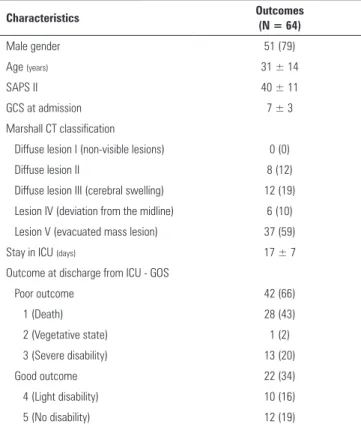
A total of 64 patients with severe TBI who underwent a DC were studied. he clinical characteristics of the population studied are shown in table 1. he prolonged stay in the ICU and days on mechanical ventilation (mean 12 ± 4 days) relect the degree of severity of our patients. Tracheostomies were performed for 28 patients (43%).
Marshall’s CT classiication of patients studied is summarized in table 1. As observed, subtypes III (difuse lesion with compression of basal cisterns) and V (evacuated space-occupying lesion) predominated. Among the space-occupying lesions, the most common were acute subdural hematoma (44%) and brain contusions (19%).
DC was performed as a primary intervention in 36 patients (56%) and as a secondary intervention in all other patients, which coincides with reports from other
authors.(20) he mean DC procedure time, measured
from the time of trauma, was 5 ± 2 hours for primary DC and 37 ± 28 hours for secondary DC. No statistically signiicant associations were detected between the mean DC procedure time and the patient outcome. he topography of the DC was lateral in 51 cases (81%) and bifrontal in 13 (19%).
ICP was measured in 58 patients (89%). In patients in which secondary DC was performed, post-DC ICP values were signiicantly lower than pre-DC values, when each patient was compared to himself (mean diference of 14 ± 4mmHg, with p = 0.021). A total of 27 patients (46%) presented post-DC ICH. he incidence rates of post-decompressive ICH were similar for secondary and primary DCs.
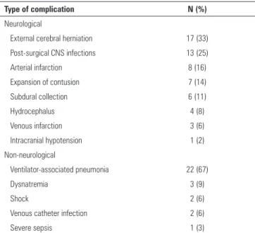
he global incidence of complications (neurological and non-neurological) was high at 90%, which has also been indicated in diferent clinical studies.(13,21) A total of 50 patients (78%) presented neurological complications, among which the most common were external cerebral herniation through the decompressive procedure, infections of the post-surgical CNS, cerebral infarction, expansion of a hemorrhagic contusion, subdural collections and hydrocephalus. he neurological complications observed and their incidence rates are shown in table 2.
A total of 33 patients (52%) presented non-neurological complications, with ventilator-associated pneumonia being the most frequent: it was present in more than two-thirds of patients. Dysnatremia, shock, severe sepsis and catheter-associated infections were common (Table 2).
he overall mortality in the study was 43% (27/64). Of the patients who survived, 14 (38%) were discharged from the ICU with severe neurological lesions (GOS 2 or 3), while 22 (62%) had good neurological outcomes (GOS 4 or 5). Of all patients studied, 22 (34%) had good neurological outcomes and were discharged from the ICU without neurological lesions or with light lesions. In table 1, the neurological outcomes of patients being discharged from the unit are summarized using the GOS reference scale.
Table 1 - Clinical characteristics of the study population
Characteristics Outcomes
(N = 64)
Male gender 51 (79)
Age (years) 31 ± 14
SAPS II 40 ± 11
GCS at admission 7 ± 3
Marshall CT classification
Diffuse lesion I (non-visible lesions) 0 (0)
Diffuse lesion II 8 (12)
Diffuse lesion III (cerebral swelling) 12 (19)
Lesion IV (deviation from the midline) 6 (10)
Lesion V (evacuated mass lesion) 37 (59)
Stay in ICU (days) 17 ± 7
Outcome at discharge from ICU - GOS
Poor outcome 42 (66)
1 (Death) 28 (43)
2 (Vegetative state) 1 (2)
3 (Severe disability) 13 (20)
Good outcome 22 (34)
4 (Light disability) 10 (16)
5 (No disability) 12 (19)
SAPS II - Simplified Acute Physiology Score II; GCS - Glasgow Coma Scale; CT - computed tomographic; GOS - Glasgow Outcome score; ICU - intensive care unit. Results are expressed as means ± standard deviations, numbers and percentages.
DISCUSSION
Historically, the removal of diferent parts of the skull has been utilized in the management of severe TBI after the irst reports of this surgical technique directed at controlling ICH were published by Kocher and Cushing.(22,23) Although this surgical procedure does not have any efect on primary brain damage, it can reduce the deleterious consequences of secondary lesions, such as the elevation of ICP and cerebral displacements or distortions.
Since the nineties, advances in imaging diagnostics and the neuro-intensive management of severe TBI have been able to revive interest in the utilization of DC for this condition. However, controversial aspects of this technique exist (precise indications, timing and long-term functional results), as does a growing need to study the complications and costs associated with the
DC procedure.(12,20,24,25) Although DC is known to be
a simple surgical technique, complications commonly occur, sometimes with signiicant clinical impacts on patient outcome.(26) Some complications are inherent in the physiological process that follows the removal of an important part of the cranial vault, which determines alterations in the dynamics of CSF circulation and
cerebral blood low.(27,28) here are also complications
linked to cranioplasty and to the lesions caused by severe TBI itself.(21,29,30)
Until now, there has been no class I clinical evidence related to DC for the management of refractory intracranial hypertension in severe TBI in adults.(9,11,12) It should also be emphasized that no evidence exists for the execution of primary DC.(31-33) However, DC seems to have a place in the management of patients with refractory ICH, which perhaps can be shown by the European study
RESCUE-ICP, currently in development.(34)
Table 2 - Complications in the study population
Type of complication N (%)
Neurological
External cerebral herniation 17 (33)
Post-surgical CNS infections 13 (25)
Arterial infarction 8 (16)
Expansion of contusion 7 (14)
Subdural collection 6 (11)
Hydrocephalus 4 (8)
Venous infarction 3 (6)
Intracranial hypotension 1 (2)
Non-neurological
Ventilator-associated pneumonia 22 (67)
Dysnatremia 3 (9)
Shock 2 (6)
Venous catheter infection 2 (6)
Severe sepsis 1 (3)
CNS - Central nervous system.
Regarding the factors associated with poor outcome, in the univariate analysis, it was found that the higher the SAPS II score, the lower the GCS at admission and the presence of post-DC intracranial hypertension. hese factors were signiicantly associated with poor outcome (death or severe disability), with p-values of 0.024, 0.01 and 0.001, respectively. In the multivariate analysis, only the presence of post-DC intracranial hypertension was signiicantly associated with poor neurological outcome (Table 3).
We emphasize the absence of a statistically signiicant association between the presence of complications and neurological outcome. Likewise, we did not detect any association between the appraisal criteria analyzed and the location or type of DC (Table 3).
Table 3 - Factors associated with poor neurological outcome
Associated factors Good outcome N = 22
Poor outcome N = 42
Univariate analysis p value
Multivariate analysis p value
Age 32 ± 2 30 ± 2 ns ns
SAPS II 38 ± 2 41 ± 3 0.024 ns
GCS at admission 8 ± 3 6 ± 1 0.01 ns
Primary/secondary DC 13/9 23/19 ns ns
Lateral/bifrontal 18/4 34/8 ns ns
Post-decompressive ICH 3 (14) 23 (55) 0.001 0.003
Neurological complications 15 (68) 36 (85) ns ns
Non-neurological complications 13 (59) 20 (48) ns ns
Our study constitutes the greatest number of patients reported at the present time in our setting. A high index of primary DC (56%) was noted in the cases analyzed. his practice has often been utilized by neurosurgeons in our setting in recent years and is based on the acting surgeon’s intraoperatory decision. his decision is often made according to the cerebral characteristics indings at the time, and at times, a pre-operatory decision is made according to the initial CT indings, as commented. In this sense, DC can be a strategy for avoiding the development of ICH as an outcome. However, it is an aggressive therapeutic measure, there is only a small body of evidence for it, and it is not free of risk for the patient.(32,33)
here was also a high incidence of post-DC ICH. he presence of post-decompressive ICH, a factor linked in part to surgical technique, plays a predominant role in the mortality of these patients, which was signiicantly associated with poor neurological outcome in the multivariate study.(35) Given the retrospective character of our study, it was not possible to measure the size of the DC with precision, knowing that the majority of authors agree that it should extend at least 12cm and should include the base of the temporal bone to consider it to be of adequate size. his unmeasured size could have been too small, which can be a factor that explains the high incidence of neurological complications. For example, this situation could be the case with external cerebral herniation and post-DC ICH, thus contributing to the negative outcomes in our patients.
he low incidence of hydrocephalus in our study is also noteworthy (8%), which is perhaps due to underdiagnosis
or to the utilization of diferent diagnostic criteria according to international references.(36,37)
In our study, we were unable to show a statistically signiicant association between the presence of complications (neurological or systemic) or the type of DC (primary or secondary) in relation to mortality, which can be explained in part by the small number in our sample.
Our study presents several limitations. It is a descriptive, retrospective study with a relatively small number of patients, conducted in a single center, which reduces statistical power and adds selection bias. Our study population is heterogeneous in terms of their clinical characteristics, indications and opportunity for DC, and technique used, which limits the precision of our results. As mentioned previously, the inability to measure the size of the DC does not allow us to evaluate the impact of this factor on the clinical outcomes of our patients. Finally, clinical results on discharge from the ICU were measured, and no long-term follow-up, such as at 3 to 6 months, was performed.
CONCLUSION
his group of very severe patients was analyzed for systemic and neurological factors, showing very severe physiological scores at admission and high mortality. Post-decompressive craniectomy intracranial hypertension, which can be due in part to surgical factors, was the main factor associated with poor outcome. his study demonstrates the complexity in managing this type of patient in our setting and the need for a protocol-driven and multidisciplinary treatment with the objective of improving patient prognosis.
Objetivo: Análisis de las características clínicas, las
complicaciones y los factores asociados al pronóstico de los pacientes con trauma encefalocraneano grave en los que se realizó craniectomía descompresiva.
Métodos: Estudio retrospectivo de los pacientes asistidos en una Unidad de Cuidados Intensivos, con trauma encefalocraneano grave en los que se realizó craniectomía descompresiva, entre los años 2003 y 2012. Se siguieron los pacientes hasta el egreso de la unidad de cuidados intensivos, analizándose sus características clínico-tomográicas, las complicaciones y los factores asociados al pronóstico (análisis uni y multivariado).
Resultados: Se estudiaron 64 pacientes. Se realizó
craniectomía descompresiva primaria y lateral en la mayoría de los pacientes. Se halló una alta incidencia de complicaciones (78% neurológicas y 52% no neurológicas). 42 pacientes (66%)
presentaron mala evolución y 22 (34%) tuvieron una buena evolución neurológica. De los pacientes que sobrevivieron, el 61% tuvo una buena evolución neurológica. En el análisis univariado, los factores asociados signiicativamente con mala evolución neurológica fueron: la hipertensión intracraneana post-craniectomía descompresiva, la mayor gravedad y el peor estado neurológico al ingreso. En el análisis multivariado, solo la hipertensión intracraneana post-craniectomía descompresiva se asoció signiicativamente con mala evolución.
Conclusión: Se trata de un grupo de pacientes muy grave, de difícil manejo, con elevada morbimortalidad, donde la hipertensión intracraneana es un factor principal de mala evolución.
RESUMEN
REFERENCES
1. Rosenfeld JV, Maas AI, Bragge P, Morganti-Kosmann MC, Manley GT, Gruen RL. Early management of severe traumatic brain injury. Lancet. 2012;380(9847):1088-98.
2. Faul M, Wald M, Rutland-Brown W, Sullivent EE, Sattin RW. Using a cost-benefit analysis to estimate outcomes of a clinical treatment guideline: testing the Brain Trauma Foundation guidelines for the treatment of severe traumatic brain injury. J Trauma. 2007;63(6):1271-8.
3. Juul N, Morris GF, Marshall SB, Marshall LF. Intracranial hypertension and cerebral perfusion pressure: influence on neurological deterioration and outcome in severe head injury. The Executive Committee of the International Selfotel Trial. J Neurosurg. 2000;92(1):1-6.
4. Miller JD, Becker DP. Secondary insults to the injured brain. J R Coll Surg Edinb. 1982;27(5):292-8.
5. Sahuquillo J, Biestro A, Mena MP, Amorós S, Lung M, Poca MA, et al. [First tier measures in the treatment of intracranial hypertension in the patient with severe craniocerebral trauma. Proposal and justification of a protocol]. Neurocirugia (Astur). 2002;13(2):78-100. Spanish.
6. Brain Trauma Foundation; American Association of Neurological Surgeons; Congress of Neurological Surgeons; Joint Section on Neurotrauma and Critical Care, AANS/CNS, Bratton SL, Chestnut RM, Ghajar J, McConnell Hammond FF, Harris OA, Hartl R, et al. Guidelines for the management of severe traumatic brain injury. J Neurotrauma. 2007;24: Suppl 1:S1-95. Erratum in: J Neurotrauma. 2008;25(3):276-8.
7. Stocchetti N, Zanaboni C, Colombo A, Citerio G, Beretta L, Ghisoni L, et al. Refractory intracranial hypertension and “second-tier” therapies in traumatic brain injury. Intensive Care Med. 2008;34(3):461-7.
8. Kolias AG, Kirkpatrick PJ, Hutchinson PJ. Decompressive craniectomy: past, present and future. Nat Rev Neurol. 2013;9(7):405-15.
9. Sahuquillo J, Martínez-Ricarte F, Poca MA. Decompressive craniectomy in traumatic brain injury after the DECRA trial. Where do we stand? Curr Opin Crit Care. 2013;19(2):101-6.
10. Vashu R, Sohail A. Decompressive craniectomy is indispensible in the management of severe traumatic brain injury. Acta Neurochir (Wien). 2011;153(10):2065-6.
11. Sahuquillo J, Arikan F. Decompressive craniectomy for the treatment of refractory high intracranial pressure in traumatic brain injury. Cochrane Database Syst Rev. 2006;(1):CD003983. Review.
12. Cooper DJ, Rosenfeld JV, Murray L, Arabi YM, Davies AR, D’Urso P, Kossmann T, Ponsford J, Seppelt I, Reilly P, Wolfe R; DECRA Trial Investigators; Australian and New Zealand Intensive Care Society Clinical Trials Group. Decompressive craniectomy in diffuse traumatic brain injury. N Engl J Med. 2011;364(16):1493-502.
13. Stiver SI. Complications of decompressive craniectomy for traumatic brain injury. Neurosurg Focus. 2009;26(6):E7. Review.
14. Le Galle JR, Lemeshow S, Saulnier F. A new Simplified Acute Physiology Score (SAPS II) based on a European/North American multicenter study. JAMA. 1993;270(24):2957-63. Erratum in: JAMA 1994;271(17):1321. 15. Teasdale G, Jennet B. Assessment of coma and impaired consciousness.
A practical scale. Lancet. 1974;2(7872):81-4.
16. Teasdale GM, Pettigrew LE, Wilson JT, Murray G, Jennett B. Analyzing outcome of treatment of severe head injury: a review and update on advancing the use of the Glasgow Outcome Scale. J Neurotrauma. 1998;15(8):587-97.
17. Marshall LF, Marshall SB, Klauber MR, van Berkum Clark M. A new classification of head injury based on computerized tomography. J Neurosurg. 1991;75 (1 Suppl):S14-20.
18. Honeybul S. Complications of decompressive craniectomy for head injury. J Clin Neurosci. 2010;17(4):430-5.
19. Dellinger RP, Levy MM, Rhodes A, Annane D, Gerlach H, Opal SM, Sevransky JE, Sprung CL, Douglas IS, Jaeschke R, Osborn TM, Nunnally ME, Townsend SR, Reinhart K, Kleinpell RM, Angus DC, Deutschman CS, Machado FR, Rubenfeld GD, Webb SA, Beale RJ, Vincent JL, Moreno R;
Surviving Sepsis Campaign Guidelines Committee including the Pediatric Subgroup. Surviving sepsis campaign: international guidelines for management of severe sepsis and septic shock: 2012. Crit Care Med. 2013;41(2):580-637.
20. Tagliaferri F, Zani G, Iaccarino C, Ferro S, Ridolfi L, Basaglia N, et al. Decompressive craniectomies, facts and fiction: a retrospective analysis of 526 cases. Acta Neurochir (Wien). 2012;154(5):919-26.
21. Honeybul S, Ho KM, Lind CR, Gillett GR. Observed versus predicted outcome for decompressive craniectomy: a population-based study. J. Neurotrauma. 2010;27(7):1225-32.
22. Kocher T. [Hirnerschütterung, Hirndruck und chirurgische Eingriffe bei Hirnkrankheitende]. In: Hölder A, editor. Die Therapie des hirndruckes. Vienna: A. Hölder; 1901. p. 262-6. Germany
23. Cushing H. The establishment of cerebral hernia as a decompressive measure for inaccessible brain tumors: with the description of intramuscular methods of making the bone defect in temporal and occipital regions. Surg Gynecol Obstet. 1905;1:297-314.
24. Timofeev I, Hutchinson PJ. Outcome after surgical decompression of severe traumatic brain injury. Injury. 2006;37(12):1125-32.
25. Hutchinson PJ, Timofeev I, Kolias AG, Corteen EA, Czosnyka M, Menon DK, et al. Decompressive craniectomy for traumatic brain injury: the jury is still out. Br J Neurosurg. 2011;25(3):441-2.
26. Flint AC, Manley GT, Gean AD, Hemphill JC 3rd, Rosenthal G. Post-operative expansion of hemorrhagic contusions after unilateral decompressive hemicraniectomy in severe traumatic brain injury. J Neurotrauma. 2008;25(5):503-12.
27. Fodstad H, Love JA, Ekstedt J, Fridén H, Liliequist B. Effect of cranioplasty on cerebrospinal fluid hydrodynamics in patients with the syndrome of the trephined. Acta Neurochir (Wien). 1984;70(1-2):21-30.
28. Yamakami I, Yamaura A. Effects of decompressive craniectomy on regional cerebral blood flow in severe head trauma patients. Neurol Med Chir (Tokyo). 1993;33(9):616-20.
29. Schuss P, Vatter H, Marquardt G, Imöhl L, Ulrich CT, Seifert V, et al. Cranioplasty after decompressive craniectomy: the effect of timing on postoperative complications. J Neurotrauma. 2012;29(6):1090-5. 30. Yang XF, Wen L, Shen F, Li G, Lou R, Liu WG, et al. Surgical complications
secondary to decompressive craniectomy in patients with a head injury: a series of 108 consecutive cases. Acta Neurochir (Wien). 2008;150(12):1241-7; discussion 1248.
31. Chen SH, Chen Y, Fang WK, Huang DW, Huang KC, Tseng SH. Comparison of craniotomy and decompressive craniectomy in severely head-injured patients with acute subdural hematoma. J Trauma. 2011;71(6):1632-6. 32. Kolias AG, Belli A, Li LM, Timofeev I, Corteen EA, Santarius T, et al. Primary
decompressive craniectomy for acute subdural haematomas: results of an international survey. Acta Neurochir (Wien). 2012;154(9):1563-5. 33. Li LM, Kolias AG, Guilfoyle MR, Timofeev I, Corteen EA, Pickard JD,
et al. Outcome following evacuation of acute subdural haematomas: a comparison of craniotomy with decompressive craniectomy. Acta Neurochir (Wien). 2012;154(9):1555-61.
34. Hutchinson PJ, Corteen E, Czosnyka M, Mendelow AD, Menon DK, Mitchell P, et al. Decompressive craniectomy in traumatic brain injury: the randomized multicenter RESCUEicp study (www.RESCUEicp.com). Acta Neurochir Suppl. 2006;96:17-20.
35. Bor-Seng-Shu E, Figueiredo EG, Amorim RL, Teixeira MJ, Valbuza JS, de Oliveira MM, et al. Decompressive craniectomy: a meta-analysis of influences on intracranial pressure and cerebral perfusion pressure in the treatment of traumatic brain injury. J Neurosurg. 2012;117(3):589-96. 36. De Bonis P, Pompucci A, Mangiola A, Rigante L, Anile C. Post-traumatic
hydrocephalus after decompressive craniectomy: an underestimated risk factor. J Neurotrauma. 2010;27(11):1965-70.