against
Francisella novicida In Vivo
and
In Vitro
and
Show GlpT Independent Efficacy
Elizabeth S. McKenney1, Michelle Sargent2, Hameed Khan2, Eugene Uh3, Emily R. Jackson3, Ge´raldine San Jose3, Robin D. Couch2,4, Cynthia S. Dowd3, Monique L. van Hoek1,4*
1School of Systems Biology, George Mason University, Manassas, Virginia, United States of America,2Department of Chemistry and Biochemistry, George Mason University, Manassas, Virginia, United States of America,3Department of Chemistry, George Washington University, Washington, D.C., United States of America, 4National Center for Biodefense and Infectious Diseases, George Mason University, Manassas, Virginia, United States of America
Abstract
Bacteria, plants, and algae produce isoprenoids through the methylerythritol phosphate (MEP) pathway, an attractive pathway for antimicrobial drug development as it is present in prokaryotes and some lower eukaryotes but absent from human cells. The first committed step of the MEP pathway is catalyzed by 1-deoxy-D-xylulose 5-phosphate reductoisomerase (DXR/MEP synthase). MEP pathway genes have been identified in many biothreat agents, including Francisella,Brucella,Bacillus,Burkholderia, andYersinia. The importance of the MEP pathway toFrancisellais demonstrated by the fact that MEP pathway mutations are lethal. We have previously established that fosmidomycin inhibits purified MEP synthase (DXR) fromF. tularensisLVS. FR900098, the acetyl derivative of fosmidomycin, was found to inhibit the activity of purified DXR fromF. tularensisLVS (IC50= 230 nM). Fosmidomycin and FR900098 are effective against purified DXR from
Mycobacterium tuberculosisas well, but have no effect on whole cells because the compounds are too polar to penetrate the thick cell wall. Fosmidomycin requires the GlpT transporter to enter cells, and this is absent in some pathogens, includingM. tuberculosis. In this study, we have identified the GlpT homologs inF. novicidaand tested transposon insertion mutants of glpT. We showed that FR900098 also requires GlpT for full activity againstF. novicida. Thus, we synthesized several FR900098 prodrugs that have lipophilic groups to facilitate their passage through the bacterial cell wall and bypass the requirement for the GlpT transporter. One compound, that we termed ‘‘compound 1,’’ was found to have GlpT-independent antimicrobial activity. We tested the ability of this best performing prodrug to inhibitF. novicidaintracellular infection of eukaryotic cell lines and the caterpillarGalleria mellonellaas anin vivoinfection model. As a lipophilic GlpT-independent DXR inhibitor, compound 1 has the potential to be a broad-spectrum antibiotic, and should be effective against most MEP-dependent organisms.
Citation:McKenney ES, Sargent M, Khan H, Uh E, Jackson ER, et al. (2012) Lipophilic Prodrugs of FR900098 Are Antimicrobial againstFrancisella novicida In Vivo
andIn Vitroand Show GlpT Independent Efficacy. PLoS ONE 7(10): e38167. doi:10.1371/journal.pone.0038167 Editor:Fatah Kashanchi, George Mason University, United States of America
ReceivedNovember 22, 2011;AcceptedMay 4, 2012;PublishedOctober 15, 2012
Copyright:ß2012 McKenney et al. This is an open-access article distributed under the terms of the Creative Commons Attribution License, which permits unrestricted use, distribution, and reproduction in any medium, provided the original author and source are credited.
Funding:MVH was supported by the Defense Threat Reduction Agency HDTRA1-11-1-054. CSD was funded by National Institutes of Health/National Institute of Allergy and Infectious Diseases (NIH/NIAID) grant RC1AI086453 and the GWU Department of Chemistry. RDC was supported by U.S. Army Medical Research and Materiel Command W23RYX1291N601. The funders had no role in study design, data collection and analysis, decision to publish, or preparation of the manuscript.
Competing Interests:The authors have declared that no competing interests exist.
* E-mail: [email protected]
Introduction
1.1 Methylerythritol phosphate pathway
Isoprenoids are involved in many critical cellular functions. They participate in electron transport, signal transduction, and maintenance of cell wall and membrane structural integrity. All isoprenoids are formed through either the mevalonic acid (MVA) or the methylerythritol phosphate (MEP) pathways [1]. Plants, algae, and bacteria utilize the MEP pathway to generate isopentenyl pyrophosphate (IPP) and dimethylallyl pyrophosphate (DMAPP) from pyruvate and glyceraldehyde-3-phosphate [2]. The MVA pathway is the only pathway used by animals, making enzymes of the MEP pathway attractive targets for novel therapeutics [1].
The first committed step of the MEP pathway is catalyzed by 1-deoxy-D-xylulose 5-phosphate reductoisomerase (DXR or MEP synthase) [2]. DXR catalyzes the reaction that generates MEP
from 1-deoxy-D-xylulose 5-phosphate (DXP) (Figure 1A) [1]. MEP pathway genes have been identified in many biothreat agents, including Francisella, Brucella, Bacillus, Burkholderia, and Yersinia [1,3,4]. DXR has been cloned from many different bacteria, including Escherichia coli, Pseudomonas aeruginosa, and Francisella tularensis [1,2]. The importance of the MEP pathway to F. tularensis is demonstrated by the fact that MEP pathway mutations are lethal [5]. It has also been demonstrated that the DXR gene is essential forMycobacterium tuberculosis[6].
fosmidomycin against the malaria parasitein vitroandin vivo[10]. Two functional groups of these antibiotics are important for their binding efficacy and inhibition of DXR: the hydroxymate moiety that chelates with a divalent metal ion (Mn2+
, Mg2+
, or Co2+
) and a negatively charged phosphonate group [11]. Fosmidomycin and FR900098 are both effective antimalarial agents, but they are limited in their effect due to re-emergence of an active infection after completing treatment [9], and low bioavailability, likely due to low lipophilicity [10]. Fosmidomycin requires the glycerol-3-phosphate transporter (GlpT) to enter cells [12]. Both compounds are well tolerated in mice up to 300 mg/kg and demonstrate antimalarial efficacy following both intraperitoneal and oral administration [13].
We have previously established that fosmidomycin inhibits purified DXR fromF. tularensisLVS with half maximal activity of 247 nM [1]. This is comparable to its effect against DXR fromM. tuberculosis(310 nM) [14] and less active against the same enzyme fromE. coli(35 nM) [2]. Jawaidet al.suggest that the difference in concentration required for half-maximal activity may be due to structural differences of the DXR homologs [1].
1.2 Lipophilic FR900098 prodrugs
Fosmidomycin and FR900098 are effective against purified DXR fromM. tuberculosis[6,14,15] but have no effect on whole cells of this bacterium because the compounds are too polar to penetrate the thick cell wall [6]. In addition, fosmidomycin requires the GlpT transporter to enter cells, and this is absent in M. tuberculosis [14]. This data prompted us to question whether lipophilic analogs of fosmidomycin and FR900098 might better penetrate bacteria with thick cell walls and/or no GlpT transporter. Recently, Ortmann et al. generated a series of acyloxyalkyl ester prodrug derivatives of FR900098, including compound 1 (Figure 1B), which demonstrated improved in vivo antimalarial activity [10]. These compounds are considered prodrugs of FR900098, and are metabolized to FR900098 in bacteria [10]. We have shown that these analogs have
antimicro-bial activity against a broad range of bacteria [16] and may also be better at penetrating the cell membranes of eukaryotic cells, which is important for access to intracellular pathogens. For example, bothF. tularensisand M. tuberculosiscolonize host cells during the course of infection. We tested the ability of some of these compounds to inhibitF. novicidaintracellular infection, using both cultured eukaryotic cell lines, and the caterpillarGalleria mellonella as anin vivoinfection model.
1.3Francisella tularensis
F. tularensis is a highly infectious Gram-negative facultative intracellular bacterium. Inhalation of as few as ten organisms can cause disease in humans [17]. It is of particular interest due to its historical use as a bioweapon, and is on the Centers for Disease Control’s list of Category A select agents [18].F. tularensiscauses the disease tularemia in mammals, including humans, which can be spread via arthropod vectors, such as ticks [19], or by aerosol. F. tularensiscan cause a pneumonic disease if it is inhaled, but more commonly causes the ulceroglandular form of the disease that occurs via skin contact.F. tularensiscan replicate in many different cell types of mammalian hosts (for example, dendritic cells, neutrophils, hepatocytes, and lung epithelial cells), but macro-phages appear to be the main target of this bacterium [19,20]. There are four closely related subspecies ofF. tularensis, known as tularensis, holarctica, mediasiatica, and novicida [19]. F. novicida is a model organism of the more virulentF. tularensisspecies [21].F. novicida is attenuated for disease in humans, but can still cause disease in small mammals, such as mice [19].F. tularensisNIH B38 is classified as the type strain for F. tularensis tularensis, but is attenuated for virulence [22–24], and thus is a good model forF. tularensisSchu S4, the fully virulent strain.F. tularensisis of concern due to its historical use as a bioweapon in an aerosolized form [18]. Such an event could cause severe pulmonary disease in thousands of individuals and would impose a severe strain and high costs on the health care and public safety systems [20]. Prompt treatment would be important in decreasing the impact of Figure 1. The reaction catalyzed by DXR (MEP synthase) and compounds used in this study. A. MEP synthase pathway: DXR catalyzes the formation of methylerythritol phosphate (MEP) from 1-deoxy-D-xylulose 5-phosphate (DXP) in an NADPH-dependent mechanism with the formation of the intermediate 2-C-methyl-D-erythrose 4-phosphate. Modified from Jawaidet al.[1].B: The structures of the compounds used in this study.Shown are the structures for Fosmidomycin, FR900098, Compound 1, Compound 2 and Compound 3.
such an attack. The potential of engineered antibiotic resistant strains suggests that new classes of antibiotics with different modes of action from the standard antibiotics, such as ciprofloxacin, should be developed againstF. tularensis.
1.4 Identification of the GlpT homolog inFrancisella
The uptake of fosmidomycin into many bacteria is an active process dependent on the 12 transmembrane-spanning protein, glycerol-3-phosphate transporter, GlpT [12]. GlpT is a member of the organophosphate:phosphate antiporter family that is part of the major facilitator superfamily (MFS) [12]. E. coli mutants defective in theglpTgene are resistant to fosmidomycin [12].M. tuberculosis lacks a GlpT homolog, partially accounting for its resistance to fosmidomycin [4,6].Brucella acquires fosmidomycin sensitivity when it expressesE. coliGlpT [4]. We identified a gene in F. tularensis (FTT0725c) and in F. novicida (FTN_0636) as a potential GlpT homolog (Table 1), and transposon insertion mutants in this locus (Table 2) were tested for their ability to be inhibited by fosmidomycin and analogs.
1.5 Hypothesis
The aim of this research is to test the effectiveness of lipophilic FR900098 prodrugs against F. novicidaas potential platforms for novel antibiotic development. Our hypothesis is that these new compounds will be more effective at crossing biological mem-branes than FR900098 or fosmidomycin, and thus may be more effective antimicrobial compounds against Francisella. Thus, we determined the minimum inhibitory concentration (MIC) and EC50for fosmidomycin, FR900098, and compounds 1–3 against F. novicida. In addition, the in vitroinhibition of F. tularensis LVS DXR by FR900098 was determined in comparison to fosmido-mycin [1]. The ability of the lipophilic compounds to cross the Francisella membrane was examined using a glpT mutant of F. novicida, the transporter required for fosmidomycin activity in many bacteria. We then assessed the efficacy of fosmidomycin, FR900098, and compound 1 in treating intracellularF. novicidain A549 human alveolar epithelial cells and RAW264.7 mouse macrophages, as well as intracellularglpTmutants in A549 cells. The cytotoxicity of the compounds was determined by measuring the LDH released from the infected cells. Finally,G. mellonellawas used as a model system to test the efficacy of fosmidomycin, FR900098, and compound 1 againstF. novicidainfectionin vivo. As a lipophilic GlpT-independent DXR inhibitor, compound 1 has the potential to be a broad-spectrum antibiotic, and should be effective against many MEP-dependent organisms [16].
Results
2.1 Susceptibility ofF. novicida to fosmidomycin, FR900098, and analogs
2.1.1 MICs and EC50s of fosmidomycin, FR900098, and
lipophilic analogs. The susceptibility of F. novicida to the FR900098 analogs was determined in a 96-well plate assay. An initial screening of a panel of compounds demonstrated that compound 1 and compound 2 were the best inhibitors of the analogs tested (data not shown). Compound 3 did not effectively inhibit F. novicida, but its structure, along with the others, is included in Figure 1B for comparison.
The MIC values of fosmidomycin, FR900098, compound 1, and compound 2 were determined against F. novicida(Table 3). The MIC was best for fosmidomycin at 136mM. The MIC of FR900098 was 254mM. The MICs of compound 1 and compound 2 were 202mM and 1.1 mM, respectively. Thus, compound 1 had approximately 2-fold weaker activity compared with fosmidomycin, but was more active than FR900098.
These compounds were assessed for the number of surviving bacteria at increasing concentration of compound, from which we calculate the EC50, a value we use to compare activities of the different compounds. This value was determined for fosmidomy-cin, FR900098, compound 1, and compound 2. The EC50 of fosmidomycin against F. novicida was determined to be 3.6mM (Figure 2A). The EC50 of FR900098 was 23.2mM (Figure 2B). The EC50of compound 1 was 45.2mM (Figure 2C) and that of compound 2 was 481mM (Figure 2D). While compound 1 had a better MIC than FR900098, it had a slightly poorer EC50.
2.1.2 Inhibition of purified F. tularensis LVS DXR by FR900098. The IC50of FR900098 against the DXR ofE. coliis reported to be 62 nM [25]. The activity of FR900098 was determined by monitoring the enzyme-catalyzed oxidation of NADPH, and using a plot of enzyme fractional activity as a function of inhibitor concentration, as we previously reported, to determine the IC50(Table 4) [1]. The half maximal inhibition ofF. tularensisLVS DXR by fosmidomycin was previously determined to be 247 nM [1]. FR900098 was found to have an IC50 of 230 nM (Figure S1). Since compound 1 is metabolized to FR900098 in bacteria [10], we did not test the activity of compound 1 against DXR in vitro.DXR fromFrancisella novicida (FTN_1483),F. tularensisLVS (FTL_0534), andF. tularensisSchu S4 (FTT1574) share.99% homology (Figure S2). The highlight-ed differences are not in critical enzymatic residues, thus we conclude that DXR from all Francisella species will have similar sensitivities.
2.1.3 Susceptibility of F. novicida glpT mutants to fosmidomycin, FR900098, and lipophilic analogs. Fosmidomycin requires the glycerol-3-phosphate transporter (GlpT) to enter bacterial cells [6]. Bioinformatic analysis suggests that allFrancisellaspecies contain GlpT homologs (Table 1). One of the goals of this work was to test compounds that were more efficient at crossing biological membranes independent Table 1.GlpT homologs identified inFrancisella spp.
FrancisellaSpecies Locus Accession Number
F. tularensis Schu S4 FTT0725c YP_169738.1
F. tularensis LVS FTL_1510 YP_514159.1
F. novicida FTN_0636 YP_898283.1
F. mediasiatica FTM_1358 YP_001891977.1
F. philomiragia Fphi_0200 YP_001676919.1
doi:10.1371/journal.pone.0038167.t001
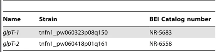
Table 2.F. novicida glpTtransposon insertion mutants used in this study.
Name Strain BEI Catalog number
glpT-1 tnfn1_pw060323p08q150 NR-5683
glpT-2 tnfn1_pw060418p01q161 NR-6558
Table 3.Inhibition of bacterial growth by selected compounds.
Compound Molecular Weight (g/mol) MIC (mM) EC50(mM)
EC5095% Confidence Interval (mM)
Wild-typeF. novicida
Fosmidomycin 183.10 g/mol 136mM 3.660.2mM 3.1–4.1mM
FR900098 196.12 g/mol 254mM 23.261.2mM 21.0–25.5mM
Compound 1 493.40 g/mol 202mM 45.263.7mM 38.4–53.2mM
Compound 2 365.45 g/mol 1094mM 481644mM 401–578mM
F. novicida glpTmutant
Fosmidomycin 183.10 g/mol .1 mM nd nd
FR900098 196.12 g/mol .1 mM nd nd
Compound 1 493.40 g/mol 200mM nd nd
Compound 2 365.45 g/mol .1 mM nd nd
MICs and EC50s of selected compounds against wild-type andglpTmutantF. novicidawere determined. (nd = not determined). doi:10.1371/journal.pone.0038167.t003
Figure 2. The antimicrobial effect of fosmidomycin, FR900098, compound 1 and compound 2 againstF. novicida.The EC50s (the concentration at which 50% of bacterial growth is inhibited) of fosmidomycin (A), FR900098 (B), compound 1 (C), and compound 2 (D) were determined as previously described. Briefly, serial dilutions of the antibiotic were performed in a 96-well plate format to a defined concentration of bacteria. The percent growth compared to wells with no antibiotic was graphed using GraphPad Prism 4.0. The EC50 of fosmidomycin was 3.660.2mM. The EC50of FR900098 was 23.261.2mM. The EC50of compound 1 was 45.263.7mM and the EC50of compound 2 was 481644mM.
of the GlpT transporter, presumably by increased penetration of the membrane due to increased lipophilicity. The data for the glpT-1mutant strain is shown here (Figure 3), and the data for the glpT-2mutant strain was very similar. Even at a concentration of 1 mM, fosmidomycin was not able to inhibit F. novicida glpT growth at all (Table 3). At this same concentration, FR900098 was only able to inhibit 50% of F. novicida glpT growth. These concentrations inhibited 100% of wild-type F. novicida growth. Since inhibition of bacterial growth was reduced against theglpT mutant, FR900098 is at least partially dependent on this transporter to enter the bacterial cell. It is possible that FR900098 uses another transporter system, accounting for its 50% inhibition of theglpTmutant. Alternatively, FR900098 may be slightly lipophilic and have some ability to cross the membrane independent of GlpT. Compound 2 demonstrated reduced bacteriostatic activity against the glpT mutant than wild-type F. novicida, suggesting that this analog may also be partially dependent on this transporter to cross the membrane, or a small amount of compound 2 is able to diffuse across the membrane. Both FR900098 and compound 2 are more lipophilic compared with fosmidomycin. In contrast, the MIC of compound 1 was the same for both wild-typeF. novicidaand theglpTmutant. This indicates that compound 1 entry into the bacteria is independent of the
transporter, and is most likely due to the increased lipophilicity of the acyloxyalkyl ester and its ability to cross the membrane.
2.2 Cell line infections and treatments
2.2.1 Infection and treatment of human type II alveolar epithelial (A549) cells. When tested at an equal concentration (250mM), all three antibiotics significantly inhibited the growth of intracellular F. novicida (p,0.05) (Figure 4A). Untreated cells contained 3.8860.726103CFU/well of F. novicida. At this concentration, fosmidomycin treated cells contained 0.0760.036103CFU/well ofF. novicida(p,0.0001), representing a 2-log reduction of intracellular bacteria. Cells treated with FR900098 and compound 1 contained 0.5660.266103CFU/ well and 0.1360.036103CFU/well, respectively (p,0.0001). In this experiment, fosmidomycin was significantly more effective than both FR900098 and compound 1 (p = 0.005 and 0.006, respectively), likely due to the fact that it was dosed at twice its MIC in the extracellular media. Compound 1 was significantly more effective at inhibiting intracellularF. novicidathan FR900098 (p = 0.009), likely due to compound 1’s extracellular concentration being slightly higher than its MIC (202mM), while FR900098’s extracellular concentration was just at its MIC (254mM). Although the penetration rate of these compounds across the eukaryotic cell membrane is not precisely known, these results suggest that it was high enough for compound 1 to cross the eukaryotic membrane to achieve intracellular concentrations that approach the MIC.
In a second experiment, each compound was tested at twice the MIC (26MIC) to encourage maximal diffusion of compound across the eukaryotic cell membrane (Figure 4A). At 26MIC, compound 1 was as effective as fosmidomycin at inhibiting intracellularF. novicida, and was more effective than FR900098. Fosmidomycin at 250mM was already at 26 MIC, thus its performance did not change. Under these 26MIC conditions, compound 1 (400mM) inhibitedF. novicidaintracellular growth to a much greater extent (0.0560.036103CFU/well intracellular bacteria), and was as effective as fosmidomycin. FR900098 (500mM) did not significantly inhibit more intracellular bacteria (0.5960.406103CFU/well) than at 250mM, and was the worst performing compound in this assay, suggesting that FR900098 may have some relative inability to penetrate the eukaryotic cell membrane at high efficiency, unlike compound 1. These results demonstrate the antimicrobial activity of compound 1 againstF. novicidawas better than FR900098 and as effective as fosmidomy-cin.
2.2.2 Infection and treatment of mouse macrophage RAW264.7 cells. The infection and treatment of RAW264.7 cells was carried out as previously described for the A549 cells. Untreated cells contained 84.00613.86105CFU/well of F. novicida. Cells treated with 250mM fosmidomycin contained 0.9360.206105CFU/well (p,0.0001), and those treated with 250mM FR900098 contained 2.8760.706105CFU/well (p,0.0001). Cells treated with compound 1 had 1.6860.286105CFU/well of intracellular F. novicida remaining (p,0.0001). Fosmidomycin was significantly more effective than both FR900098 and compound 1 (p,0.001), and compound 1 was significantly more effective than FR900098 (p,0.01) (Figure 4B).
At 26the MIC, cells treated with FR900098 (500mM) had 1.4160.086105CFU/well of intracellular bacteria remaining, which was a significant decrease from 250mM (p,0.01). Cells treated with 26 MIC of compound 1 (400mM) contained 0.9260.186105CFU/well. At this concentration, compound 1 Table 4.In vitrodetermination of IC50of selected compounds
againstF. tularensisLVS DXR.
Compound IC50Inhibition of DXR
Fosmidomycin 247 nM
FR900098 230 nM
doi:10.1371/journal.pone.0038167.t004
Figure 3. Susceptibility ofF. novicida glpTmutants to antibiot-ics. Fosmidomycin, FR900098, compound 1, and compound 2 were tested at concentrations of 200mg/ml.F. novicida glpTmutants were resistant to fosmidomycin and partially resistant to FR900098, but not at all resistant to compound 1. Compound 2 was less effective against the glpT mutant than wild-type F. novicida. Percent inhibition was calculated by comparing OD600between treated and untreated wells. Fosmidomycin inhibited 99.660.2% of wild-typeF. novicida, but did not inhibit the growth of the glpT mutant at all. FR900098 inhibited 97.160.8% of F. novicida, but only 5565% of the glpT mutant. Compound 1 inhibited 100% of the growth of both wild-type F. novicidaand theglpT mutant. Compound 2 inhibited 27614% ofF. novicida, and 1167% of theglpTmutant.
was as effective as fosmidomycin (p = 0.94) and significantly more effective than FR900098 (p,0.001) (Figure 4B).
2.2.3 Infection of A549 cells with theglpT mutant of F. novicidaand treatment with fosmidomycin, FR900098, and compound 1. To determine if theglpTmutant could be used as an intracellular model for these compounds, A549 cells were infected with F. novicida and the glpT mutant as previously described. TheglpTmutant was able to infect cells as efficiently as wild-type (p = 0.10). Fosmidomycin and FR900098 had no effect on the intracellular bacteria (p = 0.12) (Figure 4C). Compound 1 demonstrated a significant effect on the intracellularglpTmutant. Untreated cells infected with the glpT mutant contained 514.006206.476103CFU/well, while cells treated with com-pound 1 contained 22.3262.236103CFU/well (p,0.01) (Figure 4C).
2.2.4 LDH assay for determination of cytotoxicity of antibiotics. The release of lactate dehydrogenase (LDH), a stable cytoplasmic protein, can be visualized and quantified in the cellular supernatants when it interacts with a tetrazolium salt (INT) to form a red formazan product. This can be used to measure the percent cytotoxicity of antibiotics against eukaryotic cells. None of the three antibiotics (fosmidomycin, FR900098, compound 1) causes significant cytotoxicity of human lung epithelial A549 cells or mouse macrophage RAW264.7 cells (p.0.05). Treatment did not induce cytotoxicity in either infected or uninfected cells (1.5–4% LDH release, compared with 2.8% LDH release for the control).
2.3G. mellonella infection and treatment
G. mellonellalarvae were infected with 36104CFU ofF. novicida by injection into the left proleg. After incubation for 2 hours at 37uC, the larvae were treated with the appropriate antibiotic by injection of 10ml into the right proleg, as previously described [26,27]. Fosmidomycin, FR900098, and compound 1 were tested at 30 mg/kg (9mg per caterpillar), which is the dose that has been used in mice to treat malaria [13]. Ciprofloxacin was used at 20 mg/kg (6mg per caterpillar) as a positive control [26]. All antibiotics were effective at prolonging the survival of F. novicida infected larvae (Figure 5). The mean time to death of untreated larvae was 59 hours. The mean times to death of caterpillars treated with fosmidomycin, FR900098, or compound 1 were 102 hours (p = 0.001), 84 hours (p = 0.009), and 93 hours (p = 0.0006), respectively. The mean time to death for caterpillars treated with ciprofloxacin was 103 hours (p,0.0001). There was no significant difference between the survival of fosmidomycin, compound 1, or ciprofloxacin treated larvae.
Discussion
The two phosphonate antibiotics, fosmidomycin and FR900098, are both able to effectively inhibit F. novicida. Fosmidomycin is able to inhibitF. novicidareplicationin vitrowith
an MIC of 136mM and an EC50 of 3.5mM. The action of fosmidomycin was confirmed to be absolutely GlpT-dependent in F. novicida. FR900098 also inhibitedF. novicidareplicationin vitro with an MIC of 254mM and an EC50 of 23mM. However, the action of FR900098 is at least partially GlpT-dependent. Fosfomycin, an antibiotic related to fosmidomycin and FR900098, uses both GlpT and its close homolog, the glucose-6-phosphate transporter (UhpT) inE. coli[28]. A BLAST searched revealed that the UhpT transporter is not found inFrancisella.
The benzoyloxyethyl ester prodrug of FR900098 (compound 1) inhibitedin vitroreplication ofF. novicidawith an MIC of 202mM and an EC50of 45mM. Compound 1 was found to be entirely GlpT-independent in its activity, illustrating a significantly improved and novel property of this compound for antimicrobial activity againstFrancisella. This property of compound 1 could be particularly important for bacteria that lack GlpT, such as M. tuberculosis. This compound gets metabolized to FR900098 once inside the bacteria.
Compound 2 is similar to compound 1, with a hexyl ester replacing the benzoyloxyethyl ester. Compound 2 was not as effective againstF. novicida in vitro(MIC,1 mM, EC50481mM), and its action was partially GlpT-dependent. Thus, in vitro compound 2 is less effective than compound 1 and does not demonstrate improved antimicrobial properties againstF. novicida compared to compound 1. Compound 3 was ineffective againstF. novicida.
Fosmidomycin is an effective inhibitor of DXR, but may not be an optimal drug candidate because it does not penetrate bacterial membranes without a transporter. Fosmidomycin requires the GlpT transporter to carry it across the membrane to the internal cytoplasm of the bacteria, where it can target DXR. Some bacteria that do not have the GlpT transporter, such as Brucella, are relatively unaffected by fosmidomycin [4]. Sangariet al.showed that by introducing the GlpT transporter to this organism, the recombinantB. abortusbecame sensitive to fosmidomycin [4].M. tuberculosis(TB) does not have the GlpT transporter, and this may be one of the reasons that fosmidomycin is ineffective againstM. tuberculosis in vitro [6], in addition to the impenetrability of the mycolic acid layer. Indeed, forF. novicida, the GlpT mutants were completely resistant to 1 mM fosmidomycin, indicating that the efficacy of this compound is significantly GlpT dependent. The acetyl analog, FR900098, had better performance in this regard againstF. novicida, with approximately 50% inhibition against the glpT mutant, compared to 100% inhibition of the wild-type F. novicida. The lipophilic prodrug of FR900098, compound 1, demonstrated complete GlpT independence, inhibiting 100% of both the wild-type and glpT mutant F. novicida. Although its efficacy againstFrancisellain the mouse model is not yet known, our results inG. mellonellasuggest that compound 1 is as effective as FR900098. This finding is similar to those from Ortmannet al., who found that this same compound (compound 1) had Figure 4. Inhibition of intracellularF. novicidareplication in two cell lines following treatment with selected compounds.Cell lines were first infected withF. novicidaat an MOI of 500. The cells were treated with the following concentrations of antibiotics. The cells were lysed after 20 hours of treatment and the intracellular bacteria were enumerated. A) Inhibition of intracellular F. novicida in A549 cells with fosmidomycin (Fos), FR900098 (FR), or compound 1 (C1) for 20 hours.Each compound was tested at 250mM and at 26MIC (26MIC for fosmidomycin = 250mM, 26MIC for FR900098 = 500mM and 26MIC for compound 1 = 400mM). All three compounds significantly inhibited
intracellularF. novicidagrowth. At 250mM, intracellular growth was inhibited 98.060.7% by fosmidomycin, 8666% by FR900098, and 97.060.8% by compound 1. At 26MIC, FR900098 (500mM) inhibited 85610% of intracellular growth, while compound 1 inhibited 99.060.7% of intracellular
growth.B)Inhibition ofF. novicidain RAW264.7 cells with fosmidomycin, FR900098, and compound 1 for 20 hours.Similar results were seen for the RAW264.7 cells as were seen for the A549 cells.C)Inhibition of theF. novicida glpTmutant intracellular replication in A549 cells.The intracellular-replication inhibition experiment was performed using theglpTmutant as previously described for wild-typeF. novicida. Replication of intracellularglpTmutant was not affected by fosmidomycin (250mM) and FR900098 (500mM), but was susceptible to compound 1
(400mM).
Figure 5. Treatment ofFrancisella-infected wax moth caterpillars with selected compounds.G. mellonellawere injected with 36104CFU ofF. novicidaand treated with 9mg of antibiotics (or 6mg of ciprofloxacin). Surviving larvae were counted daily.A)Treatment ofG. mellonella with FR900098 and ciprofloxacin.The mean time to death for untreated caterpillars was 59 hours, and for FR900098 (9mg)- or ciprofloxacin
(6mg)-treated caterpillars it was 84 hours and 103 hours, respectively.B)Treatment ofG. mellonellawith fosmidomycin and compound 1.
The mean time to death for caterpillars treated with fosmidomycin or compound 1 (both 9mg) was 102 hours and 93 hours, respectively.
antimalarial activity in the mouse model that was similar to, but not better than, FR900098 [10].
A useful result of this study is the development of the glpT mutant of F. novicida as a model for testing the bacterial-cell penetrating ability of lipophilic fosmidomycin and FR900098 analogs. The glpT mutant of F. novicida may serve as a model organism to easily screen compounds to identify those analogs with improved membrane penetrating ability (both eukaryotic and prokaryotic membranes). GlpT has not previously been identified as a significant virulence factor ofF. tularensisusing various screens for virulence, intracellular replication, or in vivo infection [29]. Other comprehensive studies of genes involved in Francisella pathogenesis or intracellular replication have not identified GlpT homologs in any species of Francisella as playing a role in these processes [29,30]. We have also shown that glpT mutants can infect and replicate within human lung epithelial A549 cells. Thus, there is no overt role of GlpT in Francisella pathogenesis or intracellular replication, supporting the usefulness of this model as a potential screening tool for lipophilic DXR inhibitors.
Our studies with human lung epithelial cells (A549) and mouse macrophages (RAW264.7) demonstrate that fosmidomycin, FR900098 and compound 1 can cross eukaryotic cell membranes efficiently. The LDH assay demonstrates that these compounds are not cytotoxic, suggesting that reduced bacterial counts are due to the antibiotics inhibiting intracellular bacteria, and not due to eukaryotic cell death. Although we did not quantitatively measure the intracellular concentration of these compounds, we can conclude that when concentrations greater than or equal to the MIC are applied extracellularly, the MIC was achieved inside of the eukaryotic cells, as indicated by the complete (fosmidomycin, compound 1) or significant (FR900098) reduction in intracellular bacterial growth. This information suggests that our model using F. novicida glpTmutants to screen membrane-penetrating analogs of fosmidomycin can be expanded to be an intracellular infection model, as illustrated in Figure 6.
Our results with the in vivo G. mellonella model suggest that fosmidomycin, FR900098, and compound 1 could be effective at treating Francisella infections in mammals. The G. mellonella infection system has been used to study many human pathogens, includingFrancisella[26,27].G. mellonellademonstrates a humoral immune response similar to that of mammals, and this response is carried out by hemocytes within the hemolymph of the caterpillar [26]. The host defense response includes phagocytosis, nodulation, and melanization [26,31]. In the mouse model, 30 mg/kg of fosmidomycin has been used to treatPlasmodium infections [13]. This translates to approximately 9mg per caterpillar. At these concentrations, fosmidomycin, FR900098, and compound 1 were able to prolong the survival ofF. novicida-infected caterpillars. This suggests that in a mammalian model, these antibiotics could be used to treatFrancisellainfections.
Although the activity of compound 1 against wild-type F. novicidais relatively similar to that of fosmidomycin and FR900098, its GlpT-independent activity is of particular interest, especially when considering the problem of antibiotic resistance [32]. For many species of bacteria that cause significant infectious disease, there exists strains that are resistant to commonly used antibiotics [33]. Despite an increase in multiple drug resistant (MDR) pathogens, there has been a dramatic decrease in antibiotic research performed by pharmaceutical companies [34]. No successful discovery of a novel antibacterial agent has occurred since 1987 [35]. Furthermore, resistance to every main class of antibiotic arises within one to ten years of their introduction to clinical use [36], indicating that antibiotic resistance must be planned for.
Antibiotic resistance can occur through many different mech-anisms, including the inactivation or modification of drugs, the alteration of the drug target, or efflux pumps that remove drugs from the cytoplasm [37]. Studies suggest that for naturally occurring antibiotics, the rate of resistance is higher than that of synthetic compounds, but horizontal gene transfer can spread resistance for both types of compounds [35,38]. Thus there is a need for constant antibiotic discovery and improvement on current antibiotics to combat the resistance problem.
The importance of the GlpT-independent activity of compound 1 becomes apparent when considering the possible resistance mechanisms to fosmidomycin and FR900098 [32]. In organisms that utilize the MEP pathway, DXR is an essential enzyme, and mutations are lethal [5,6] so it is considered a validated drug target. This indicates that antibiotics that target DXR, such as fosmidomycin and FR900098, could potentially be excellent candidates for broad-spectrum use. DXR is an essential enzyme, and while mutations could theoretically occur in the active site that would alter drug activity, loss of this enzyme is unlikely. However, the same cannot be said for the GlpT transporter, as demonstrated by our use of a glpT mutant [5,32]. GlpT is an inorganic phosphate/glycerol-3-phosphate antiporter that depends on the phosphate gradient, so any environmental conditions that affect this gradient could also affect the activity of the transporter [39]. As stated previously, organisms such asB. abortusandM. tuberculosis use DXR, but lack GlpT [4,14], thus these compounds may also have utility against these organisms.
Figure 6. Model for screening method to identify lipophilic, fosmidomycin-derived analogs effective against intracellular pathogens.This is the model for a new screening method for a library of fosmidomycin/FR900098 analogs against host cells infected with intracellular bacteria to identify lipophilic derivatives that can cross both eukaryotic and prokaryotic membranes. In this example, the mamma-lian cell (orange line) is infected with intracellular bacteria,F. novicida (green GlpT) or F. novicida glpT mutant (red GlpT) separately. If intracellular bacterial growth ofF. novicida glpTmutant is inhibited by a compound (inhibition of the DXR enzyme), the fosmidomycin analog is likely able to cross both eukaryotic (orange) and prokaryotic (blue) cell membranes. Such analogs would be good candidates for further testing in other models such as host cells infected with TB. If the analog does not inhibit growth, it may be GlpT-dependent for bacterial cell entry, and thus could not reach the intra-bacterial DXR enzyme in theglpT mutant. This would be verified by further testing against wild-typeF. novicidainfected host cells. Thus, by screening eukaryotic cells infected withF. novicidaglpT mutants, we are able to simultaneously screen for the three critical functional properties of the desired compound (eukaryotic & prokaryotic membrane penetration and GlpT indepen-dence).
A different gene has been identified in E. coli that confers resistance to fosmidomycin. It has been labeled fsr and is homologous to other drug efflux proteins, such as those that export tetracycline and chloramphenicol [40]. Interestingly, this gene did not confer resistance to fosfomycin, another phosphonate antibiotic dependent on GlpT [40]. A BLAST search revealed that this protein is not present inF. tularensis.
Drug targets can be upregulated as a mechanism of resistance. P. falciparumthat are resistant to fosmidomycin have been shown to upregulate the target enzyme, DXR [41]. This has also been seen as a resistance mechanism of bacteria to fosfomycin, which is in the same class of antibiotic [35]. However, it has recently been shown that fosmidomycin also targets the enzyme following DXR in the MEP pathway, 2-C-methylerythritol-4-phoshpate cytidyl-transferase (or IspD) in E. coli and P. falciparum [42]. This is a promising discovery, as there is a lower frequency of resistance to antibiotics that have multiple targets [35].
Another challenge with both fosmidomycin and FR900098 as therapeutics is their low bioavailability in the serum, probably as a result of their low lipophilicity [10]. The ionization of the phosphonate groups of fosmidomycin and FR900098 at physio-logical pH is believed to contribute to the low bioavailability of these antibiotics [10,43]. The plasma concentration of FR900098 was more than twice as high in mice after the administration of an FR900098 prodrug, as opposed to FR900098 on its own [10]. Analogs with higher bioavailability in the serum may be important if bacteria become resistant by upregulating the target enzyme. An antibiotic that has fosmidomycin- or FR900098-like activity, but can freely cross cell walls and membranes, with increased bioavailability, would be an excellent contribution to current therapeutics.
In conclusion, we have shown that the FR900098 prodrug, compound 1, is effective at inhibiting the growth ofF. novicida, both in vitroandin vivo. Compound 1 can inhibit intracellularF. novicida growth as well as fosmidomycin, and better than FR900098. This prodrug enters the bacterial cell entirely independent of GlpT. Our studies with thein vivomodel organism,G. mellonella, indicate that compound 1 may be effective at clearing mammalian Francisella infections. Its effectiveness at treating mice infected with P. falciparum further supports this hypothesis [10]. Our collaborators have recently demonstrated the effectiveness of compound 1 against a variety of pathogens [16]. Thus, compound 1 is an effective lipophilic, GlpT-independent prodrug of FR900098 with the potential to be used as a broad-spectrum antibiotic.
Materials and Methods
3.1 Growth of bacteria and tissue culture cells
F. novicida(#NR13, BEI Resources, Manassas, VA) was grown in Trypticase Soy Broth with 0.1% cysteine HCl (TSB-C), on TSB-C agar plates, or Chocolate II Agar plates (BD Biosciences) as noted for each assay.Francisellaon plates was grown in a 37uC incubator with 5% CO2. F. novicida was grown from bacterial stocks stored in 20% glycerol in a280uC freezer.
Human epithelial cells A549 (#CCL-185, American Type Culture Collection, Manassas, VA) were grown in Ham’s F12 with 10% fetal bovine serum (FBS) in a 37uC incubator with 5% CO2. The cells were passed every 3–5 days using trypsin-EDTA and a 1:3 dilution. RAW 264.7 (#TIB-71, American Type Culture Collection, Manassas, VA) were grown in Roswell Park Memorial Institute (RPMI) media with 10% FBS in a 37uC incubator with 5% CO2. The cells were passaged every 3–5 days by scraping at a 1:3 ratio.
3.1.1 Identification of the GlpT homolog in
Francisella. The GlpT coding region (GlpT) was identified in theF. tularensis SchuS4 (FTT0725c) and F. novicida(FTN_0636) genome (accession numbers YP_169738.1 and YP_898283.1) via a BLAST search using the E. coli K12 homologous sequence (accession number NP_416743.1) as the query [10]. Transposon insertion mutants in this locus (FTN_0636, Table 1) were obtained through the NIH Biodefense and Emerging Infectious Disease Research Resources Repository, NIAID, NIH:Francisella tularensis subsp.novicida, ‘‘Two-Allele’’ Transposon Mutant Library. TheF. novicida glpTmutants were grown as above, with the addition of 10mg/ml of kanamycin to select for the transposon mutant.
3.2 Susceptibility ofF. novicida to Fosmidomycin, FR900098, and analogs
3.2.1 Synthesis of FR900098 analogs. Compounds 1–3 were made as reported [16]. Synthesis of these compounds was facilitated by referencing strategies in the literature [44–48].
3.2.2 Stock solutions of Fosmidomycin, FR900098, and compounds 1–3. All antibiotic stocks were made to have a high starting concentration between 10 and 20 mg/ml. Fosmidomycin (Invitrogen#F-23103) and FR900098 (Sigma-Aldrich #F8307) were obtained as dry powders and were dissolved in water. Compounds 1–3 were dissolved in 100% DMSO to the final concentrations indicated. Gentamicin (Cellgro#61-098-RF), used for cell infection assays, was dissolved in water to a final concentration of 50 mg/ml and was diluted either in Ham’s F12 media or RPMI.
3.2.3 Determination of MIC and EC50. The minimum
inhibitory concentration of an antibiotic compound is the minimum concentration that will completely inhibit all visible growth of bacteria in culture media (bacteriostatic concentration). The EC50is the concentration of compound that will inhibit 50% of bacterial growth. Bacteria were grown in a 96-well flat bottom plate to determine both of these values. To begin the assay, the appropriate amount of compound (or antibiotic) was added to 16105CFU/150ml of F. novicida in TSB-C to obtain a final volume of 1 ml and final concentration of compound to 200mg/ ml. The DXR inhibitors were first screened for bacterial growth inhibition by adding 150ml of the bacteria/antibiotic mixture to a 96-well plate in three separate wells. The plate was incubated at 37uC for 48 hours and read for the MIC assay. Those compounds that inhibited growth were further tested. For the EC50 assay, 300ml of the bacteria/compound mixture were added to three wells. In all other wells there was 16105CFU/150ml ofF. novicida. 1:2 serial dilutions were performed in the plate. The plate was wrapped in tin foil to protect it from the light and placed at 37uC with 5% CO2for 24–48 hours. The OD600is read before the plate was placed in the incubator and after 24 or 48 hours.
3.2.4 Data analysis. Data were analyzed using the following equation and GraphPad Prism 4 (GraphPad Software Inc., San Diego, CA).
Y~Bottomz((TopBottom)=
(1z10^½(LogEC50{X)Hill Slope):
ð1Þ
curve (Equation 1) with a Hill slope of 1. Errors were reported based on the standard deviation from the mean of the log EC50 values.
3.3 FR900098 IC50 determination
The F. tularensis LVS DXR was cloned, expressed in E. coli, purified, and assayed essentially as described previously [1]. All assays were performed in duplicate, with the 1-deoxy-D-xylulose 5-phosphate (DXP; Echelon Biosciences, Salt Lake City, UT) concentration fixed at the KM (104mM) and a saturating concentration of NADPH (150mM). A plot of enzyme specific activity as a function of inhibitor concentration was nonlinear regression fit to a sigmoidal dose-response curve to determine the half-maximal inhibition value (IC50) of FR900098. Because fosmidomycin is known to be a slow, tight binding inhibitor of DXR [2], the enzyme was pre-incubated with FR900098 for 10 minutes prior to addition of substrate.
3.4 Quantification of intracellular F. novicida
3.4.1 Seeding a 96-well plate. A549 and RAW264.7 cells were grown to 75–90% confluence, and then passaged as previously described. Three T75 flasks of cells were combined and the cells were counted using a hemocytometer [49]. They are then spun down (2506g) and resuspended in media for a final concentration of 16105cells/100ml. Cells are added to a 96-well plate so that each well contained 16105 cells. The plate was incubated overnight (less than 24 hours) at 37uC with 5% CO2, allowing the cells to adhere to the plate.
3.4.2 Infection of cells with F. novicida and treatment with antibiotics. After overnight growth, A549 or RAW264.7 cells were infected with an overnight culture of F. novicida at a multiplicity of infection (MOI) of 500. The MOI is the number of infectious agents divided by the number of cells to be infected. AfterF. novicidais added to the cells, the plate was incubated for 2 hours at 37uC with 5% CO2. Extracellular bacteria were then removed by washing the plate 2 times with the appropriate cell culture media. Cells were then incubated for 1 hour with 50mg/ ml of gentamicin to eliminate extracellular bacteria. After this incubation period, cells were again washed twice with media and treated with fosmidomycin, FR900098, or compound 1 at the indicated concentrations. This was the 0 hour time point. The antibiotic solutions also contained 2mg/ml of gentamicin to prevent extracellular bacterial growth, ensuring that only intra-cellular bacteria would be counted. The controls (untreated and uninfected) received the 2mg/ml gentamicin solution with an appropriate amount of DMSO, to account for any effect of the DMSO in the compound 1 solution.
3.4.3 Quantification of intracellular F. novicida. Intracellular F. novicida was quantified at 0 and 20 hours. For the 0 hour time point the concentration of intracellular F. novicida was determined immediately after the addition of the antibiotics. For 20 hour time point, the cells were incubated for an additional 20 hours at 37uC, 5% CO2. The media was aspirated and the cells were washed twice with 16PBS. Cells were lysed to release the intracellular bacteria by the addition of 100ml of sterile distilled water and serially diluted in 16PBS. Dilutions were plated on TSB-C agar plates and incubated at 37uC with 5% CO2 for 24 hours. Colonies were counted to determine the CFU/well of intracellular bacteria that remained following each treatment.
3.4.4 LDH assay for determination of cytotoxicity of antibiotics. The cytotoxicity of antibiotics against eukaryotic cells can be determined by measuring the release of cytoplasmic proteins, which is a sign of apoptosis. Lactate dehydrogenase
(LDH) is a stable cytoplasmic protein that is released when the cell is lysed. LDH can be visualized and quantified in the cellular supernatants when it interacts with a tetrazolium salt to form a red formazan product, which has absorbance at 490 nm. The CytoTox-96HNon-radioactive Cytotoxicity Assay kit (Promega) was used to quantitatively measure lactate dehydrogenase (LDH) release at 22 hours, following the manufacturer’s instructions. Absorbance values were recorded at 490 nm by spectrophotom-eter (mQuant, BioTek). Background values were subtracted from sample readings. The percent cell death was determined using the formula inEquation 2.
%cytotoxicity~ðExperimental LDH release{
Spontaneous LDH releaseÞ=
Maximum LDH release{
ð
Spontaneous LDH releaseÞ:
ð2Þ
3.5Galleria mellonellainfection and treatment
G. mellonella(wax moth caterpillars) larvae were used to test the in vivoeffect of fosmidomycin, FR900098, and compound 1 against F. novicida infection. G. mellonella larvae were used in their final larval stage and stored at room temperature in the dark. Ten caterpillars of 0.30–0.35 g were used in each group. The following control groups were used for each experiment: caterpillars that received no injection, caterpillars injected with 16 PBS to determine any mortality associated with the trauma of injection, caterpillars injected with PBS and antibiotics to determine any mortality caused by antibiotics alone, and caterpillars injected with bacteria and ‘‘treated’’ with 16PBS. All injected caterpillars received two injections. A 1 cc tuberculin syringe was used for all injections. The syringe was used to inject 10ml of 36106CFU/ml ofF. novicidainto the hemocoel via the left proleg of all caterpillars to be infected. Control caterpillars were injected with 10ml of 16 PBS in the left proleg. Caterpillars were then incubated at 37uC for 2 hours. After the incubation period, the next round of injections was performed using the right proleg. Control caterpil-lars received either 10ml of 16PBS or 10ml of antibiotic. Treated caterpillars received 10ml of antibiotic. Fosmidomycin, FR900098, and compound 1 were tested at the concentrations indicated in the results section. Live caterpillars were counted daily. The mean time to death was determined for each group.
3.6 Statistical analysis
Two tailed T-tests were performed to determine statistical significance of bacterial infection and LDH release data, assuming unequal variance in the different populations. Forin vivotesting in G. mellonella, Kaplan-Meier survival analysis was used (http:// www.medcalc.be/).
Supporting Information
Figure S1 IC50 of FR900098 against DXR. Nonlinear
regression fitting the sigmoidal dose-response curve resulted in an IC50 of 230 nM, slightly more potent than fosmidomycin (EC50= 247 nM) [1]. All assays were performed in duplicate. The R2value is indicated.
(TIFF)
share .99% homology. The highlighted differences are not in critical enzymatic residues.
(TIFF)
Author Contributions
Conceived and designed the experiments: ESM MVH RDC CSD. Performed the experiments: ESM MKS HAK. Analyzed the data: ESM MVH. Contributed reagents/materials/analysis tools: MVH CSD RDC. Wrote the paper: ESM MVH. Synthesized compounds: EU ERJ GSJ.
References
1. Jawaid S, Seidle H, Zhou W, Abdirahman H, Abadeer M, et al. (2009) Kinetic characterization and phosphoregulation of the Francisella tularensis 1-deoxy-D-xylulose 5-phosphate reductoisomerase (MEP synthase). PLoS One 4: e8288. 2. Koppisch AT, Fox DT, Blagg BS, Poulter CD (2002) E. coli MEP synthase:
steady-state kinetic analysis and substrate binding. Biochemistry 41: 236–243. 3. Singh N, Cheve G, Avery MA, McCurdy CR (2007) Targeting the methyl
erythritol phosphate (MEP) pathway for novel antimalarial, antibacterial and herbicidal drug discovery: inhibition of 1-deoxy-D-xylulose-5-phosphate re-ductoisomerase (DXR) enzyme. Curr Pharm Des 13: 1161–1177.
4. Sangari FJ, Perez-Gil J, Carretero-Paulet L, Garcia-Lobo JM, Rodriguez-Concepcion M (2010) A new family of enzymes catalyzing the first committed step of the methylerythritol 4-phosphate (MEP) pathway for isoprenoid biosynthesis in bacteria. Proc Natl Acad Sci U S A 107: 14081–14086. 5. Gallagher LA, Ramage E, Jacobs MA, Kaul R, Brittnacher M, et al. (2007) A
comprehensive transposon mutant library of Francisella novicida, a bioweapon surrogate. Proc Natl Acad Sci U S A 104: 1009–1014.
6. Brown AC, Parish T (2008) Dxr is essential in Mycobacterium tuberculosis and fosmidomycin resistance is due to a lack of uptake. BMC Microbiol 8: 78. 7. Okuhara M, Kuroda Y, Goto T, Okamoto M, Terano H, et al. (1980) Studies
on new phosphonic acid antibiotics. I. FR-900098, isolation and characteriza-tion. J Antibiot (Tokyo) 33: 13–17.
8. Okuhara M, Kuroda Y, Goto T, Okamoto M, Terano H, et al. (1980) Studies on new phosphonic acid antibiotics. III. Isolation and characterization of FR-31564, FR-32863 and FR-33289. J Antibiot (Tokyo) 33: 24–28.
9. Borrmann S, Issifou S, Esser G, Adegnika AA, Ramharter M, et al. (2004) Fosmidomycin-clindamycin for the treatment of Plasmodium falciparum malaria. J Infect Dis 190: 1534–1540.
10. Ortmann R, Wiesner J, Reichenberg A, Henschker D, Beck E, et al. (2003) Acyloxyalkyl ester prodrugs of FR900098 with improved in vivo anti-malarial activity. Bioorg Med Chem Lett 13: 2163–2166.
11. Zingle C, Kuntz L, Tritsch D, Grosdemange-Billiard C, Rohmer M (2010) Isoprenoid biosynthesis via the methylerythritol phosphate pathway: structural variations around phosphonate anchor and spacer of fosmidomycin, a potent inhibitor of deoxyxylulose phosphate reductoisomerase. J Org Chem 75: 3203– 3207.
12. Sakamoto Y, Furukawa S, Ogihara H, Yamasaki M (2003) Fosmidomycin resistance in adenylate cyclase deficient (cya) mutants of Escherichia coli. Biosci Biotechnol Biochem 67: 2030–2033.
13. Jomaa H, Wiesner J, Sanderbrand S, Altincicek B, Weidemeyer C, et al. (1999) Inhibitors of the nonmevalonate pathway of isoprenoid biosynthesis as antimalarial drugs. Science 285: 1573–1576.
14. Dhiman RK, Schaeffer ML, Bailey AM, Testa CA, Scherman H, et al. (2005) 1-Deoxy-D-xylulose 5-phosphate reductoisomerase (IspC) from Mycobacterium tuberculosis: towards understanding mycobacterial resistance to fosmidomycin. J Bacteriol 187: 8395–8402.
15. Andaloussi M, Henriksson LM, Wieckowska A, Lindh M, Bjorkelid C, et al. (2011) Design, synthesis, and X-ray crystallographic studies of alpha-aryl substituted fosmidomycin analogues as inhibitors of Mycobacterium tuberculosis 1-deoxy-D-xylulose 5-phosphate reductoisomerase. J Med Chem 54: 4964– 4976.
16. Uh E, Jackson ER, San Jose G, Maddox M, Lee RE, et al. (2011) Antibacterial and antitubercular activity of fosmidomycin, FR900098, and their lipophilic analogs. Bioorg Med Chem Lett 21: 6973–6976.
17. McLendon MK, Apicella MA, Allen LA (2006) Francisella tularensis: taxonomy, genetics, and Immunopathogenesis of a potential agent of biowarfare. Annu Rev Microbiol 60: 167–185.
18. Dennis DT, Inglesby TV, Henderson DA, Bartlett JG, Ascher MS, et al. (2001) Tularemia as a biological weapon: medical and public health management. JAMA 285: 2763–2773.
19. Santic M, Al-Khodor S, Abu Kwaik Y (2010) Cell biology and molecular ecology of Francisella tularensis. Cell Microbiol 12: 129–139.
20. Pechous RD, McCarthy TR, Zahrt TC (2009) Working toward the future: insights into Francisella tularensis pathogenesis and vaccine development. Microbiol Mol Biol Rev 73: 684–711.
21. Liu J, Zogaj X, Barker JR, Klose KE (2007) Construction of targeted insertion mutations in Francisella tularensis subsp. novicida. Biotechniques 43: 487–490, 492.
22. Nano FE, Zhang N, Cowley SC, Klose KE, Cheung KK, et al. (2004) A Francisella tularensis pathogenicity island required for intramacrophage growth. J Bacteriol 186: 6430–6436.
23. Biegeleisen JZ Jr, Moody MD (1960) Sensitivity in vitro of eighteen strains of Pasteurelia tularensis to erythromycin. J Bacteriol 79: 155–156.
24. Olsufjev NG, Meshcheryakova IS (1982) Infraspecific taxonomy of tularemia agent Francisella tularensis McCoy et Chapin. J Hyg Epidemiol Microbiol Immunol 26: 291–299.
25. Haemers T, Wiesner J, Giessmann D, Verbrugghen T, Hillaert U, et al. (2008) Synthesis of beta- and gamma-oxa isosteres of fosmidomycin and FR900098 as antimalarial candidates. Bioorg Med Chem 16: 3361–3371.
26. Aperis G, Fuchs BB, Anderson CA, Warner JE, Calderwood SB, et al. (2007) Galleria mellonella as a model host to study infection by the Francisella tularensis live vaccine strain. Microbes Infect 9: 729–734.
27. Ahmad S, Hunter L, Qin A, Mann BJ, van Hoek ML (2010) Azithromycin effectiveness against intracellular infections of Francisella. BMC Microbiol 10: 123.
28. Lemieux MJ, Huang Y, Wang DN (2004) Glycerol-3-phosphate transporter of Escherichia coli: structure, function and regulation. Res Microbiol 155: 623– 629.
29. Weiss DS, Brotcke A, Henry T, Margolis JJ, Chan K, et al. (2007) In vivo negative selection screen identifies genes required for Francisella virulence. Proc Natl Acad Sci U S A 104: 6037–6042.
30. Meibom KL, Barel M, Charbit A (2009) Loops and networks in control of Francisella tularensis virulence. Future Microbiol 4: 713–729.
31. Kavanagh K, Reeves EP (2004) Exploiting the potential of insects for in vivo pathogenicity testing of microbial pathogens. FEMS Microbiol Rev 28: 101– 112.
32. Mackie RS, McKenney ES, van Hoek ML (2012) Resistance of francisella novicida to fosmidomycin associated with mutations in the glycerol-3-phosphate transporte. Front Microbiol 3: 226.
33. Cirz RT, Chin JK, Andes DR, de Crecy-Lagard V, Craig WA, et al. (2005) Inhibition of mutation and combating the evolution of antibiotic resistance. PLoS Biol 3: e176.
34. Arias CA, Murray BE (2009) Antibiotic-resistant bugs in the 21st century–a clinical super-challenge. N Engl J Med 360: 439–443.
35. Silver LL (2011) Challenges of antibacterial discovery. Clin Microbiol Rev 24: 71–109.
36. Walsh C (2003) Where will new antibiotics come from? Nat Rev Microbiol 1: 65–70.
37. Li XZ, Nikaido H (2009) Efflux-mediated drug resistance in bacteria: an update. Drugs 69: 1555–1623.
38. D’Costa VM, McGrann KM, Hughes DW, Wright GD (2006) Sampling the antibiotic resistome. Science 311: 374–377.
39. Huang Y, Lemieux MJ, Song J, Auer M, Wang DN (2003) Structure and mechanism of the glycerol-3-phosphate transporter from Escherichia coli. Science 301: 616–620.
40. Fujisaki S, Ohnuma S, Horiuchi T, Takahashi I, Tsukui S, et al. (1996) Cloning of a gene from Escherichia coli that confers resistance to fosmidomycin as a consequence of amplification. Gene 175: 83–87.
41. Dharia NV, Sidhu AB, Cassera MB, Westenberger SJ, Bopp SE, et al. (2009) Use of high-density tiling microarrays to identify mutations globally and elucidate mechanisms of drug resistance in Plasmodium falciparum. Genome Biol 10: R21.
42. Zhang B, Watts KM, Hodge D, Kemp LM, Hunstad DA, et al. (2011) A second target of the antimalarial and antibacterial agent fosmidomycin revealed by cellular metabolic profiling. Biochemistry 50: 3570–3577.
43. Tsuchiya T, Ishibashi K, Terakawa M, Nishiyama M, Itoh N, et al. (1982) Pharmacokinetics and metabolism of fosmidomycin, a new phosphonic acid, in rats and dogs. Eur J Drug Metab Pharmacokinet 7: 59–64.
44. Fokin AA, Yurchenko AG, Rodionov VN, Gunchenko PA, Yurchenko RI, et al. (2007) Synthesis of the antimalarial drug FR900098 utilizing the nitroso-ene reaction. Org Lett 9: 4379–4382.
45. Zwierzak AO, Krystyna (1984) A Convenient Method for the Protection of Amino Groups as Diethyl Phosphoramidates Synthesis 1984: 223–224. 46. Blazewska K, Gajda, Tadeusz (2003)
N-(Diethoxyphosphoryl)-benzylhydrox-ylamine—a convenient substrate for the synthesis of N-substituted O-benzylhydroxylamines. Tetrahedron 59: 10249–10254.
47. Ortmann R, Wiesner J, Reichenberg A, Henschker D, Beck E, et al. (2005) Alkoxycarbonyloxyethyl ester prodrugs of FR900098 with improved in vivo antimalarial activity. Arch Pharm (Weinheim) 338: 305–314.
48. Kurz T, Behrendt C, Pein M, Kaula U, Bergmann B, et al. (2007) gamma-Substituted bis(pivaloyloxymethyl)ester analogues of fosmidomycin and FR900098. Arch Pharm (Weinheim) 340: 661–666.