Renal Tumor Cryoablation Planning. The Efficiency of
Simulation on Reconstructed 3D CT Scan
Ciprian Valerian LUCAN
1 Department of Endourology, Clinical Institute of Urology and Renal Transplantation, 4-6
Clinicilor Str, 400006 Cluj-Napoca, Romania. E-mail: [email protected]
* Author to whom correspondence should be addressed; Tel./Fax: +4-0264-591827.
Received: 1 October 2010 /Accepted: 1 November 2010 / Published online: 15 December 2010
Abstract
Introduction & Objective: Nephron-sparing surgical techniques risks are related to tumor relationships with adjacent anatomic structures. Complexity of the renal anatomy drives the interest to develop tools for 3D reconstruction and surgery simulation. The aim of the article was to assess the simulation on reconstructed 3D CT scan used for planning the cryoablation. Material & Method: A prospective randomized study was performed between Jan. 2007 and July 2009 on 27 patients who underwent retroperitoneoscopic T1a renal tumors cryoablation (RC). All patients were assessed preoperatively by CT scan, also used for 3D volume rendering. In the Gr.A, the patients underwent surgery planning by simulation on 3D CT scan. In the Gr.B., patients underwent standard RC. The two groups were compared in terms of surgical time, bleeding, postoperative drainage, analgesics requirement, hospital stay, time to socio-professional reintegration. Results: Fourteen patients underwent preoperative cryoablation planning (Gr.A) and 13 patients underwent standard CR (Gr.B). All parameters analyzed were shorter in the Gr.A. On multivariate logistic regression, only shortens of the surgical time (138.79±5.51 min. in Gr.A. vs. 140.92±5.54 min in Gr.B.) and bleeding (164.29±60.22 mL in Gr.A. vs. 215.38±100.80 mL in Gr.B.) achieved statistical significance (p<0.05). The number of cryoneedles assessed by simulation had a 92.52% accuracy when compared with those effectively used. Conclusions: Simulation of the cryoablation using reconstructed 3D CT scan improves the surgical results. The application used for simulation was able to accurately assess the number of cryoneedles required for tumor ablation, their direction and approach.
Keywords:Cryosurgery; Renal tumor; Nephron-sparing surgery.
Introduction
Assessment of the risks related to nephron-sparing surgical techniques should consider besides the tumor size, also the tumor relationship with anatomic structures from the renal sinus [1].
Although, open partial nephrectomy is considered the golden standard for small renal tumors (T1a), guidelines stress that this nephron-sparing technique may not be recommended in every case due to technical particularities [2].
Minimal invasive techniques have limitations due to smaller working space and reduced degrees of freedom characteristic for the design of the laparoscopic instruments. Thus, the laparoscopic or retroperitoneoscopic partial nephrectomy is multifactorial related to tumor size but also to its location, relationship with anatomic renal structures and availability of a direct approach [3].
PADUA ("preoperative aspects and dimensions used for anatomical") classification was intended to assess the main preoperative factors related to the surgical risks associated with the partial nephrectomy for small renal tumors [1].
The aim of the article was to assess the simulation on reconstructed three dimensional CT scan used for planning the renal tumor cryoablation.
Material and Method
Study design
A prospective study was performed between January 2007 and July 2009 on 27 patients who underwent retroperitoneoscopic T1a renal tumors cryoablation. All patients were assessed preoperatively by enhanced spiral CT scan (Siemens 64 slice Somatom Sensation). The CT slices were used for three dimensional (3D) volume rendering. The patients were randomly distributed in two groups. In the Gr.A, the patients underwent planning of the surgery by simulation of the cryoablation on individualized reconstructed 3D CT scan. Renal tumor cryoablation was done without prior planning for the patients from Gr.B. The two groups were compared in terms of surgical time, bleeding, postoperative drainage, analgesics requirement, hospital stay, time to socio-professional reintegration. For all patients, the number of cryoneedles was assessed by simulation on reconstructed 3D CT scan and compared with the number effectively used.
Renal tumor cryoablation procedure and its simulation on reconstructed 3D CT
The retroperitoneoscopic cryoablation procedure was described in a previous paper [5].
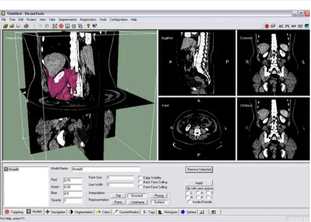
Cryoablation simulation was done using an application developed as part of the CrioLapSim research project in partnership between Clinical Institute of Urology and Renal Transplantation and the Computer Science Department of the Technical University of Cluj-Napoca. The simulation was done using an implementation of the DicomTools application which allows qualitative and quantitative assessment of the cryoneedles setting, evolution of the temperature map. The application allows using of a oblique plan, which could be set perpendicular to the direction of the cryoneedles.
After processing the CT slices, the region of interest is selected to include the affected kidney and the tumor. A 3D surface of the reconstructed volume is created and visualized in the whole body CT scan (Figure 1.).
Following identification of the ideal oblique plan, the cryoablation volumes corresponding to the -20°C isotherm and a 20 mm size, generated by every cryoneedle, are placed to cover the whole tumor volume.
The planning of the cryoablation is presented in Figure 2.
The 3D reconstruction was also used for planning the surgical approach, the extension of the perirenal dissection, the direction and number of the cryoneedles required for the procedure.
Statistical analysis
Parametric continuous variables are given as the mean plus or minus standard deviation and categorized according to the median value. The efficiency of the simulation on reconstructed 3D CT scan was assessed by logistic regression. A p < 0.05 was considered statistically significant. All data were analyzed with the EpiInfo™, Version 3.5.1 (National Center for Public Health Informatics, USA).
Results
Fourteen patients underwent preoperative cryoablation planning using reconstructed 3D CT model (Gr.A) and 13 patients underwent renal tumor cryoablation without prior surgery planning (Gr.B).
Figure 1. The region of interest has been used to generate the 3D reconstructed surface, displayed in the thoraco-abdominal CT scan.
Table 1. Main results
Gr.A Gr.B
Number of patients 14 13
Surgical time (min.) 138.79±5.51 140.92±5.54
Bleeding (mL.) 164.29±60.22 215.38±100.80
Postoperative drainage (mL.) 80.36±22.31 98.08±18.99
Analgesics requirements (mg equivalent morphine sulphate) 10.36±4.55 11.53±3.67
Hospital stay (days) 5.5±2.38 5.69±2.25
Socio-professional reintegration (days) 13.93±4.34 14.85±4.10
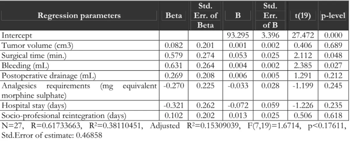
Table 2. Multifactorial logistic regression
Regression parameters Beta
Std. Err. of Beta B Std. Err. of B
t(19) p-level
Intercept 93.295 3.396 27.472 0.000
Tumor volume (cm3) 0.082 0.201 0.001 0.002 0.406 0.689
Surgical time (min.) 0.579 0.274 0.053 0.025 2.112 0.048
Bleeding (mL) 0.631 0.264 0.004 0.002 2.385 0.027
Postoperative drainage (mL) 0.269 0.208 0.006 0.005 1.291 0.212 Analgesics requirements (mg equivalent
morphine sulphate)
-0.270 0.225 -0.033 0.028 -1.199 0.245
Hospital stay (days) -0.321 0.262 -0.072 0.059 -1.226 0.235
Socio-profesional reintegration (days) 0.102 0.202 0.013 0.025 0.506 0.618 N=27, R=0.61733663, R2=0.38110451, Adjusted R2=0.15309039, F(7,19)=1.6714, p<0.17611,
Std.Error of estimate: 0.46858
The number of cryoneedles was assessed by simulation on reconstructed 3D CT scan with a 92.52% accuracy when compared with those effectively used. The average tumor size, in regard with the number of cryoneedles used, is presented in Table 3.
Table 3. Relation between tumor size and number of cryoneedles planned preoperatively.
Tumor size (cm) Tumor volume (cm3)
Nr. cryoneedles Nr. Patients
average range average range
1 8 1.49±0.36 1-2 8.98±5.94 2.4-18.8
3 5 2.38±0.16 2.2-2.5 32.12±6.42 25.1-36.8
4 5 2.72±0.13 2.6-2.9 47.68±6.93 41.4-57.5
5 4 3.13±0.05 3.1-3.2 71.95±3.51 70.2-77.2
6 1 3.8 129.29
7 4 4±0.17 3.8-4.2 148.6±19.41 129.3-174.6
Total 27 2.58±0.91 54.9±49.35
Discussion
Minimal invasive nephron sparing surgery is challenging regardless the specific technique involved. Experience is a key point to feasibility and efficiency but knowing the specific anatomic characteristic allows proper indications and may improve the surgery and the patient's outcome.
Considering 6 studies on laparoscopic partial nephrectomy [3,6-11], renal hilar vessels clamping was required only in four studies in a proportion varying between 45% to 99%, while average tumor size vary between 1.9 cm to 2.9 cm. Also, the complication rate varies largely from 10% to 33%. An explanation for a such variability of the results may be due to other factors than simply tumor size [11].
At present, there are many software and hardware solutions for 3D CT reconstruction but such instruments are used mainly by those involved in imaging diagnosis and only for limited purposes by those who treat. Many applications are dedicated to various types of radiotherapy and less to surgical treatment.
A specific attention was paid to renal vasculature in the case of planning the partial nephrectomy. For such purpose, 3D CT imaging proves utility but vessels are not the only aspects of interest [13]. The correct approach to the kidney and tumor, the tumor relation to the calyx may also contribute to less complications and a better outcome after nephron sparing surgery.
The application used in this study allows specifically planning the surgery with significantly reduced surgical time and bleeding. The software allows establishing the extension of the perirenal dissection, the direction and approach points for cryoneedles placement and assessing the particular number of cryoneedles required for a specific tumor.
The application was customized for different cryoneedle in regard with the producer specifications. The number of cryoneedles required was related to tumor size: one for tumors less than 2 cm, 3 for tumor size between 2 to 2.5 cm, 4 cryoneedles for tumors up to 3 cm, 5 for tumors 3 to 3.5 cm and 6-7 cryoneedles for tumors up to 4 cm.
Conclusions
Simulation of the cryoablation using reconstructed 3D CT scan improves the surgical results. The application used for simulation was able to accurately assess the number of cryoneedles required for tumor ablation, their direction and approach.
Ethical Issues
The study was approved by the local comity of ethics and considered beneficial to the patient since the use of simulation may add valuable data for procedure planning, and the surgical protocol was not changed in the absence of simulation.
Conflict of Interest
The patients give the informed consent for participating in the study, with respect of the confidentiality. The author declares that he has no conflict of interest.
Acknowledgements
The software used for cryoablation simulation was produced by the Computer Science Department of the Technical University of Cluj-Napoca, as partner in the research project CrioLapSim conducted by Clinical Institute of Urology and Renal Transplantation.
References
1. Ficarra V, Novara G, Secco S, Macchi V, Porzionato A, De Caro R, et al. Preoperative Aspects and Dimensions Used for an Anatomical (PADUA) Classification of Renal Tumours in Patients who are Candidates for Nephron-Sparing Surgery. Eur Urol 2009;56:786-793.
2. Novick AC, Campbell SC, Belldegrun A, Blute ML, Chow GK, Derweesh IH, et all. Guideline for management of the clinical stage 1 renal mass. [online] ©2009 [Accessed September 2010]. American Urological Association Web site. http://www.auanet.org/ content/guidelinesand-quality-care/clinical-guidelines/main-reports/renalmass09.pdf.
3. Link RE, Bhayani SB, Allaf ME, Varkarakis I, Inagaki T, Rogers C, et all. Exploring the learning curve, pathological outcomes and perioperative morbidity of laparoscopic partial nephrectomy performed for renal mass. J Urol 2005;173:1690-1694.
visualization of renal masses: impact on diagnosis and patient treatment., AJR Am J Roentgenol 1997;169(5):1331-4.
5. Ghervan L, Lucan V, Elec F, Suciu M, Bologa F, Iacob G, Lucan M. Retro-peritoneoscopic assisted cryoablation for small renal tumors: the first cases treated in Romania. Chirurgia 2007;102(5):557-62.
6. Ramani AP, Desai MM, Steinberg AP, Ng CS, Abreu SC, Kaouk JH, et al. Complications of laparoscopic partial nephrectomy in 200 cases. J Urol 2005;173:42-47.
7. Guillonneau B, Bermudez H, Gholami S, El Fettouh H, Gupta R, Adorno Rosa J, et al. Laparoscopic partial nephrectomy for renal tumor: single center experience comparing clamping and no clamping techniques of the renal vasculature. J Urol 2003;169:483-486.
8. Hafez KS, Novick AC, and Butler BP. Management of small solitary unilateral renal cell carcinomas: impact of central versus peripheral tumor location. J Urol 1998;159:1156-1159.
9. Jeschke K, Peschel R, Wakonig J, Schellander L, Bartsch G, Henning K. Laparoscopic nephron-sparing surgery for renal tumors. Urology 2001;58:688-692.
10. Janetschek G, Jeschke K, Peschel R, Strohmeyer D, Henning K, Bartsch G. Laparoscopic surgery for stage T1 renal cell carcinoma: radical nephrectomy and wedge resection. Eur Urol 2000;38:131-135.
11. Venkatesh R, Weld K, Ames CD, Figenshau SR, Sundaram CP, Andriole GL, et all. Laparoscopic partial nephrectomy for renal masses: effect of tumor location. Urology 2006;67(6):1169-74.
12. Dighe M, Takayama T, Bush WH Jr. Preoperative planning for renal cell carcinoma-benefits of 64-slice CT imaging. Int Braz J Urol 2007;33(3):305-12.
13. Coll DM, Herts BR, Davros WJ, Uzzo RG, Novick AC. Preoperative use of 3D volume rendering to demonstrate renal tumors and renal anatomy. Radiographics 2000;20(2):431-8.