Sea: Evidence from Bone Microstructure
Alexandra Houssaye1,2*, Paul Tafforeau3, Christian de Muizon4, Philip D. Gingerich5
1UMR 7179 CNRS/Muséum National d’Histoire Naturelle, Département Ecologie et Gestion de la Biodiversité, Paris, France,2Steinmann Institut für Geologie, Paläontologie und Mineralogie, Universität Bonn, Bonn, Germany,3European Synchrotron Radiation Facility, Grenoble, France,4Sorbonne Universités, CR2P—CNRS, MNHN, UPMC-Paris 6, Département Histoire de la Terre, Muséum National d’Histoire Naturelle, Paris, France,5Department of Earth and Environmental Sciences and Museum of Paleontology, University of Michigan, Ann Arbor, Michigan, United States of America
Abstract
Cetacea are secondarily aquatic amniotes that underwent their land-to-sea transition during the Eocene. Primitive forms, called archaeocetes, include five families with distinct degrees of adaptation to an aquatic life, swimming mode and abilities that remain difficult to estimate. The lifestyle of early cetaceans is investigated by analysis of microanatomical features in postcranial elements of archaeocetes. We document the internal structure of long bones, ribs and vertebrae in fifteen specimens belonging to the three more derived archaeocete families—Remingtonocetidae, Protocetidae, and Basilosauridae—using microtomogra-phy and virtual thin-sectioning. This enables us to discuss the osseous specializations ob-served in these taxa and to comment on their possible swimming behavior. All these taxa display bone mass increase (BMI) in their ribs, which lack an open medullary cavity, and in their femora, whereas their vertebrae are essentially spongious. Humeri and femora show opposite trends in microanatomical specialization in the progressive independence of ceta-ceans from a terrestrial environment. Humeri change from very compact to spongious, which is in accordance with the progressive loss of propulsive role for the forelimbs, which were used instead for steering and stabilizing. Conversely, hind-limbs in basilosaurids be-came strongly reduced with no involvement in locomotion but display strong osteosclerosis in the femora. Our study confirms that Remingtonocetidae and Protocetidae were almost exclusively aquatic in locomotion for the taxa sampled, which probably were shallow water suspended swimmers. Basilosaurids display osseous specializations similar to those of modern cetaceans and are considered more active open-sea swimmers. This study high-lights the strong need for homologous sections in comparative microanatomical studies, and the importance of combining information from several bones of the same taxon for im-proved functional interpretation.
a11111
OPEN ACCESS
Citation:Houssaye A, Tafforeau P, de Muizon C, Gingerich PD (2015) Transition of Eocene Whales from Land to Sea: Evidence from Bone Microstructure. PLoS ONE 10(2): e0118409. doi:10.1371/journal.pone.0118409
Academic Editor:Brian Lee Beatty, New York Institute of Technology College of Osteopathic Medicine, UNITED STATES
Received:October 27, 2014
Accepted:January 14, 2015
Published:February 25, 2015
Copyright:© 2015 Houssaye et al. This is an open access article distributed under the terms of the
Creative Commons Attribution License, which permits unrestricted use, distribution, and reproduction in any medium, provided the original author and source are credited.
Data Availability Statement:All relevant data are within the paper.
Introduction
Many amniote groups (e.g. sauropterygians, squamates, cetaceans, sirenians, pinnipeds) made the evolutionary transition from a fully terrestrial to a semi- to fully aquatic life. This required major morphological and physiological changes that are best developed in the most specialized aquatic forms, like extant cetaceans and sirenians, which now live totally independent of the terrestrial environment. Several lineages are known with transitional fossil forms, but it re-mains difficult to determine both their degree of physiological adaptation to an aquatic milieu and their locomotor ability in water. Better knowledge of these intermediate forms is essential for understanding the process of secondary adaptation to life in water.
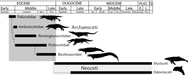
Here we focus on the transition of cetaceans from land to sea. Cetaceans arose in the early Eocene (about 50 Myr ago), when the earliest fossils are known in Indo-Pakistan.‘Archaic’or ‘primitive’cetaceans, called archaeocetes, include five families illustrating various modes of ad-aptation to an aquatic life (Fig. 1). The degree of aquatic adaptation and swimming modes of these taxa are debated (e.g. [1–11]). Here we address the lifestyle of early cetaceans by analysis of microanatomical features in postcranial elements of the three more derived archaeocete families, Remingtonocetidae, Protocetidae, and Basilosauridae, extending research by Buffrénil et al. [12], Madar [13,14] and Gray et al. [15].
(a) Remingtonocetidae
Early middle Eocene Remingtonocetidae have skeletons indicating that they were long-bodied, with a long cranial rostrum, short limbs, fused sacral vertebrae, and a powerful tail [16,17]. They are considered an early aquatic radiation with distinct specializations [6,11], and are sometimes interpreted as amphibious with an otter-like or gavial-like mode of swimming [6,18]. Bebej et al. [19] showed that terrestrial abilities were limited in remingtonocetids, and that propulsion during swimming was powered by the hindlimbs rather than undulation of the lumbar region. This is consistent with their sense organs being poorly compatible with terres-trial locomotion (small eyes, small semicircular canals; [20]). Both sedimentological and isoto-pic evidence suggests that remingtonocetids lived in coastal marine environments [6,10,21]. The specimens ofRemingtonocetus domandaensisGingerich et al., [22] that we analyze here came from the early middle Eocene (middle Lutetian) of Pakistan.
Fig 1. Phylogenetic relationships of early cetaceans showing the temporal ranges and general relationships of Pakicetidae, Ambulocetidae, Remingtonocetidae, Protocetidae, and Basilosauridae discussed here.Modified from [75,76].
doi:10.1371/journal.pone.0118409.g001
(b) Protocetidae
Middle Eocene Protocetidae are a parallel radiation of early cetaceans evolving independently of Remingtonocetidae. Protocetids are found in Indo-Pakistan [2,16,22–24], but also in North Africa [25], West Africa [26], North America [27–29], and South America [30]. Protocetids were the first cetaceans to disperse widely through the world’s oceans.
Kellogg ([31], p. 277) regardedProtocetusas being“far advanced”in the transition to life in water, and“well adapted for a pelagic life.”This was partially confirmed when more complete protocetid skeletons were found [2,22,24,27,32]. Protocetids have a short lumbar region of the vertebral column, short ilium of the pelvis, and short femur, combined with relatively long manual and pedal phalanges. However, retention of well developed and powerful hind limbs connected to the vertebral column is an indication that protocetids were not yet fully aquatic. The characteristics of protocetids, taken together, indicate foot-powered swimming in a rela-tively aquatic mammal [7]. The pedal phalanges of protocetids are long and delicate relative to the size of the animal. While protocetids could still come out on land to give birth [24], they could not have moved far from a shoreline.
Protocetids have small semicircular canals in accordance with their limited terrestrial loco-motion [33]. Early protocetids have a true pelvis with the innominates attached to a sacrum of 3 or 4 co-ossified vertebrae and functional hind limbs well articulated to the innominate. This arrangement provided the stable platform required for foot-powered swimming. Although the attachment of the innominates to a solid sacrum has been reduced (e.g. inNatchitochia, [34]) or possibly even lost in later members of the family (Georgiacetus, [27]), no protocetids are known to have been fully aquatic like later basilosaurids.
Here we analyse specimens ofRodhocetus kasraniiGingerich et al. [32],Qaisracetus arifi
Gingerich et al. [22], andMaiacetus inuusGingerich et al. [24], all from the early middle Eo-cene (Lutetian) of Pakistan.
(c) Basilosauridae
Middle and late Eocene Basilosauridae are morphologically similar to modern cetaceans, with forelimbs modified into flippers retaining a mobile elbow, reduced hind limbs, and a powerful vertebral column with a tail fluke adapted for undulatory or oscillatory tail-powered swimming [11,35]. Basilosaurids had reduced hind limbs articulated to a pelvis lacking any bony connec-tion to the vertebral column, and were undoubtedly fully aquatic. From the known fossil re-cord, they were also fully marine. Basilosaurids likeDorudonandCynthiacetushad body proportions close to those of recent dolphins or porpoises, butBasilosaurushad an exception-ally long body and tail, differing greatly from the other two genera in being more serpentine.
Basilosaurushad a tail fluke, but the tail was probably not the only source of propulsion.
Basilo-saurusprobably swam by undulation of the whole body (an anguilliform swimming mode),
and Gingerich [7] even suggested that the propulsion may have included lateral as well as dorsoventral undulation.
Here we analyze specimens ofBasilosaurus isisBeadnell in Andrews [36] andDorudon
atroxAndrews [37] from the late Eocene of Egypt, and ofCynthiacetus peruvianus
Martínez-Cáceres & Muizon [38] from the late Eocene to early Oligocene of Peru.
(d) Bone microanatomical features
Two alternative microanatomical specializations are found in virtually all strongly or exclu-sively aquatic amniotes that forage below the water surface [44]. These specializations of bone architecture involve either an increase in bone mass or the development of a spongy inner or-ganization and are related to swimming ability through the control of buoyancy (see [45,46]). Bone mass increase (BMI) is a specialization found in various groups of slow and relatively inactive, but essentially or even exclusively aquatic, subsurface swimmers like sirenians and various aquatic fossil reptiles (see [45] for a review). The latter display compact bone organiza-tion (osteosclerosis), which makes the bones brittle, with possible addiorganiza-tional cortical bone de-posits (pachyostosis). BMI by itself confers hydrostatic regulation of buoyancy and body trim. A spongious inner organization is found in highly aquatic active swimmers (modern ceta-ceans, derived mosasaurs, ichthyosaurs, plesiosaurs). The latter display a spongy bone organi-zation, with much reduced compact bone and a tight network of osseous trabeculae oriented in the direction of maximal stress, probably associated with a more even distribution of forces during active locomotion, in order to prevent breakage in a milieu of reduced gravity (see [46–48]). Osteoporosis requires hydrodynamic regulation of buoyancy and body trim.
Neither of these divergent specializations is considered compatible with terrestrial locomo-tion and neither is found with any frequency in terrestrial taxa [45].
Very little microanatomical information is available for archaeocete whales spanning the transition from land to sea. The relative distribution of compact and spongious bone has been reconstructed hypothetically for various archaeocete long bones based on radiographs reflect-ing essentially density differences [13]. However, bone compactness alone is not as good an in-dicator of behavior and ecology as bone compactness combined with the internal organization of bone (see [49]). Rib sections have been described in archaeocetes [12,15], as well as some fracture sections of long bones of pakicetids [14,50]. Buffrénil et al.’s [12] and Gray et al.’s [15] studies are based on broken rib fragments, whose position along the vertebral column or within a rib could not be specified, meaning that their observations have to be interpreted
with caution.
Here we document much more of the internal structure of bone from various parts of the skeleton in archaeocete specimens belonging to three of the five known families. This enables a more substantial discussion of skeletal specialization observed in these taxa and offers greater constraint when discussing behavioral and ecological implications.
Materials and Methods
We are thankful to C. Sagne and S. Sanchez for the loan and transport of theCynthiacetus ma-terial. We thank the ESRF (Grenoble, France) and Steinmann Institut (University of Bonn, Germany) for providing beamtime and support, the ESRF in the framework of the proposal EC-774 on the beamline ID17.
(a) Materials
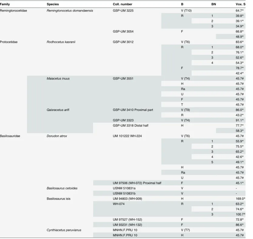
We focused our study on archaeocetes from the three more derived archaeocete families— Remingtonocetidae, Protocetidae, Basilosauridae (seeTable 1)—illustrating a wide spectrum of the diversity of this group after its earliest stages.
Table 1. List of material analyzed in this study.
Family Species Coll. number B BN Vox. S
Remingtonocetidae Remingtonocetus domandaensis GSP-UM 3225 V (T10) 64.7*
R 1 39.8*
2 39.1*
3 34.9*
GSP-UM 3054 F 66.8*
48.9*
Protocetidae Rodhocetus kasranii GSP-UM 3012 V (T6) 83.6*
R 1 68.0*
2 76.1*
3 52.6*
4 54.3*
F 78.7*
42.4*
Maiacetus inuus GSP-UM 3551 V (T4) 45.7#
H 45.7#
Ra 45.7#
U 45.7#
F 45.7#
T 45.7#
Qaisracetus arifi GSP-UM 3410 Proximal part V (T9) 86.0*
R 43.2*
GSP-UM 3323 V (T4) 91.1*
GSP-UM 3318 Distal half H 77.7*
58.3*
Basilosauridae Dorudon atrox UM 101222 WH-224 V (T6) 45.7#
R 1 55.9*
2 75.5*
3 65.2*
4 42.6*
5 49.1*
H 45.7#
Ra 45.7#
U 45.7#
UM 97506 (WH-072) Proximal half F 45.1*
Basilosaurus cetoides USNM 510831a V
-USNM 510831b V
-Basilosaurus isis UM 94803 (WH-009) H 169.0*
WH-074 R 1 63.2*
2 74.6*
3 100.7*
UM 97527 (WH-152) F 72.8*
UM 93231 (WH-132) F 86.6*
Cynthiacetus peruvianus MNHN.F.PRU 10 V (T7) 45.7#
MNHN.F.PRU 10 H 45.7#
Abbreviations: B–bone, F–femur, H–humerus, R–rib, Ra–radius, T–tibia, U–ulna, V–vertebra, and Vox. S–voxel size. BN is the block number, with numbering increasing proximodistally.
*: scanned at the Steinmann Institut (Bonn, Germany). # scanned at the ESRF (Grenoble, France);—not scanned.
GSP-UM: Geological Survey of Pakistan-University of Michigan, specimens archived in Quetta, Pakistan; MNHN: Muséum national d’Histoire naturelle, Paris, France; UM: University of Michigan Museum of Paleontology, USA.
adults, based on tooth eruption and/or epiphyseal fusion, except the holotype ofCynthiacetus
peruvianusthat is proposed to be a young adult [38].
(b) Methods
The fossils analyzed here are rare and parts of exceptionally complete skeletons, meaning that destructive sampling was precluded. No permits were required for the described study. Osteo-logical cross sections were obtained from microscale computed tomography (CT), allowing non-destructive imaging of the three-dimensional outer and inner structure of the samples. Both conventional and synchrotron X-ray micro-CT (seeTable 1) were used: (1) high-resolu-tion computed tomography (GEphoenixjX-ray vjtomejxs 240) was used at the Steinmann-Institut, University of Bonn (Germany), with reconstructions performed using datox/res soft-ware; and (2) third generation synchrotron propagation phase-contrast micro-CT [52] at the European Synchrotron Radiation Facility (ESRF, Grenoble, France), on beamline ID 17. The scans were performed with 5 meters of propagation, using a detector giving an isopetric voxel size of 45.71 µm. The energy was set at 100 keV using a double Laue Laue Si 111 bendable crys-tal monochromator. Most of the specimens being very large and dense, a specific protocol to optimize the X-ray transmission profile through the sample was used [53,54], allowing high quality scans despite transmission lower than 1%. Reconstructions were performed using a fil-tered back-projection algorithm with ESRF PyHST software.
Complete bone shafts could be scanned in conventional microtomography but only a short mid-diaphyseal section was scanned via synchrotron microtomography due to limited access to beam time. Image segmentation and visualization of resulting data were performed using Avizo 6.3. (VSG, Burlington MA, USA) and VGStudioMax 2.0. and 2.2. (Volume Graphics Inc., Heidelberg, Germany).
Virtual thin-sections were made in cross-sectional planes of interest that serve as a reference for comparative studies. These were longitudinal and mid-diaphyseal transverse sections for long bones, mid-sagittal and neutral transverse sections for vertebrae (see [55]). Following ini-tial analyses, additional transverse virtual sections were made for long bones (see below). Rib transverse and longitudinal virtual thin sections were made at different positions along the bone (with the number of sections depending on rib preservation). Two lumbar or anterior caudal vertebrae ofBasilosauruswere sectioned along their mid-sagittal and mid-transverse planes respectively. Finally, for long bone and rib sections, a compactness index (CI) was calcu-lated representing the sectional area occupied by bone as a percentage of total cross-sectional area.
The histological terminology is based primarily on Francillon-Vieillot et al. [56].
Results
(a) Remingtonocetus
RibThere is no open medullary cavity. The rib displays a spongious organization. Cavities are fairly large in the medullary area and smaller in the cortex, which is much more compact. The latter displays some circumferential lines, which probably correspond to lines of arrested growth (LAGs—illustrating the cyclical growth), indicating that it is only feebly remodelled (as these primary structures are not remodelled). The relative thickness of the cortex decreases dis-tally, while the tightness of the spongiosa slightly increases, trabeculae and intertrabecular spaces becoming slightly thinner and smaller respectively. Compactness is fairly high in the proximal part of the rib (CI~80) but lower in the distal one (CI~ 67).
The vertebra is spongious but a layer of compact cortex surrounds the bone periphery and the neural canal. Trabeculae are sagittally oriented in the longitudinal section. The spongiosa is much looser in the periosteal than in the enchondral territory.
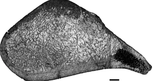
Femur
The femur displays a thick cortex rather compact in its inner part and extremely compact in its periphery, and an off-center open medullary cavity (Fig. 2). The compactness index is rather high (82) proximal to the mid-diaphysis (Fig. 2).
(b) Rodhocetus
RibThe rib lacks any open medullary cavity (Fig. 3). It displays a spongious organization but is highly compact. The first two thirds of the rib show a distinct surrounding layer of more com-pact periosteal bone, cavities being more numerous and larger in the medullary area (Fig. 3A-C). Some LAGs are observed in the cortex, which appears thus poorly remodelled. Compact-ness is high in the first two thirds of the rib (89.4<CI<91.8 in the sections analyzed). In the distal part of the rib the spongiosa becomes much tighter and occupies almost the whole sec-tion (Fig. 3D), so that compactness decreases (CI = 73.9).
Vertebra
The vertebra is cancellous (Fig. 4). It is similar to that ofRemingtonocetus, except in the ab-sence of layers of compact bone. The transverse section illustrates a circumferential orientation of the trabeculae in the outer part of the centrum.
Femur
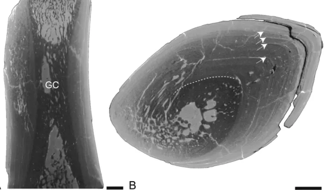
The femur ofRodhocetusresembles that ofRemingtonocetus, although it is more compact. The longitudinal section shows that the inner organization of the bone changes markedly along the diaphysis (Fig. 5A). The growth center, i.e., the point where growth originated, corre-sponds to the point of the transverse section displaying the thicker remains of the original Fig 2. Left femur ofRemingtonocetus domandaensis, GSP-UM 3054, virtual diaphyseal cross section.
Section located just below the lesser trochanter, about one third of the length of the bone from the proximal end. MC: medullary cavity. The contrast between bone and the infilling sediment shows that the MC is open. Scale bar equals 5 mm.
cones of primary periosteal bone where the cones of endochondral and periosteal origin inter-sect (Fig. 5B). The growth center is usually located close to the mid-diaphysis, but here it ap-pears clearly proximal (Fig. 5A). Around this point, the bone is strongly compact (CI = 83.6 and 87.4 on two different sections). The open medullary cavity is clearly off-center and sur-rounded by a cortex displaying numerous fairly small cavities, although they are larger posteri-orly (Fig. 5C). Proximal and distal to the open medullary cavity, the micro-organization changes rapidly to more spongious bone.
(c) Maiacetus
MostMaiacetuslong bones show cracks or slight distortion so that compactness indices
(mea-sured at mid-shaft) are difficult to calculate and can only be estimates. Vertebra
The vertebral microanatomical features are similar to those observed in the vertebra of
Rodhocetus.
Humerus
The humerus ofMaiacetushas, at mid-shaft, a rather thick layer of compact cortex sur-rounding an entirely spongious medullary area, with the contrast between the two being very sharp (Fig. 6A). Compactness is estimated at around 69%.
Radius and ulna
These bones display a microanatomical organization similar to that of the humerus. Howev-er, the layer of compact cortex at mid-diaphysis is proportionally thicker in the radius, so that Fig 3. Left rib 9 ofRodhocetus kasraniiGSP-UM 3012, in anterior view.A-D, virtual transverse sections (left) and corresponding binary images (right; in black: bone; in white: cavities) following the positions labelled on the rib. Scale bars equal 1 mm.
compactness is higher in the latter than in the ulna (about 73% in the radius versus about 63% in the ulna). The distal section of the radius presents a thick compact cortex surrounding a small medullary area with a few large cavities separated by thick short trabeculae. It shows very high compactness (about 85%).
Femur
The femur transverse section, slightly distal to the mid-shaft, is very compact (Fig. 6B). A thick layer of compact cortex surrounds a relatively compact medullary area. Because of Fig 4. Vertebral virtual sections.A,Rodhocetus kasraniiGSP-UM 3012, transverse virtual section of thoracic vertebra T6. B,Qaisracetus arifiGSP-UM 3410, mid-sagittal section of centrum of thoracic vertebra T9. Scale bars equal: A, 10 mm; B, 5 mm.
doi:10.1371/journal.pone.0118409.g004
Fig 5. Left femur ofRodhocetus kasraniiGSP-UM 3012.A-B, partial longitudinal section in lateral view; proximal is at the top. Limits of the compact cortex (dotted lines), as well as the position of the growth center (GC), are indicated on B; C, transverse section cutting the growth center. Scale bars equal 5mm. Cavities are either filled by sediment (light grey) or by epoxy (black) resulting from bone preparation.
breakage and of the limited area scanned, it is difficult to determine whether there was a med-ullary cavity. If present, it must have been much reduced. Compactness is estimated at about 82%.
Tibia
The tibia is also highly compact. A transverse section at about two thirds (distally) of its length shows a very thick and compact cortex and a reduced medullary area with only a few trabeculae, an organization similar to that observed in the distal third of the radius (see above). Fig 6.Maiacetus inuusGSP-UM 3551.Virtual transverse sections. A- Right humerus; B- Left femur. Scale bars equal 5mm.
Compactness is estimated at about 88%. A quite proximal section shows a compact cortical layer surrounding a spongiosa. Compactness remains relatively high (66%).
(d) Qaisracetus
RibThe proximal rib fragment ofQaisracetusshows a microanatomical organization similar to that observed in theRodhocetusrib.
Vertebrae
Vertebral microanatomical features are again similar to those observed in the
Rodhocetusvertebra.
Humerus
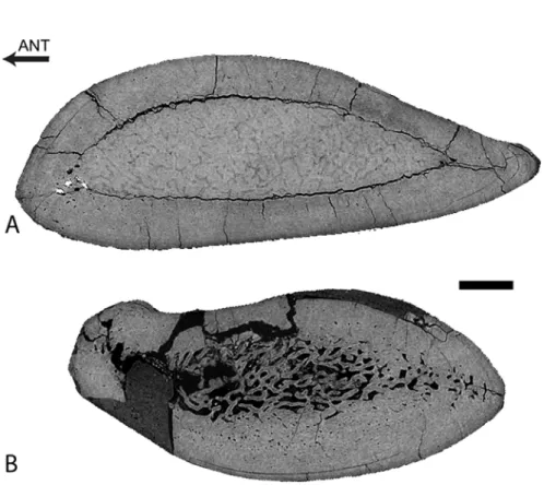
Only the distal half of a humerus is available. The mid-diaphysis is nevertheless well pre-served. In longitudinal section, important variations in micro-organization occur along the shaft (Fig. 7A), as in the femur ofRodhocetus(see above). Around the center of growth, there is a small and off-center open medullary cavity in a spongious rather small medullary area that is surrounded by a thick layer of compact bone (Fig. 7B). Differences in grey levels (seeFig. 7) seem to indicate the transition between primary periosteal bone (light grey) and secondary bone of both periosteal and endochondral origin (dark grey), as suggested by the observation of LAGs in the light grey area. Periosteal bone appears thus only slightly remodelled. Compact-ness is very high around the growth center (CI = 91.7 and 92.5) and remains high at some dis-tance from this point. However, it then strongly decreases proximally and distally towards the metaphyses because of thinning of the compact cortical layer and also transformation of the medullary area from more compacted to looser spongiosa (seeFig. 7A).
Fig 7. Left humerus ofQaisracetus arif.GSP-UM 3318 in virtual longitudinal (A) and transverse (B) sections. The longitudinal section is in posterior view. Scale bars equal 5 mm. Arrows point to LAGs. GC: growth center.
(e) Dorudon
RibThere is a significant variation in microanatomical organization along the rib (Fig. 8). One constant feature is the absence of an open medullary cavity; instead, the medullary area is spon-gious. The most proximal part of the rib is very compact (CI = 96.1;Fig. 8A), with a thick com-pact cortex surrounding a rather small spongious medullary area. The latter increases
proportionally in size distally. Compactness remains high at about one-third (CI = 91.1;
Fig. 8B), one-half (CI = 85.8;Fig. 8C), and two-thirds (CI = 79.2;Fig. 8D) of rib length, but compactness decreases progressively distally while the spongious area becomes the widest. The most distal part of the rib is conspicuously more spongious (CI = 61.3;Fig. 8E) and displays a much thinner compact cortical layer.
Vertebra
The vertebra ofDorudonis made of a tight spongiosa. Endochondral and periosteal territo-ries are distinct in longitudinal section; the spongiosa is much tighter in the former, with very numerous thin trabeculae and reduced intertrabecular spaces. There is no surrounding com-pact layer of periosteal bone.
Humerus
The mid-diaphyseal section of the humerus is almost exclusively a relatively loose spon-giosa, with only a very thin layer of compact cortex (Fig. 9).
Radius and ulna
These two bones have, at mid-diaphysis, a thick cortical layer surrounding an entirely spon-gious medullary area (Fig. 10). Both areas are very clearly distinct (Fig. 10). The spongiosa is loose with rather large trabeculae and intertrabecular spaces. The compactness index of the ulna is difficult to estimate because of the weak contrast between osseous trabeculae and sedi-ment filling intertrabecular spaces (Fig. 10A). The cortex is slightly thicker in the radius (Fig. 10B), making it more compact (CI~82).
Femur
The femur ofDorudonis incomplete. Only the proximal shaft is preserved. The most distal part of the fragment, the mid-diaphysis, is extremely compact (CI = 98.8) and consists only of compact bone with a few small cavities in the core of the section (Fig. 11A). Compactness de-creases proximally. In the metaphysis, a loose spongiosa occupies half of the section and is sur-rounded by a rather thick compact cortex (CI = 64.2).
(f) Cynthiacetus
VertebraTheCynthiacetusvertebra is similar to that ofDorudon, i.e., spongious with a tight network
of numerous thin trabeculae and reduced intertrabecular spaces, notably in the endochondral territory. There is no compact layer of cortical bone in the bone periphery.
Humerus
The mid-diaphysis of the humerus ofCynthiacetusis mainly a loose spongiosa (Fig. 12). However, the thin peripheral layer of compact bone is thicker than inDorudon.
(g) Basilosaurus
RibFig 8. Left rib 4 ofDorudon atroxUM 101222 (WH-224).A photo of the rib with scanned segments (1 to 5) and positions of the transverse sections (A to E) labelled is shown on the left in posterior view. Corresponding virtual transverse and longitudinal sections are shown on the right (in center and right columns, respectively). Scale bars equal: A-E, 5 mm; 1–5, 10 mm.
Fig 9. Proximal portion of the left humerus ofDorudon atroxUM 101222 (WH-224) in virtual transverse section.Scale bar equals 5mm.
doi:10.1371/journal.pone.0118409.g009
Fig 10.Dorudon atroxUM 101222 (WH-224).Virtual transverse sections of the left ulna (A) and radius (B). Scale bar equals 5 mm.
longitudinal sections (Fig. 13B, D). In the most distal part of the rib, the spongiosa occupies most of the section but is dense and surrounded by a thick layer (especially laterally) of com-pact cortical bone (Fig. 13C), so that the rib remains strongly compact (CI = 84.9). LAGs are observed in the compact cortex, where remodelling is thus probably limited.
Vertebrae
The vertebraeof Basilosaurusare spongious and rather similar to that ofCynthiacetus, ex-cept that thick layers of compact cortical bone, with successive cycles of deposition, are visible along the dorsal and ventral borders of the centrum at its core (Fig. 14A), and along the dorsal and ventral borders of the centrum anterior and posterior to the core (Fig. 14B).
Humerus
A longitudinal section of the humerus ofBasilosaurus isisshows that the center of growth is clearly distal in this taxon, being located near the distal end of the deltopectoral crest
Fig 11. Basilosaurid femora virtual sections.A, Proximal right femur ofDorudon atroxUM 97506 (WH-072), longitudinal section in lateral view. B, Distal left femur of femaleBasilosaurus isisUM 97527 (WH-152), in medial view; C, Proximal left femur of maleBasilosaurus isisUM 93231 (WH-132), in medial view. For all sections, anterior is at the left and proximal at the top. In each panel there is a longitudinal partial section on the left and three transverse sections. Scale bars equal 10 mm for longitudinal and 5 mm for transverse sections.
doi:10.1371/journal.pone.0118409.g011
Fig 12. Virtual transverse section of the humerus ofCynthiacetus peruvianusMNHN.F.PRU 10.Scale bar equals 5mm.
(Fig. 15A). The humerus shows a thick layer of compact cortical bone that surrounds a spon-gious medullary area (Fig. 15B). LAGs are observed in this compact bone that probably repre-sents primary periosteal bone. Around the center of growth, the spongiosa is rather open, but its tightness increases proximally and distally (i.e., intertrabecular spaces become smaller and trabeculae thinner).
Femora
Two femora ofBasilosauruswere analyzed. Both show a strongly compact mid-diaphysis (CI = 98.9 and 90.4 for UM 97527 and UM 93231, a female and a male femur, respectively, see [57]) with an off-center medullary area (Fig. 11B-C), corresponding to an open medullary cavi-ty in the larger male specimen. The center of growth seems located almost at mid-diaphysis, slightly proximally. Compactness decreases away from the growth center, both distally (Fig. 11B) and proximally (Fig 11C). The smaller female specimen (Fig. 11B) shows a rather long part of the diaphysis to be extremely compact, whereas this compactness is more reduced in the male one (Fig. 11C). However, the spongious medullary area away from the compact area is much greater in diameter in the female specimen and the metaphyses are thus much more spongious (see the loose spongiosa in the most distal section of the female specimen in
Fig. 11B).
Fig 13. Left rib 4 ofBasilosaurus isisWH-074.A-C, transverse sections from the proximal, middle, and distal portions of the rib; medial is at the left and posterior at the top. D, longitudinal section from the middle of the rib, in anterior view; medial is at the left and posterior at the top. Scale bars equal 1 cm.
Discussion
(a) Bone microanatomical features
RibsAll ribs lack an open medullary cavity and have unusually high compactness, as compared to other amniotes (see [58];Table 2). If theRemingtonocetusrib is reminiscent ofEnhydra
lutris(see [59]), the other archaeocetes analyzed show a thicker compact cortex and smaller
inner cavities. Compactness increases from remingtonocetids to protocetids and from protoce-tids to basilosaurids and the contrast between a thick compact cortex and an inner spongiosa is much sharper in basilosaurids than it is in protocetids.
The proximal halves ofBasilosaurusandDorudonribs are notably compact, with compact-ness indices for the most proximal parts close to those observed in some desmostylians
(Ashoroa,PaleoparadoxiaandBehemotops; see [56]) and in the semi-aquatic sloth
Thalassoc-nus[60]. The microanatomical organization is also generally similar to that of these taxa: a thick layer of compact bone surrounding a reduced spongious medullary area, whereas such an area is not distinguishable in sirenians that show even stronger compactness [59].
There is important change in bone microanatomy along the ribs. The proximal part of the rib is usually the most compact part, with a particularly thick layer of cortical bone. Compact-ness remains important as far as the midshaft and then decreases distally, the distal portion of the rib usually consisting only of spongiosa. This is however not the case inBasilosaurus, where even the distalmost portion of the rib shows a thick layer of compact cortex. This thick-ening was interpreted as resulting from pachyostosis (see references in [12]). As in sirenians displaying pachyostosis,Basilosaurusribs show a clearly off-center medullary area, the lateral part of the rib growing faster, which might thus be associated with this osseous specialization being more intense laterally.
The marked change in bone microanatomy along the shaft makes homologous comparisons of ribs difficult because there are fewer landmarks to define a reference plane than, for example, in long bones.Dorudon’s rib for example resembles ribs of different taxa depending on the re-gion analyzed. From the most proximal rere-gion to the most distal one, the rib ofDorudon
evokes 1) the desmostyliansPaleoparadoxiaandAshoroa, and the nothrotheriid sloths
Thalas-socnus littoralisandT.carolomartini, 2) the desmostylianBehemotops, 3) a modern dolphin, 4)
the rorqualBalaenoptera, 5) the sirenianPezosiren(see [58,59]). Comparisons must thus be made very cautiously.
Fig 14. Scanned polished sections of lumbar or anterior caudal vertebrae ofBasilosaurus cetoides.A- USNM 510831a, transverse section figured in Fordyce & Watson, 1998; dorsal is at the top. B, USNM 510831b, longitudinal section. Scale bars equal 2 cm.
It can nevertheless be observed that these archaeocete ribs are all less compact than those of sirenians (except for non-osteosclerotic sirenians, see [59]). However, the archaeocete ribs ana-lyzed all display a clear increase in compactness when compared to extant terrestrial amniotes (see [58]). Similarities, notably for basilosaurid ribs, are observed with the desmostylians
Behe-motops,PalaeoparadoxisandAshoroa(see [58]).
Pachyostosis has been mentioned for various archaeocete ribs [12,14]. However, it is neither described nor illustrated in Gray et al. [15], who rely on observation of a thick compact layer of primary periosteal bone in sections without evidence of clear morphological thickening of the bone. TheZyghorizaandBasilosaurusribs illustrated in Buffrénil et al. [12] and theDorudon
andBasilosaurusribs studied here show some bulging in their distal halves in anterior ribs
bound to sternebrae. The thickening evokes what is observed in the desmostylianAshoroa[56] or the youngest species of aquatic sloths,Thalassocnus littoralisandT.yaucensis[60] but it is not comparable to the strong thickening observed in pachyosteosclerotic sirenians. A
Fig 15. Left humerus ofBasilosaurus isisUM 94803 (WH-9).A, Longitudinal section of the specimen in lateral view. B, transverse section. Arrows point to LAGs. Scale bar equals 10 mm.
quantitative anatomical study would be required to clearly determine if this is a common ana-tomical feature within cetaceans or if it really corresponds to pachyostosis.
The thick peripheral layer of compact cortical bone observed alongBasilosaurusrib and the osseous drift would be in accordance with the occurrence of pachyostosis (and not only osteo-sclerosis) with a clear increase in intensity laterally (and not medially as suggested by Buffrénil et al. [12]). Asymmetrical cortical growth also occurs in the pachyostotic ribs of the manatee with also much thicker deposits on the lateral side (see [12]), consistently with the rib morphol-ogy and the maintenance of rib curvature during growth. In our sample, if onlyBasilosaurus
might display pachyostosis, all other archaeocetes analyzed show only osteosclerosis. By comparison,Ichthyolestes(Pakicetidae) ribs show a tubular structure with a medullary cavity that is clearly open [14], although relatively small (as compared to other amniotes). The cortex is extremely compact and thick.Pakicetus andAmbulocetusalso display compact ribs with an extremely compact cortex and dense trabecular struts; unfortunately no large scale im-ages of the sections are available so that the occurrence and size of an open medullary cavity re-main unclear [14].Kutchicetus(Remingtonocetidae) ribs are strongly compact. They appear more similar to those of the protocetids here sampled than to that ofRemingtonocetus([14]; see above). Resorption seems not to have occurred in the outer cortex and remodelling in the inner cortex, and medullary area appears characterized by excessive secondary bone deposits [14], which thus confers on the bone an extremely high compactness. Among Protocetidae,
Gaviacetusribs are also very dense, butGeorgiacetusones are less compact and show more
nu-merous thinner struts [14].Zygorhizaribs show a wide cancellous medullary area [12,14]. Fur-ther investigations would be required to determine the degree of osteosclerosis inZygorhiza. Table 2. Summary of the microanatomical features observed.
Remingtonocetus Rodhocetus Maiacetus Qaisracetus Dorudon Cynthiacetus Basilosaurus
Rib No OMC—Spongious organization CIp~80. CId~67 Highly compact.
89.4<CIp/m <91.8. CId = 73.9
X Highly compact.
CIp high
Highly compact. CIp = 96.1,91.1, 85.8. CIm = 79.2. CId = 61.3.
X Highly compact
CIp = 95.2. CIm = 87.9. CId = 84.9
Vertebra Spongious. Layer of compact cortex surrounding the bone periphery and the neural canal
Spongious Spongious Spongious Tight
spongiosa
Tight spongiosa Tight spongiosa. Thick layer of compact cortex surrounding all the centrum around its core
Humerus X X Medullary area
entirely spongious. Thick compact cortex. CIm = 69 Small off-center OMC Spongious small medullary area Thick compact cortex. CId~92 Relatively loose spongiosa Very thin compact cortex Loose spongiosa. Very thin compact cortex. Spongious medullary area. Thick compact cortex
Femur Thick cortex Rather compact inner part Extremely compact in periphery. Off-center OMC. CId = 82
Compact. Off-center OMC. CIp = 83.6 CIm = 87.4. Distally becomes quickly spongious
Very compact. OMC? CIm = 82
X Very compact.
CIp = 98.8
X Very compact.
Off-center medullary areaCI = 98.9 & 90.4.
OMC: open medullary cavity; CI: compactness index (CIp: proximal, CIm; mid-diaphysis, CId: distal).
Vertebrae
All archaeocete vertebrae analyzed are spongious (Table 2). The variations observed in archaeocete vertebrae, except forRemingtonocetusandBasilosaurus(see below), are variations in tightness of the spongiosa (i.e. of the trabecular network), which increases with specimen size (trabeculae becoming more numerous and thinner with smaller intertrabecular spaces). This positive (qualitative) correlation was already quantitatively highlighted in various amni-otes [49,61].Remingtonocetusdisplays a circumferential layer of compact cortex, like the extant polar bear but not to the extent of the hippopotamus and manatee (see [58]). It evokes a condi-tion intermediate between those of the desmostyliansBehemotopsandAshoroa, respectively (see [58]). Comparisons with diverse amniotes (cf. [58,61,62]) show that the other (more de-rived) archaeocetes have a vertebral micro-organization similar to that of modern cetaceans.
OnlyBasilosaurusdiffers from this condition, with a thick layer of compact cortical bone
sur-rounding the middle of the centrum (the centrum being the only part of the vertebra available for this study) but also the neural arch and transverse processes (PDG. pers. obs.). This layer is thicker than in the extantHippopotamus,ChoeropsisandTrichechus(cf. [58,61]). To our knowledge, such a structure (engendering local bone mass increase [BMI]) has not been ob-served in any other (extant or fossil) taxon. This peculiarity is, moreover, not associated with any morphologically observable bulging and thus does not correspond to pachyostosis. It thus differs from the condition observable inBasilotritusvertebrae, which show laterally swollen neural arches and robust zygapophyses [63].Basilosauruscondition seems related to the very peculiar morphology of its vertebrae and might reflect a structural requirement for these large spongious vertebrae related to muscle insertion and locomotion.
Humeri
The longitudinal sections clearly show an important change in bone microanatomy along the diaphysis. This condition is unusual in amniotes, whatever their ecology. It has so far only been observed inEnhydra lutrisand in turtles and fossil ichthyosaurs and plesiosaurs [64,65]. For this reason, homologous comparisons require a precisely cut transverse“perfect diaphyseal plane”(sensu [64]), i.e., the plane cutting the point where growth originated (see [65] for more details about this sectional plane). In order to locate such a cut, a longitudinal section of a rath-er long part of the central diaphysis is required. Unfortunately, because only vrath-ery short mid-diaphyseal portions were imaged inMaiacetus,DorudonandCynthiacetus, perfectly homolo-gous comparisons cannot be made. The longitudinal sections ofQaisracetusandBasilosaurus
show that the center of growth is not located at mid-shaft but much more distally. This suggests that growth was much faster proximally than distally in the humerus in these taxa. Because of the weak remodelling of compact cortical bone (as suggested by the grey-level differences re-flecting density differences, and, especially, by the observation of LAGs), the area around the center of growth is the more compact one. The spongiosa is extended much farther away from the center of growth.
The transverse section of the humerus ofQaisracetusevokes the condition observed in some fossil marine sauropterygians (e.g.Cymatosaurus,Anarosaurus,Placodus; [66]). It is the only humerus analyzed in which a medullary cavity, though very small, is observed (Table 2). The section ofMaiacetus, from the mid-diaphysis, evokes what is observed in some otariids and placodonts (AH, pers. obs.); however this section is probably at some distance from the perfect diaphyseal plane and it cannot be determined whether a medullary cavity was present around the centre of growth. Distance from the diaphyseal plane would also explain the rela-tively large spongious medullary area in theMaiacetussection. However, even theMaiacetus
section clearly shows an increase in bone compactness as compared to extant amniotes, with a thick layer of compact cortex and a spongious medullary area but lacking an open
The humerus ofBasilosaurusshows a thick cortex. However, it is distinctly thinner in
Cynthiacetusand even more reduced inDorudon, whose section is almost entirely spongious.
The sections of these last two taxa resemble the condition in modern cetaceans, with the layer of compact cortex inCynthiacetusbeing thinner than inPlatanista, and that ofDorudonbeing similar to those ofDelphinusorLagenorhynchus(AH, pers. obs.; [67]). Conversely, the propor-tional thickness of compact cortex ofBasilosaurussections evokes what is observed inEnhydra,
LutraorLeptonychotes. However, the medullary area ofBasilosaurusis spongious, whereas it is
almost open with only a few trabeculae in these taxa.
The humerus ofIchthyolestes(Pakicetidae) was studied by Thewissen et al. [68]. It shows an extremely compact tubular structure with a very thick compact cortex and a reduced (also off-centered) open medullary cavity, and thus appears rather similar to theQaisracetustransverse section described above.
Femora
RemingtonocetusandRodhocetusfemora show a similar microanatomical organization
(Table 2), although the inner cortex appears more compact inRodhocetus. The variation in inner bone structure along the diaphysis is rather similar to what was described in the humerus but the growth center is located proximally in the femur. Unfortunately it cannot be deter-mined whether theMaiacetustransverse section is close to the growth center. Moreover, be-cause of breakage, it is difficult to determine whether a medullary cavity was present. If it was, it would have been smaller than those inRemingtonocetusandRodhocetus. The bone was prob-ably much more compact.
Basilosaurid femora clearly differ from the others (Table 2).Dorudon’s femur is strongly compact with no medullary cavity. Away from the metaphysis, the diaphysis is extremely com-pact.Basilosaurusfemora are also strongly compact. The growth center appears also rather proximal in these taxa. The differences between the twoBasilosaurusspecimens studied here are associated with an important bone size difference. This variation does not result from on-togeny, as the smaller specimen is clearly not a juvenile from an anatomical perspective and as suggested by the multiple possible growth marks observable on the sections. It could rather re-sult from sexual dimorphism, as suggested by Gingerich et al. [69] and Antar et al. [56].
RemingtonocetusandRodhocetusfemoral sections resemble those of some fossil marine
rep-tiles (e.g.,Nothosaurus,Simosaurus) and of the modernAlligatorandTrichechus manatus
(manatee). Basilosaurid femora are more compact, to our knowledge, than in any extant amni-ote. It evokes very compact femora of some fossil sauropterygians (e.g.,Paraplacodus, Pisto-saurus; [70]).
Femora ofAmbulocetus,Rodhocetus,RemingtonocetusandBasilosauruswere analyzed by Madar [13], based on radiographs, in order to document the distribution of compact and spon-gious bone and the possible occurrence of an open medullary cavity. Our observations for the femora ofRemingtonocetusandRodhocetus, based on the same specimens, differ substantially. Madar [13] found the cortical bone inRemingtonocetusto be extremely thin near mid-shaft. The compact cortex is indeed thin but not as extremely as indicated by Madar. If Madar did not note the occurrence of an open medullary cavity, she nevertheless observed a difference in compactness in the medullary area (see [13]:Fig. 4) corresponding to the contrast between the inner cortex (a very compact spongiosa) and the medullary cavity. Madar [13] described a thin cortex all along the diaphysis inRodhocetusand did not observe any medullary cavity. Dense mineralization ofRodhocetus(cf. [13]) undoubtedly affected her radiographs.
some desmostylian (Ashoroa,PaleoparadoxiaandBehemotops), the aquatic slothThalassocnus
carolomartini[60] and sirenian long bones [58,67]. However, no longitudinal section is
avail-able for these taxa, so that the variations along the diaphysis observed inBasilosauruscannot be compared. They are however clearly distinct from the pattern usually observed in amniote long bones ([71]; A.H. pers. obs.), which have a tubular diaphysis with a rather homogeneous thickness of compact cortical bone all along the diaphysis.
Zeugopod bones
Bones of the zeugopodium (ulna-radius and/or tibia-fibula) were only analyzed for
Maiace-tusandDorudonand, unfortunately on a very short section of the diaphysis. Thus, it is unknown
whether the microanatomical differences observed in the archaeocete stylopodium (humerus and/or femur) also occur in the zeugopod. However, the distal section of theMaiacetusradius shows a more compact structure than the mid-diaphyseal section. This suggests a rather distal location of the growth center and a high compactness around this point as in stylopod bones. Sections ofDorudonsuggest greater compactness in stylopod bones, with a thicker layer of com-pact cortex. However, longitudinal sections will be required to confirm this.
(b) Swimming behaviour
RemingtonocetusRemingtonocetusrib is spongious. The femur displays a tubular structure but the open
med-ullary cavity is rather small. These two bones generally show high compactness values, as com-pared to other amniotes (see [58]). The microanatomy of the rib, vertebra and femur evokes that of the sea otter, polar bear, and of some not actively swimming semi-aquatic to aquatic reptiles (see above). These microanatomical features are thus in accordance with anatomical and geological data to assume an amphibious, though essentially aquatic, lifestyle. The compact femur indeed suggests a difficult hind-limbs-supported terrestrial locomotion. Indeed, such a thick compact cortex is observed either in semi-aquatic taxa with a rather poorly active terres-trial locomotion or in graviportal ones (A.H. pers. obs.). Based on its morphological and micro-anatomical features,Remingtonocetusis thus assumed to have displayed extremely limited terrestrial locomotion. Bebej et al. [19] highlighted a lack of mobility between functional series of vertebrae, as opposed to the condition observed in protocetids and basilosaurids. In accor-dance with this result, adaptations to an aquatic life at the microanatomical level appear more limited as compared to these other archaeocetes (see below). However, Bebej et al. [19] consid-ered the rest of the morphology as indicating active foot-powconsid-ered swimming, which appears only slightly compatible with the occurrence of BMI, based on our current knowledge. The re-cent suggestion of this mode of swimming in the semiaquatic dinosaurSpinosaurus aegyptiacus
displaying strong BMI in its hindlimbs [72] would nevertheless agree with the previous hy-pothesis. Further comparisons with extant semi-aquatic taxa would be required to see if active foot-powered swimming can be consistent with the microanatomical features of Remingtonoce-tusand to propose more precise inferences about its possible swimming style.
Protocetids
Rodhocetusmicroanatomical features are very similar to those ofRemingtonocetus. However,
protocetids. The very small medullary cavity observed in the humerus shows a high degree of BMI. These protocetids, with their compact long bones and ribs, probably had considerable dif-ficulties withlocomotion on land. However, contrary to some previous assumptions (see intro-duction), the occurrence of BMI in the long bones and ribs of the protocetids sampled is more compatible with suspended swimming in shallow waters than with a pelagic life.
Basilosaurids
Dorudonshows a spongious, rather lightly built humerus but compact ribs, in at least their
proximal half, and a strongly compact femur. Femora inDorudonare greatly reduced bones not involved in locomotion. A similar BMI is observed inBasilosaurusfemora. Such regressed limb elements with a supposedly similar function also occurred in Late Cretaceous hind-limbed snakes. However, if the latter display BMI in much of their skeleton [46], their femora are de-prived of this osseous specialization [73]. The occurrence of BMI in regressed hind-limbs re-mains unexplained. Despite the femur microanatomy, the other bones ofDorudonanalyzed show microanatomical features very similar to those of modern dolphins. This is not the case
forBasilosauruswhose ribs show a higher inner compactness (osteosclerosis) and what seems
to correspond to increased periosteal bone deposits (pachyostosis). Only a humerus and a ver-tebra ofCynthiacetuswere analyzed. Both bones show a microanatomy more similar to Doru-donthan toBasilosaurus.Basilosaurusthus appears as peculiar among basilosaurids. In addition to its peculiarly long vertebrae characterized by the occurrence of a yoke of compact bone surrounding the mid-centrum, neural arches and transverse processes,Basilosaurusalso displays 1) probable pachyostosis in its ribs and 2) osteosclerosis in its humerus. The occur-rence of pachyostosis in the ribs was also documented inZygorhizaand the expanded distal ex-tremities of ribs 3 to 7 ofCynthiacetus peruvianus(CdM; pers. obs.) also suggests pachyostosis in this taxon. However, rib general morphology and osseous microstructure need to be further investigated in basilosaurids, and more generally in cetaceans in order to clearly determine whether pachyostosis really occurs inBasilosaurus,Zygorhiza, andCynthiacetus. Moreover, rib microanatomical features, and notably the variation along the bone, need to be further investi-gated inZygorhizaand no data are available concerning other bones. The occurrence of BMI in
Basilosaurusis surprising as this taxon is generally considered an active predator. BMI was
as-sumed to be associated with its particularly long (serpentine) post-thoracic region to assist in body trim control [12]. However, this argument cannot be used forZygorhizaand Cynthiace-tus, which show a length of the post-thoracic region similar to those of modern mysticetes and odontocetes. Moreover, body trim control is not compatible with BMI in lumbar, and thus rather posterior, vertebrae. The occurrence of this specialization, at various degrees of intensity, in several bones of this taxon remains mysterious. Further comparisons among basilosaurids and with large modern whales are required to better understand its significance.
Fordyce and Watson [74] described some“archaic fossil mysticeti”from New Zealand as showing osteosclerosis or“peripheral osteosclerosis”, after describing the vertebra USNM 510831a (Fig. 14) as itself osteosclerotic. Further investigations and comparisons would be re-quired in order to determine whether thespecialization inBasilosaurusresembles that of these early mysticetes.
Conclusions
Analysis of the microanatomical features of various bones of three of the five archaeocete fami-lies enables us to discuss evolutionary trends in the progressive adaptation to an exclusively aquatic life in the cetacean lineage, and to make paleoecological inferences for the taxa studied.
while in contrast having essentially or exclusively spongious vertebrae. In the protocetids studied humeri and femora are essentially compact with a small open medullary cavity around the growth center. In this respect, protocetid humeri resemble humeri of the
pakice-tidIchthyolestes, but they differ markedly from the essentially spongious humeri of
basilo-saurids. As opposed to Remingtonocetidae and Protocetidae, basilosaurids display very compact femora. Anterior and posterior long bones thus show clearly distinct trends in mi-croanatomical specialization in the progressive independence from a terrestrial environ-ment, which is naturally associated with the functional role of these bones. Forelimbs progressively lost any propulsive role and became used for steering and stabilization, consis-tent with acquisition of a spongious organization, whereas hind limbs became strongly re-duced and lost any involvement in locomotion. The occurrence of strong osteosclerosis in these reduced appendages remains unexplained.
2. Our observations are in accordance with previous geological and anatomical data that sug-gest an amphibious lifestyle with very limited terrestrial locomotion for both the remingto-nocetids and the protocetids sampled. Basilosaurids, on the other hand, show
specializations similar to modern cetaceans and were clearly more actively swimming in the open sea.Basilosaurusitself is unusual among basilosaurids in displaying bone mass in-crease in its ribs and long bones, although with various intensities, with a yoek of compact bone surrounding the mid-centrum, neural arches, and transverse processes of most verte-brae, which are unusually long. The observation of BMI in posteriorly-located bones shows that this specialization occurs for reasons other than body trim control. BMI in posteriorly located bones is poorly compatible withBasilosaurusmorphology in general and with its presumed behavior and ecology, and BMI inBasilosaurusremains to be explained.
3. This study also highlights the significant variation in bone microanatomy observable along the shaft of the ribs and the diaphysis of long bones, showing that comparisons have to be made with caution in order to deal with homologous regions.
4. The previous works by Madar [13] and Gray et al. [14] were the most substantial contribu-tions available previously on archaeocete bone microanatomical features. Both studies cov-ered the five archaeocete families. However, they focused on a single bone (the femur and the rib, respectively). It is important to combine information from various bones to get a better idea of the variation in degrees of adaptation to aquatic life during the land to sea transition.
Acknowledgments
We thank D.P. Domning (Howard University, Washington, D.C., USA) and O. Lambert (Royal Belgian Institute of Natural Sciences, Belgium) for fruitful comments that improved the quality of the manuscript, and to B. Beatty for editorial help. Specimens from Pakistan and Egypt were collected with multiple grants from the National Geographic Society and the U. S. National Science Foundation. The holotype ofCynthiacetus peruvianuswas collected with funds of the Institut Français d’Études Andines (Lima, Peru).
Author Contributions
References
1. Gingerich PD, Wells NA, Russell DE, Shah SMI (1983) Origin of whales in epicontinental remnant seas: new evidence from the early Eocene of Pakistan.Science220: 403–406. PMID:17831411 2. Gingerich PD, Haq MU, Zalmout IS, Khan IH, Malkani MS (2001) Origin of whales from early
artiodac-tyls: hands and feet of Eocene Protocetidae from Pakistan. Science 293: 2239–2242. PMID:11567134 3. Thewissen JGM, Hussain ST, Arif M (1994) Fossil evidence for the origin of aquatic locomotion in
archaeocete whales. Science 263: 210–212. PMID:17839179
4. Thewissen JGM, Fish FE (1997) Locomotor evolution in the earliest cetaceans: functional model, mod-ern analogues, and paleontological evidence. Paleobiology 23: 482–490.
5. Thewissen JGM, Williams EM, Roe LJ, Hussain ST (2001) Skeletons of terrestrial cetaceans and the relationships of whales to artiodactyls. Nature 413: 277–281. PMID:11565023
6. Thewissen JGM, Williams EM (2002) The early radiations of Cetacea (Mammalia): Evolutionary pattern and development correlations. Annual Review of Ecology, Evolution, and Systematics 33: 73–90.
7. Gingerich PD (2003) Land-to-sea transition in early whales: evolution of Eocene Archaeoceti (Cetacea) in relation to skeletal proportions and locomotion of living semiaquatic mammals. Paleobiology 29: 429–1454.
8. Gingerich PD (2012) Evolution of whales from land to sea.Proceedings of the American Philosophical Society156: 309–323.
9. Muizon Cde (2009) L’origine et l’histoire évolutive des Cétacés. Comptes Rendus Palevol 8: 295–309.
10. Thewissen JGM, Cooper LN, George JC, Bajpai S (2009) From land to water: the origin of whales, dol-phins, and porpoises. Evolution Education & Outreach 2: 272–288. doi:10.1007/s00253-014-6131-7
PMID:25621864
11. Uhen MD (2010) The origin(s) of whales. Annual Review of Earth and Planetary Sciences 38: 189–219.
12. Buffrénil Vde, Ade Ricqlès, Ray CE, Domning DP (1990) Bone histology of the ribs of the archaeocetes (Mammalia: Cetacea). Journal of Vertebrate Paleontology 10: 455–466.
13. Madar SI (1998) Structural Adaptations of Early Archaeocete Long Bones. InThe Emergence of Whales, vol. 12 (ed. Thewissen J. G. M.), pp. 353–378. Plenum Press, New York.
14. Madar SI (2007) The postcranial skeleton of Early Eocene pakicetid cetaceans. Journal of Paleontolo-gy 81, 176–200.
15. Gray NM, Kainec K, Madar SI, Tomko L, Wolfe S (2007) Sink or swim? Bone density as a mechanism for buoyancy control in early cetaceans. Anatomical Record 290: 638–653. PMID:17516430 16. Gingerich PD, Arif M, Clyde WC (1995) New archaeocetes (Mammalia, Cetacea) from the middle
Eo-cene Domanda Formation of the Sulaiman Range, Punjab (Pakistan).Contributions from the Museum of Paleontology,University of Michigan, 29: 291–330.
17. Thewissen JGM, Bajpai S (2009) New skeletal material ofAndrewsiphiusandKutchicetus, two Eocene cetaceans from India. Journal of Paleontology 83: 635–663.
18. Bajpai S, Thewissen JGM (2000) A new, diminutive whale from Kachchh (Gujarat, India) and its impli-cations for locomotor evolution of cetaceans. Current Science (New Delhi) 79: 1478–1482.
19. Bebej RM, ul-Haq M, Zalmout IS, Gingerich PD (2012) Morphology and function of the vertebral column inRemingtonocetus domandaensis(Mammalia, Cetacea) from the Middle Eocene Domanda Forma-tion of Pakistan. Journal of Mammalian EvoluForma-tion 19: 77–104.
20. Spoor F, Bajpai S, Hussain ST, Kumar K, Thewissen JG (2002) Vestibular evidence for the evolution of aquatic behaviour in early cetaceans. Nature 417: 163–166. PMID:12000957
21. Roe LJ, Thewissen JGM, Quade J, O’Neil JR, Bajpai S, Sahni A, Hussain ST (1998) Isotopic ap-proaches to understanding the terrestrial-to-marine transition of the earliest cetaceans. InThe Emer-gence of Whales:Evolutionary Patterns in the Origin of Cetacea(ed. Thewissen J.G.M.), pp. 399–422, New York: Plenum Press.
22. Gingerich PD, Haq M, Khan IH, Zalmout IS (2001) Eocene stratigraphy and archaeocete whales (Mam-malia, Cetacea) of Drug Lahar in the eastern Sulaiman Range, Balochistan (Pakistan).Contributions from the Museum of Paleontology,University of Michigan, 30: 269–319.
23. Sahni A, Mishra V (1975) Lower Tertiary vertebrates from western India. Monograph of the Paleonto-logical Society of India 3: 1–48.
24. Gingerich PD, ul-Haq M, von Koenigswald W, Sanders WJ, Smith BH, Zalmout IS (2009) New protoce-tid whale from the Middle Eocene of Pakistan: birth on land, precocial development, and sexual dimor-phism. PLoS ONE 4: e4366. doi:10.1371/journal.pone.0004366PMID:19194487
26. Gingerich PD, Cappetta H (2014) A new Archaeocete and other marine mammals (Cetacea and Sire-nia) from lower Middle Eocene phosphate deposits of Togo. Journal of Paleontology 88: 109–129.
27. Hulbert RC, Petkewich RM, Bishop GA, Bukry D., Aleshire DP (1998) A new Middle Eocene protocetid whale (Mammalia: Cetacea: Archaeoceti) and associated biota from Georgia. Journal of Paleontology 72: 907–927.
28. Geisler JH, Sanders AE, Luo Z-X (2005) A new protocetid whale (Cetacea, Archaeoceti) from the late middle Eocene of South Carolina. American Museum Novitates 3480: 1–68.
29. Uhen MD (2008) New protocetid whales from Alabama and Mississippi, and a new cetacean clade, Pelagiceti. Journal of Vertebrate Paleontology 28: 589–593.
30. Uhen MD, Pyenson ND, DeVries TJ, Urbina M, Renne PR (2011) New middle Eocene whales from the Pisco Basin of Peru. Journal of Paleontology 85: 955–969.
31. Kellogg R (1936) A review of the Archaeoceti. Carnegie Institution of Washington Publications 482: 1–366.
32. Gingerich PD, Raza SM, Arif M, Anwar M, Zhou X (1994) New whale from the Eocene of Pakistan and the origin of cetacean swimming.Nature368: 844–847.
33. Bajpai S, Thewissen JGM, Sahni A (2009) The origin and early evolution of whales: macroeveolution documented on the Indian Subcontinent. Journal of Biosciences 34: 673–686. PMID:20009264 34. Uhen MD (2014) New material ofNatchitochia jonesiand a comparison of the innominata and
locomo-tor capabilities of Protocetidae. Marine Mammal Science 30: 1029–1066.
35. Uhen MD (2004) Form, function, and anatomy ofDorudon atrox(Mammalia, Cetacea): an archaeocete from the Middle to Late Eocene of Egypt. Papers on Paleontology 34: 1–222.
36. Andrews CW (1904) Further notes on the mammals of the Eocene of Egypt, Part III. Geological Maga-zine, London 1: 211–215.
37. Andrews CW (1906) A descriptive catalogue of the Tertiary Vertebrata of the Fayum, Egypt. British Mu-seum (Natural History), London. Pp. 1–324.
38. Martínez-Cáceres M., Muizon Cde (2011) A new basilosaurid (Cetacea, Pelagiceti) from the Late Eo-cene to Early OligoEo-cene Otuma Formation of Peru. Comptes Rendus Palevol 10: 517–526.
39. Turner CH (1998) Three rules for bone adaptation to mechanical stimuli. Bone 23: 399–407. PMID:
9823445
40. Ruimerman R, Hilbers P, Rietbergen Bv, Huiskes R (2005a) A theoretical framework for strain-related trabecular bone maintenance and adaptation. Journal of Biomechanics 38: 931–941. PMID:15713314 41. Ruimerman R, Rietbergen Bv, Hilbers P, Huiskes R (2005b) The effects of trabecular-bone loading
var-iables on the surface signaling potential for bone remodeling and adaptation. Annals of Biomedical En-gineering 33: 71–78. PMID:15709707
42. Chappard D, Baslé MF, Legrand E, Audran M (2008) Trabecular bone microarchitecture: A review. Morphologie 92: 162–170. doi:10.1016/j.morpho.2008.10.003PMID:19019718
43. Liu XS, Bevill G, Keaveny TM, Sajda P, Guo XE (2009) Micromechanical analyses of vertebral trabecu-lar bone based on individual trabeculae segmentation of plates and rods. Journal of Biomechanics 42: 249–256. doi:10.1016/j.jbiomech.2008.10.035PMID:19101672
44. Ricqlès Ade, Vde Buffrénil (2001) Bone histology, heterochronies and the return of Tetrapods to life in
water: where are we? InSecondary Adaptation of Tetrapods to life in Water(ed. Mazin J. M. and de Buffrénil V.), pp. 289–310, München.
45. Houssaye A (2009)“Pachyostosis”in aquatic amniotes: a review. Integrative Zoology 4: 325–340. doi:
10.1111/j.1749-4877.2009.00146.xPMID:21392306
46. Houssaye A (2013) Palaeoecological and morphofunctional interpretation of bone mass increase: an example in Late Cretaceous shallow marine squamates. Biological Reviews 88: 117–139. doi:10. 1111/j.1469-185X.2012.00243.xPMID:22943660
47. Currey JD (2003) The many adaptations of bone. Journal of Biomechanics 36: 1487–1495. PMID:
14499297
48. Houssaye A (2008) A preliminary report on the evolution of the vertebral microanatomy within mosasaur-oids (Reptilia, Squamata). InProceedings of the Second Mosasaur Meeting(ed. MJ E.), pp. 81–89. Fort Hays State University, Hays, Kansas.
49. Houssaye A, Bardet N (2012) Rib and vertebral micro-anatomical characteristics of hydropelvic mosa-sauroids. Lethaia 45: 200–209.
51. Canoville A, Laurin M (2010) Evolution of humeral microanatomy and lifestyle in amniotes, and some comments on palaeobiological inferences. Biological Journal of the Linnean Society 100: 384–406.
52. Tafforeau P, Boistel R, Boller E, Bravin A, Brunet M, Chaimanee Y, Cloetens P, Feist M, Hoszowska J, Jaeger J-J, Kay RF, Lazzari V, Marivaux L, Nel A, Nemoz C, Thibault X, Vignaud P, Zabler S (2006) Ap-plications of X-ray synchrotron microtomography for non-destructive 3D studies of paleontological specimens. Applied Physics A 83: 195–202.
53. Carlson K, Stout D, Jashashvili T, Ruiter DJde, Tafforeau P, Carlson K, Berger LR (2011) The endocast of MH1,Australopithecus sediba. Science 333: 1402–1407. doi:10.1126/science.1203922PMID:
21903804
54. Sanchez S, Fernandez V, Pierce SE, Tafforeau P (2013) Homogenization of sample absorption for the imaging of large and dense fossils with synchrotron microtomography. Nature Protocol 8: 1708–1717. doi:10.1038/nprot.2013.098PMID:23928503
55. Buffrénil Vde, Houssaye A, Böhme W (2008) Bone vascular supply in monitor lizards (Squamata: Vara-nidae): Influence of size, growth, and phylogeny. Journal of Morphology 269: 533–543. PMID:
18157866
56. Francillon-Vieillot H, Buffrénil Vde, Castanet J, Geraudié J, Meunier FJ, Sire JY, Zylberberg L, Ricqlès
Ade (1990). Microstructure and mineralization of vertebrate skeletal tissues. InSkeletal Biomineraliza-tion:Patterns,Processes and Evolutionary Trends, vol. 20 (ed. J.G. C.), pp. 471–1529, New York.
57. Antar MS, Zalmout IS, Gingerich PD (2010) Sexual dimorphism in hind limbs of late Eocene Basilo-saurus isis(Mammalia, Cetacea), Wadi Al Hitan World Heritage Site, Egypt. Journal of Vertebrate Pale-ontology, 30(Suppl 2): 54A–55A.
58. Hayashi S, Houssaye A, Nakajima Y, Kentaro C, Ando T, Sawamura H, Inuzuka N, Kaneko N, Osaki T (2013) Bone inner structure suggests increasing aquatic adaptations in Desmostylia (Mammalia, Afrotheria). PLoS ONE 8: e59146. doi:10.1371/journal.pone.0059146PMID:23565143
59. Buffrénil Vde, Canoville A, D’Anastasio R, Domning DP (2010) Evolution of sirenian pachyosteosclero-sis, a model-case for the study of bone structure in aquatic tetrapods. Journal of Mammalian Evolution 17: 101–120.
60. Amson E, Muizon Cde, Laurin M, Argot C, Buffrenil V de (2014) Gradual adaptation of bone structure to aquatic lifestyle in extinct sloths from Peru. Proceedings of the Royal Society Biological Sciences 281: 20140192. doi:10.1098/rspb.2014.0192PMID:24621950
61. Houssaye A, Tafforeau P, Herrel A (2014) Amniote vertebral microanatomy—what are the major trends? Biological Journal of the Linnean Society 112: 735–746.
62. Dumont M, Laurin M, Jacques F, Pelle E, Dabin W, Buffrenil Vde (2013) Inner architecture of vertebral centra in terrestrial and aquatic mammals: a two-dimensional comparative study. Journal of Morpholo-gy 274: 570–584. doi:10.1002/jmor.20122PMID:23400967
63. Gol’din P., and Zvonok E.. 2013. Basilotritus uheni, a new cetacean (Cetacea, Basilosauridae) from the late Middle Eocene of Eastern Europe. Journal of Paleontology 87:254–268.
64. Houssaye A, Scheyer TM, Kolb C, Fischer V, Sander PM (2014) A new look at ichthyosaur long bone microanatomy and histology: implications for their adaptation to an aquatic life. PLoS One 9: e95637. doi:10.1371/journal.pone.0095637PMID:24752508
65. Nakajima Y, Houssaye A, Endo H (2014) Osteohistology ofUtatsusaurus hataii(Reptilia: Ichthyoptery-gia): Implications for early ichthyosaur biology. Acta Palaeontologica Polonica 59: 343–352. doi:10. 2478/s11686-014-0249-8PMID:24827109
66. Klein N (2010) Long bone histology of Sauropterygia from the Lower Muschelkalk of the Germanic Basin provides unexpected implications for phylogeny. PLoS ONE 5: e11613. doi:10.1371/journal. pone.0011613PMID:20657768
67. Laurin M, Canoville A, Germain D (2011) Bone microanatomy and lifestyle: a descriptive approach. Comptes Rendus Palevol 10: 381–402.
68. Thewissen JG, Cooper LN, Clementz MT, Bajpai S, Tiwari BN (2007) Whales originated from aquatic artiodactyls in the Eocene epoch of India. Nature 450: 1190–1194. PMID:18097400
69. Gingerich PD, Smith BH, Simons EL (1990) Hind limbs of EoceneBasilosaurus: evidence of feet in whales. Science 249: 154–157. PMID:17836967
70. Klein N, Houssaye A, Neenan JM, Scheyer TM (In press) Long bone histology and microanatomy of Placodontia (Diapsida: Sauropterygia). Contributions to Zoology
71. Stein BR (1989) Bone density and adaptation in semiaquatic mammals. Journal of Mammalogy 70: 467–476.
73. Houssaye A, Xu F, Helfen L, Buffrénil Vde, Tafforeau P (2011) Three dimensional pelvis and limb anato-my of the Cenomanian Lebanese hind-limbed snakeEupodophis descouensi(Squamata, Ophidia) re-vealed by synchrotron-radiation computed laminography. Journal of Vertebrate Paleontology 31: 2–7.
74. Fordyce RE, Watson AG (1998) Vertebral pathology in an Early Oligocene whale (Cetacea,? Mysticeti) from Wharekuri, North Otago, New Zealand. Mainzer Naturwissenschaft. Archiv/Beiheft 21: 161–176.
75. Gingerich PD (2010) Cetacea. InCenozoic Mammals of Africa(eds. Werdelin L. & Sanders W. J.), pp. 873–899, University of California Press, Berkeley.