Integration of AI-2 Based Cell-Cell Signaling
with Metabolic Cues in
Escherichia coli
Arindam Mitra¤a, Christopher D. Herren¤b, Isha R. Patel¤c, Adam Coleman¤d, Suman Mukhopadhyay¤e
*
Virginia-Maryland Regional College of Veterinary Medicine, University of Maryland, College Park, Maryland, United States of America
¤a Current address: Department of Microbiology, Adamas University, Kolkata, West Bengal, India ¤b Current address: Division of Biology, Kansas State University, Manhattan, KS, United States of America ¤c Current address: Division of Molecular Biology, Office of Applied Research and Safety Assessment (HFS-025), Center for Food Safety and Applied Nutrition, Food and Drug Administration, Laurel, MD, United States of America
¤d Current address: Division of Newborn and Childhood Screening, Maryland Department of Health and Mental Hygiene, Baltimore, MD, United States of America
¤e Current address: Division of Microbiology and Infectious Diseases, NIAID/NIH, Bethesda, MD, United States of America
Abstract
The quorum sensing molecule Autoinducer-2 (AI-2) is generated as a byproduct of acti-vated methyl cycle by the action of LuxS inEscherichia coli. AI-2 is synthesized, released and later internalized in a cell-density dependent manner. Here, by mutational analysis of the genes,uvrYandcsrA, we describe a regulatory circuit of accumulation and uptake of AI-2. We constructed a single-copy chromosomalluxS-lacZfusion in aluxS+merodiploid strain and evaluated its relative expression inuvrYandcsrAmutants. At the entry of station-ary phase, the expression of the fusion and AI-2 accumulation was positively regulated by
uvrYand negatively regulated bycsrArespectively. A deletion ofcsrAaltered message sta-bility of theluxStranscript and CsrA protein exhibited weak binding to 5’luxSregulatory region. DNA protein interaction and chromatin immunoprecipitation analysis confirmed direct interaction of UvrY with theluxSpromoter. Additionally, reduced expression of the fusion inhfqdeletion mutant suggested involvement of small RNA interactions inluxS regu-lation. In contrast, the expression oflsrAoperon involved in AI-2 uptake, is negatively regu-lated byuvrYand positively bycsrAin a cell-density dependent manner. The dual role of
csrAin AI-2 synthesis and uptake suggested a regulatory crosstalk of cell signaling with car-bon regulation inEscherichia coli. We found that the cAMP-CRP mediated catabolite repression ofluxSexpression wasuvrYdependent. This study suggests thatluxS expres-sion is complex and regulated at the level of transcription and translation. The multifactorial regulation supports the notion that cell-cell communication requires interaction and integra-tion of multiple metabolic signals.
a11111
OPEN ACCESS
Citation:Mitra A, Herren CD, Patel IR, Coleman A, Mukhopadhyay S (2016) Integration of AI-2 Based Cell-Cell Signaling with Metabolic Cues in Escherichia coli. PLoS ONE 11(6): e0157532.
doi:10.1371/journal.pone.0157532
Editor:Utpal Pal, University of Maryland, College Park, UNITED STATES
Received:February 10, 2016
Accepted:June 1, 2016
Published:June 30, 2016
Copyright:This is an open access article, free of all copyright, and may be freely reproduced, distributed, transmitted, modified, built upon, or otherwise used by anyone for any lawful purpose. The work is made available under theCreative Commons CC0public domain dedication.
Data Availability Statement:All relevant data are within the paper and its Supporting Information files.
Funding:This work was in part supported by USDA-NRI-CSREES Competitive Grant 2004-35204-14749, USDA-Animal Health 2002-1106-0195318 and Maryland Agriculture Experimental Station grant from the University of Maryland. The funders had no role in study design, data collection and analysis, decision to publish, or preparation of the manuscript.
Introduction
Quorum sensing is a process of cell-to-cell communication in bacteria via freely diffusible mol-ecules called autoinducers, which modulates gene expression in a population density-dependent manner [1,2]. Many physiological processes and group behaviors such as motility, swarming, exopolysaccharide synthesis, stress survival, biofilm formation and virulence in bacteria are mediated by quorum sensing (QS) [2–8]. Interference with quorum sensing by quorum sensing inhibitors (QSI) can block infection processes and consequently have the potential to tackle infec-tious disease caused by antibiotic resistant pathogens [9].E.coliis known to synthesize at least three types of autoinducers of which autoinducer-2 (AI-2) is generated as a byproduct of the acti-vated methyl cycle and requires the action of enzyme, LuxS in a key step of the process[10,11]. The geneluxSencodes S- ribosyl homocysteinase that interconverts S-ribosyl homocysteine to homocysteine generating a furnanone borate ester, the active autoinducer, AI-2. AI-2 is thought to be a metabolic cue as it is generated from a central metabolic pathway. Homologs ofluxI, the AI-1 synthase, are not found inE.coliand consequently Acyl-homoserine lactone (AI-1) is not detected in cell-free supernatant from cultures ofE.coli, however a AI-1 receptor is present [11]. TheluxSgene is conserved across many gram-positive and gram-negative bacterial pathogens and it is thought to be acquired by horizontal transfer million years ago [12–14]. Consequently synthesis of AI-2 is thought to be universal across both gram-positive and gram-negative bacte-rial species and thus AI-2 is considered as universal signaling molecule. Accumulation of AI-2 is controlled by a homolog of ribose uptake transport system, Lsr, (luxSregulated), which imports AI-2 from the external environment. Induction of the Lsr system at high cell density minimizes the levels of AI-2 from the extracellular milieu [15,16]. Several quorum sensing circuits are well established in many clinically important pathogens, many of which integrate with two-compo-nent regulatory systems [17–21].
Two-component regulatory systems (TCS) are unique bacterial signaling systems that facili-tate adaptation in a rapidly changing environment [22–25]. TCS consists of a sensor kinase that senses and transmits external signals to its cognate response regulator by phosphorelay; the response regulator upon phosphorylation regulates gene expression usually by transcription acti-vation [26]. TCS are attractive choice as drug targets for pathogenic bacteria as several of them are strongly are associated with virulence [27].E.coliharbors more than thirty two-component regulatory systems, many of which are linked with pathogenesis [28]. The fimbrial gene regula-tions are an important contributing factor in virulence and establishing an infection. Previously, we have elucidated regulation of biofilm formation, adhesion, motility and virulence genes by an important two-component regulatory system, the BarA/UvrY/CsrA pathway in extra-intestinal pathogenicEscherichia coli. Particularly, we found that in avian pathogenicEscherichia coli (APEC) and uropathogenicEscherichia coli(UPEC),uvrYstimulates transcription of fimbrial and virulence genes [29–31]. Because the BarA/UvrY/CsrA pathway is strongly associated with virulence, we hypothesized that QS might be one of the mechanism by which virulence and other pleiotropic roles could be mediated through the pathway inE.coli.
microbes [31]. Most importantly, both the BarA/UvrY/CsrA pathway and AI-2 based quorum sensing contributes to biofilm formation inE.coli[36]. These observations led us to study the regulatory effect of the pathway onluxSbased quorum sensing. In this study, we investigated the role of theuvrYandcsrAgenes in regulation of synthesis and uptake of AI-2 andluxSgene expression. Based on our results, we propose a regulatory circuit that controls quorum sensing at the transcriptional level viauvrYand post transcriptional level via CsrA. These findings sug-gest a principal role of BarA-UvrY-CsrA system in establishing early infection in pathogenic proteobacteria via quorum sensing and mediating a switch from a planktonic state to biofilm mode of persistence.
Materials and Methods
Bacterial strains, plasmids, media and growth conditions
Bacterial strains, plasmids, and bacteriophages used in this study are listed inTable 1.E.coliwas grown in Luria-Bertani (LB) medium (10 gl-1Tryptone, 5 gl-1Yeast Extract, 10 gl-1Sodium Chlo-ride, pH 7) and strains harboringλfusions were grown in Tryptone Broth (TB) (10gl-1Tryptone, 5gl-1Sodium Chloride, pH 7). Selection of phageλlysates and plating’s were done on R medium (10 gl-1tryptone, 1 gl-1yeast extract, 5 gl-1NaCl, 1 mM CaCl2and 0.1% glucose). M9 minimal
medium with or without 0.1% Casamino acids was used for glucose induction assay.V.harveyi strains were grown in AB medium (17.5 gl-1NaCl, 12.3 gl-1MgSO4, 2 gl-1Casamino acids, pH 7.5)
supplemented with 10 mM potassium phosphate (pH 7.0), 1 mM L-arginine and 1% glycerol. The antibiotics were added in the given concentration; Ampicillin 100μg ml-1, Chloramphenicol 20μg ml-1, Kanamycin 50μg ml-1, Streptomycin 50μg ml-1and Tetracycline 10μg ml-1. For proper growth, all strains were grown in baffled flasks at 150 rpm in shaking water bath at 37°C or 30°C. For gene expression experiments, overnight cultures were diluted 1:100 and subcultured two times to an OD600of 0.30, before inoculation into fresh pre-warmed media to an initial OD600of 0.05.
Recombinant DNA techniques
Standard molecular techniques were used for cloning [37]. Amplifications for cloning were performed by Tgo polymerase and other amplifications by Taq or Pfx polymerase. PCR prod-ucts were cloned into pCR2.1 using the TOPO-TA cloning system (Invitrogen, Carlsbad, CA) and few clones were verified by sequence analysis. TheuvrYgene was cloned in pBR322 gener-ating pSM2 (p-uvrY) as described earlier [31]. Similarly theluxSgene was amplified using OSM34 and OSM35 (Table 2) and cloned into pCR2.1. A 700 bp EcoRI fragment was subse-quently cloned into EcoRI site of pBR322 creating pSM3 (p-luxS). Both theluxSanduvrY open reading frame were oriented in the same direction as thetetgene in the vector.
Construction of chromosomal deletion insertion mutants
TheuvrYandluxSgenes in MG1655 were disrupted by lambda red recombination method [38]. TheuvrYgene was deleted and replaced with a chloramphenicol cassette by using the primers OSM43 and OSM44. TheluxSgene was similarly deleted with a kanamycin cassette by using the primers OSM49 and OSM50. P1virtransductions were performed as earlier
described [39]. Mutations were transduced into relevant background whenever necessary and characterized for known phenotypes.
Construction of chromosomal
luxS-lacZ
transcriptional fusion
luxSATG codon. A 469 bp fragment incorporating 290 bp upstream regulatory sequences region and 59 codons ofluxSgene were PCR amplified with Tgo polymerase from MG1655 chromosomal DNA using primers OSM53 which includes a SalI restriction site and OSM54 which includes a SmaI restriction site. The amplified fragment was cloned within the SalI-SmaI site of promoterlesslacZtranscriptional fusion vector pSP417, a modified pRS415 vector with extended multiple cloning sites [40,41]. The clones were sequenced to check the integrity of the amplified fragment and the fusion junction. The plasmid-borne fusion was transferred to
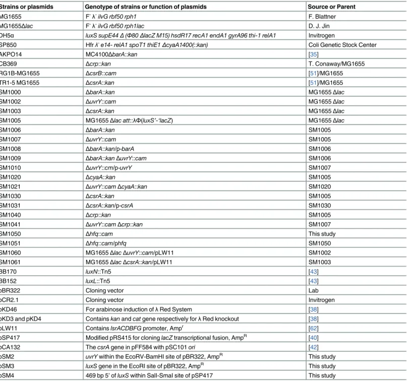
Table 1. List of bacterial strains and plasmids used in the study.
Strains or plasmids Genotype of strains or function of plasmids Source or Parent
MG1655 F-λ-ilvG rbf50 rph1 F. Blattner
MG1655Δlac F-λ-ilvG rbf50 rph1lac D. J. Jin
DH5α luxS supE44Δ(Φ80ΔlacZ M15) hsdR17 recA1 endA1 gyrA96 thi-1 relA1 Invitrogen
SP850 Hfrλ-e14- relA1 spoT1 thiE1ΔcyaA1400(::kan) Coli Genetic Stock Center
AKPO14 MC4100ΔbarA::kan [35]
CB369 Δcrp::kan T. Conaway/MG1655
RG1B-MG1655 ΔcsrB::cam [51]/MG1655
TR1-5 MG1655 ΔcsrA::kan [51]/MG1655
SM1000 ΔbarA::kan MG1655Δlac
SM1002 ΔuvrY::cam MG1655Δlac
SM1003 ΔcsrA::kan MG1655Δlac
SM1005 MG1655Δlac att::λΦ(luxS’-‘lacZ) MG1655Δlac
SM1006 ΔbarA::kan SM1005
SM1007 ΔuvrY::cam SM1005
SM1008 ΔbarA::kan/p-barA SM1006
SM1009 ΔbarA::kanΔuvrY::cam SM1006
SM1010 ΔuvrY::cm/p-uvrY SM1007
SM1020 ΔcyaA::kan SM1005
SM1021 ΔuvrY::camΔcyaA::kan SM1020
SM1030 ΔcsrA::kan SM1005
SM1031 ΔcsrA::kan/p-csrA SM1030
SM1040 Δcrp::kan SM1005
SM1041 ΔuvrY::camΔcrp::kan SM1007
SM1050 Δhfq::cam This study
SM1051 Δhfq::cam/phfq SM1050
SM1060 MG1655ΔlacΔuvrY::cam/pLW11 SM1002
SM1061 MG1655ΔlacΔcsrA::kan/pLW11 SM1003
BB170 luxN::Tn5 [43]
BB152 luxL::Tn5 [43]
pBR322 Cloning vector Lab
pCR2.1 Cloning vector Invitrogen
pKD46 For arabinose induction ofλRed System [38]
pKD3 and pKD4 Containskanandcatgene respectively forλRed knockout [38]
pLW11 ContainslsrACDBFGpromoter, Ampr [62]
pSP417 Modified pRS415 for cloninglacZtranscriptional fusion, AmpR [40]
pCA132 ThecsrAgene in pFF584 with pSC101ori [42]
pSM2 uvrYwithin the EcoRV-BamHI site of pBR322, AmpR This study
pSM3 luxSgene in the EcoRI site of pBR322, AmpR This study
pSM4 469 bp 5’ofluxSwithin SalI-SmaI site of pSP417 This study
λRS45. The resulting recombinant phage,λPluxS-lacZ(λSM001) was used to transfer the fusion into MG1655Δlac, creating a merodiploidluxS+luxS-lacZfusion (SM105). Single-copy fusions were isolated and verified by a Ter assay followed by measuringβ-galactosidase activity. A single copy fusion integrated within theλattsite of theE.colichromosome was selected to studyluxSexpression under various experimental conditions. We also observed that when a csrA::kanmutation was transduced from the parent strain TR1-5 MG1655, the resultant phe-notype was that of a very slow growing strain as reported inS.typhimurium[42]. Because nor-mal growing suppressors could be easily isolated after prolonged growth with aeration in LB broth, we selected single copyλF(luxS’-‘lacZ) (hereafter referred asluxS-lacZ) fusion in TR1-5 MG1655.
Enzymatic assays
The extracellular AI-2 in cell-free supernatant was assayed usingV.harveyistrain BB170 as described [43]. The reporter strain BB170, aluxNmutant of BB120 was chosen because of its sensitivity to AI-2 but not to AI-1. The positive controls were either BB152 (AI-1-, AI-2+) or BB120 (AI-1+AI-2+) and the negative control wasEscherichia coliDH5α, aluxSmutant which was unable to synthesize AI-2.V.harveyiwas cultured in autoinducer bioassay (AB) medium TheV.harveyireporter strains were grown overnight (~16 h) at 30°C on rotating wheels in AB medium, diluted 1:2500 into fresh medium and 180μl of the diluted cells were added to
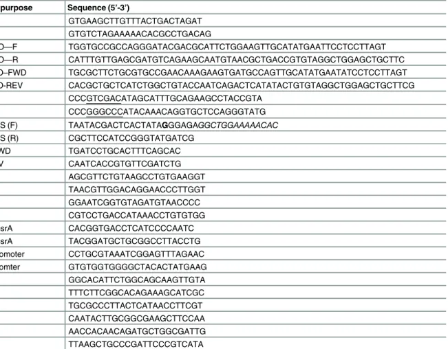
Table 2. List of oligonucleotides used in the study.
Primer Name Primer purpose Sequence (5’-3’)
OSM34 luxS–F GTGAAGCTTGTTTACTGACTAGAT
OSM35 luxS–R GTGTCTAGAAAAACACGCCTGACAG
OSM43 uvrY KO—F TGGTGCCGCCAGGGATACGACGCATTCTGGAAGTTGCATATGAATTCCTCCTTAGT
OSM44 uvrY KO—R CATTTGTTGAGCGATGTCAGAAGCAATGTAACGCTGACCGTGTAGGCTGGAGCTGCTTC
OSM49 luxS KO–FWD TGCGCTTCTGCGTGCCGAACAAAGAAGTGATGCCAGTTGCATATGAATATCCTCCTTAGT
OSM50 luxS KO-REV CACGCTGCTCATCTGGCTGTACCAATCAGACTCATATACTGTGTAGGCTGGAGCTGCTTCG
OSM53 luxS–F CCCGTCGACATAGCATTTGCAGAAGCCTACCGTA
OSM54 luxS–R CCCGGGCCCATACAAACAGGTGCTCCAGGGTATG
OSM55 T7–luxS (F) TAATACGACTCACTATAGGGAGAGGCTGGAAAAACAC
OSM56 T7–luxS (R) CGCTTCCATCCGGGTATGATCG
OSM59 luxS-FWD TGATCCTGCACTTTCAGCAC
OSM60 luxS-RV CAATCACCGTGTTCGATCTG
OSM61 rrnA-F AGCGTTCTGTAAGCCTGTGAAGGT
OSM62 rrnA-R TAACGTTGGACAGGAACCCTTGGT
OSM63 icd-F GGAATCGGTGTAGATGTAACCCC
OSM64 icd-R CGTCCTGACCATAAACCTGTGTGG
OSM 70 ChIP–csrA CACGGTGACCTCATCCCCAATC
OSM71 ChIP–csrA TACGGATGCTGCGGCCTTACCTG
OSM72 csrB promoter CCTGCGTAAATCGGAGTTTAGAAC
OSM72 csrB promter GTGTGGTGGGGCTACACTATGAAG
OSM 75 lsrK–F GGCACATTCTGGCAGCAAGTTGTA
OSM76 lsrK-R TTTCTTCGGCACAGAAAGCATCGC
OSM 77 lsrA-F TGCGCCCTTACTCATAACCTTCGT
OSM 78 lsrA-R CAATACTTGCGGCGAAGCTTCCAA
OSM 79 lsrR-F AACCACAACAGATGCTGGCGATTG
OSM 80 lsrR-R TTAAGCTGCCCGATTCCCGTCATA
microtiter wells (Nalge Nunc, Rochester, NY) alongwith 20μl of the cell-free culture superna-tants. To minimize fluctuations in luminescence and light scattering a higher volume per well was used. The microtiter plates were incubated at 30°C on a rotary shaker at 150 rp m. Light production was measured every 30 minutes using a Mediators PhL™Luminometer (ImmTech, Inc, New Windsor, MD) or by a VICTOR3™V Multilabel Counter (PerkinElmer) for 24 hours. Serial dilutions ofV.harveyiBB120 (wild type) and DH5α13h-old culture supernatant were used as a positive and negative control respectively. The relative AI-2 activity was reported in relative light units (RLU) where background reading of media and surface is subtracted from the actual reading as directly reported by the instrument for each plate.β-galactosidase assay was determined as described [39]. All assays were performed in duplicate and repeated three times.
Electrophoretic Mobility Shift Assay
Since many response regulators can be phosphorylated by acetyl phosphate, we wanted to determine whether UvrY requires phosphorylation in order to interact withluxSpromoter. TheluxSpromoter DNA was radiolabeled and various concentration of purified UvrY protein ranging from 1 to 2.5μM was used for gel-shift analysis. Purified UvrY was phosphorylated with 20 mM acetyl phosphate. Cold DNA was added to determine the strength of the interac-tion of the protein with the promoter.
Chromatin Immunoprecipitation assay
In vitro binding of UvrY protein to various promoters was determined by methods described previously [44,45]. Briefly, cultures were grown to an O. D.600of 0.8 and the expression
His6-UvrY was induced for 1 h with 1 mM IPTG at 37°C under aerobic conditions. Formalde-hyde was added at 1% final concentration to the growing cultures. After 20 min, 0.5 M glycine was added to quench the reaction, and the culture was placed on ice for another 20 minutes. The cells were subsequently washed twice with PBS (pH 7.5) and re-suspended in 1/100th vol-ume of lysis buffer (50 mM Tris HCl, pH 7.4, 150 mM NaCl, 1mM EDTA, 1% Triton X-100) with lysozyme and PMSF added to final concentrations of 4 mg/ml and 1 mM respectively. After 15 minutes of incubation at 37°C, cells were lysed and DNA sheared by sonicating in a cup horn sonicator (Misonix 3000, Farmingdale, NY). After removal of cell debris, the clear lysate was used for immunoprecipitation using anti-UvrY antibody linked to agarose beads. 300μl of clear supernatant was added to 80μl of antibody conjugated resin and mixed at room temperature with shaking for 2 h. The mixture was washed three times with 1 ml wash buffer (50 mM Tris HCl, pH 7.4, 150 mM NaCl). 100μl of elution buffer (0.1 M Glycine, pH 3.5) with 1% SDS was used to elute the antibody-captured proteins from the resin. The clear supernatant comprising of approximately 500 bp DNA fragments, was further de-crosslinked and de-pro-teinized overnight at 65°C in the wash buffer containing1 mg/ml of Proteinase K. 50μl of the de-crosslinked reaction was then cleaned using QIAquick PCR purification (Qiagen, Valencia, CA) and subsequently used to test presence of target promoters. The ChIP assay was repeated with a Flag-UvrY and anti-Flag antibody (Sigma, St. Louis, MO), confirming similar results.
Real time RT-PCR
from 5μg of total RNA using Moloney Murine Leukemia Virus Reverse Transcriptase, Super-script II RNase H-(Invitrogen, Carlsbad, CA) and 50 ng of random hexamers (Invitrogen, Carlsbad, CA). For the PCR reaction, 10 ng of first-strand cDNA was amplified separately with 10μM each of gene-specific primer and 16SrrnAgene-specific primers in a 25μl total reaction volume with Taq polymerase in a Biometra T-Gradient PCR instrument (Biometra, Horsham, PA) for 30 cycles. At the end of several cycles, a gene-specific and anrrnA-specific reaction tube was removed. Fiveμl of the reaction products were resolved separately in a 1.2% agarose gel and the product intensities were quantitated by a BioRad Gel Documentation system (BioRad, Hercules, CA). The linear range of amplification for therrnAgene was from 5–15 cycles in all backgrounds, while that of theluxSwere from 12–22 cycles in the wild-type strain, and appeared later in the mutants. A qRT-PCR reaction was performed on the above set of samples under identical reaction conditions in a LightCycler (Roche, Indianapolis, IN) with SYBR Green-1 PCR Master Mix. The fluorescence signal from SYBR Green intercalation was monitored to quantify double-stranded DNA product formed after each PCR cycle. The threshold cycle for which a statistically significant increase in the amount of the PCR product is detected is denoted as Ct. Starting with individual cDNA pools from various genetic back-grounds, Ct values were determined forrrnAandluxSamplification products. TheΔCt values between samples derived from various strains were normalized with therrnAproduct, as
ΔCt =−(CtrrnA−Ctwild-type)−(CtrrnA−Ctmutant). Since PCR products double with each
ampli-fication cycle, the fold difference in the initial concentration of each transcript is determined by 2ΔCt. The results derived from gel-based experiment indicated slightly less difference than the SYBR Green fluorescent method.
Rifampicin chase assay
RNA stability assay was performed as described earlier [31]. Briefly total RNA was isolated from cells at the entry of stationary phase whencsrAis maximally expressed. Rifampicin was added to inhibit transcription initiation and before and after 2.5, 7.5 and 10 minutes post addi-tion of rifampicin, total RNA was isolated and RT-PCR was performed. Rifmapicin was added at a final concentration of 500μg/ml. A housekeeping controlicdwas kept as the internal con-trol. Gene specific primers were used to amplifyluxSandicdmessage (Table 2).
Results
Effect of extracellular AI-2 accumulation in
uvrY
mutant
The AI-2 accumulation ofE.coligrown in LB broth was growth phase-dependent in consis-tence with previous studies [16,46]. The accumulation of AI-2 in the extracellular milieu peaks at the mid logarithmic phase, declines at the entry of stationary phase and vanishes once the cells shift deep within stationary phase of the growth cycle. No secondary peak of AI-2 was observed within a span of 24 hours. A mutation in theuvrYgene reduced AI-2 accumulation in the extracellular milieu compared to the isogenic wild-type (Fig 1A). The difference was pro-nounced in mid-exponential and in early stationary phase. In the complemented strain, the extracellular AI-2 accumulation was similar to the wild type. A very low level of AI-2 was detected in theluxS::kanmutant from cell free supernatant, and plasmid complementation in the mutant increased AI-2 levels to that of the wild type (S1 Fig).
mutant was significantly lower in mid-exponential and early stationary phase as compared to the wild type. The basal level of expression of the fusion inuvrY::cammutant was around 2-fold lower than wild type in both mid-exponential and stationary-phase and plasmid com-plementation ofuvrYin the mutant partly restores the wild type expression levels (Fig 1B).
Mutation in
uvrY
reduced expression of
luxS
transcript
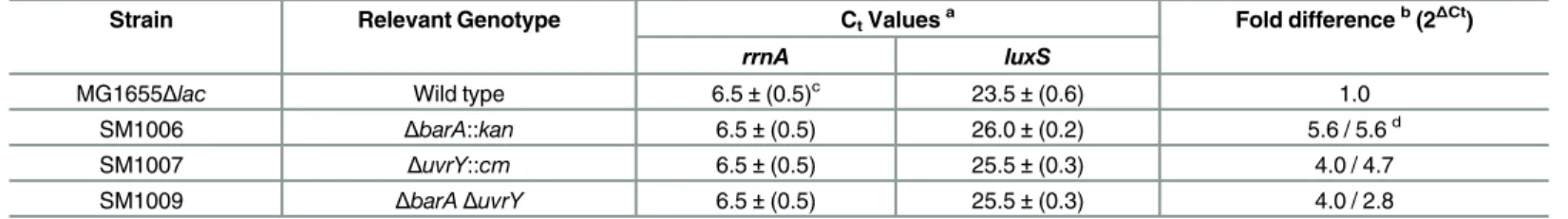
Transcription ofluxSwas evaluated by quantitative reverse transcription PCR analysis and Northern analysis (Table 3andS2 Fig). On qRT-PCR, we observed visiblerrnAamplification products by 9thcycle, and theluxStranscript in the 12thcycle in the wild-type strain. Although the accumulation ofrrnAproduct remained consistent for the mutant strains, visibleluxS amplification was observed around 16thcycles foruvrY::cmand in the double mutant strain. The experiment was repeated using a more sensitive Real Time Light Cycler (Roche, Indianap-olis, IN) using SYBR-Green to follow the simultaneous amplification of therrnAproduct with that of theluxSproducts from the same initial cDNA sample. Northern blotting was further performed to quantify the level ofluxStranscript. Loss ofuvrYresulted in reduced expression ofluxStranscript (~3.3 fold) which could be restored upon plasmid complementation (S2 Fig).
Fig 1. Effect ofuvrYon AI-2 accumulation and expression ofluxS::lacZfusion.(A) AI-2 accumulation in the supernatant was assayed byVibrio harveyireporter assay in SM1005 (luxS::lacZ), SM1007 (ΔuvrY luxS::
lacZ) and SM1010 (ΔuvrY/puvrY luxS::lacZ) for a period of 24 hours. (B) Expression of the fusion in the strains was monitored byβ-galactosidase assay for 24 hours. Dotted lines indicate growth of corresponding bacterial strains. The graphs represent mean of three experiments. Asterisk marks are provided for a representative data point used for calculation of significance.
doi:10.1371/journal.pone.0157532.g001
Table 3. The BarA-UvrY TCS regulatesluxStranscription as determined by qRT-PCR.
Strain Relevant Genotype CtValuesa Fold differenceb(2 ΔCt)
rrnA luxS
MG1655Δlac Wild type 6.5±(0.5)c 23.5±(0.6) 1.0
SM1006 ΔbarA::kan 6.5±(0.5) 26.0±(0.2) 5.6 / 5.6d
SM1007 ΔuvrY::cm 6.5±(0.5) 25.5±(0.3) 4.0 / 4.7
SM1009 ΔbarAΔuvrY 6.5±(0.5) 25.5±(0.3) 4.0 / 2.8
a
Ct values are the threshold values of PCR cycles where the SYBR Greenfluorescence was detected above background in the linear range, taken at 7.0 Relative Light Units.
b
The fold down-regulation is calculated as 2ΔCt
, WhereΔCt= (Ctwt—CtrrnA)–(Ct mutant—CtrrnA). c
Standard deviation of three independent experiments.
d
In vitro
and
in vivo
interaction of UvrY with
luxS
promoter
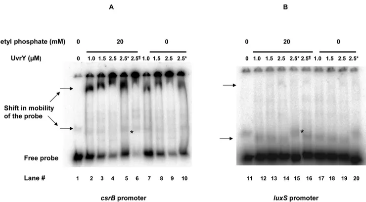
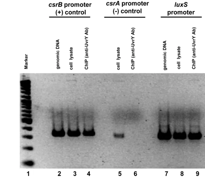
Purified UvrY interacts withluxSpromoter without and with acetyl phosphate (Fig 2). The shift in the probe was shown by an arrow. The interaction of UvrY withluxSpromoter is rela-tively weak compared to that withcsrBpromoter. Furthermore, interaction was also modulated by addition of negative and positive competitor DNA. Addition of positive competitor releases the binding whereas that of negative competitor tightens it. Since the interaction ofuvrYwith luxSpromoter was relatively weak indicating there may be yet other factors associated with the interaction, we performed a in vivo chromatin immunoprecipitation of theluxSpromoter frag-ment to determine thein vivointeraction using anti-uvrY antibody. Our result confirms the in-vivo binding of UvrY withluxSpromoter (Fig 3). For the control reactions, we found thein vivointeraction of UvrY withcsrBis relatively strong, while that withcsrAwas very weak, as expected from previous studies.
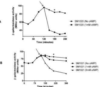
UvrY is required for cAMP-CRP repression of
luxS
-lacZ expression
We further wanted to test any additional regulator might be involved in expression of luxS-lacZfusion. An important regulation occurs through the cyclic AMP-cyclic AMP Receptor Protein (cAMP-CRP) system. To further determine the role of UvrY in the glucose repression ofluxS-lacZfusion, we deleted adenylate cyclase encoding genecyaA, which blockssynthesis of cAMP from ATP. Disruption ofcyagene led to a constitutive expression of theluxS-lacZ fusion (Fig 4A). Addition of 1mM cAMP reduced the expression ofluxSfusion to a basal level in the mutant. In case ofΔuvrYΔcyadouble mutant, the expression of the fusion is constitutive and addition of 1mM cAMP reduced the expression marginally (Fig 4B). The repression was faster in aΔcyamutant (30 min) compared to aΔuvrYΔcyamutant (~90 min). Only additionFig 2. Electrophoretic mobility shift assay (EMSA) demonstrating interaction of UvrY with A) labeledcsrBB) labeledluxSpromoter.The promoter DNA was radiolabelled with P32. Purified UvrY was added at indicated concentrations without and with 20mM acetyl phosphate. The shift in probe was indicated by an arrow. Unlabeled DNA were added as negative and positive competitors shown by*and ¶ respectively.
of 5mM reduced the expression of the double mutant to a basal level. Similarly, deletion ofcrp resulted in constitutive expression ofluxSand aΔuvrYΔcrpmutant reduced the expression of the fusion marginally (Fig 5).
Effect of extracellular AI-2 accumulation and expression of
luxS-lacZ
fusion in
csrA
mutant
In contrast touvrYmutant, mutation in thecsrAleads to elevated levels of AI-2 relative to the wild type and complementation ofcsrAin the mutant reduced extracellular accumulation of AI-2 to the wild-type level (Fig 6A). The inverse correlation of AI-2 accumulation in theuvrY andcsrAdeficient strains suggested a regulation both at the level of transcription and post tran-scription ofluxSand AI-2 uptake. The uptake of AI-2 was also evaluated in theuvrYandcsrA mutants and indicated an inverse relationship in thelsroperon expression. Similarly in case of thecsrAmutant, the expression of the fusion was significantly higher at the entry of stationary
Fig 3. Directin-vivobinding of UvrY withluxSpromoter by chromatin immunoprecipitation assay (ChIP).The occupancy of UvrY at thecsrA,csrB
andluxSpromoters inE.coliwas analyzed by ChIP using an anti-UvrY antibody. Purified DNA was PCR amplified using target specific primers. Genomic DNA and cell lysates were used as template for additional controls in PCR reaction. A 100 bp ladder marker was used for size comparison of amplicons. Expected sizes for all amplicons were between 300–400 bp.
phase as compared to the wild type while complementation of the mutant restored the expres-sion to the wild type level (Fig 6B). Interestingly, the expression pattern was similar to that of the wild-type strain, with maximum expression being observed as cells enter the stationary phase. A mutation in thecsrBgene resulted in marginally higher expression of theluxS-lacZ fusion.
Regulatory interaction of CsrA with
luxS
mRNA stability or
luxS
mRNA
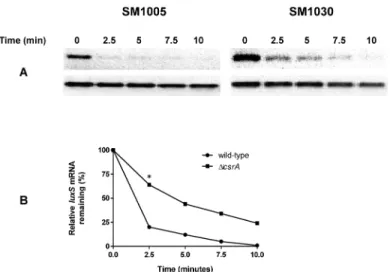
leader sequence
We evaluated the effect ofcsrAinluxStranscript stability. At the entry into stationary phase, loss ofcsrAresulted in stabilization ofluxStranscript relative to the wild-type straining harbor-ingcsrA(Fig 7A). Quantification of the DNA intensities by ImageJ suggested a three-fold
Fig 4. UvrY influences cAMP-CRP catabolite repression ofluxS.A) Expression ofluxS::lacZin SM1020 (ΔcyaA::kan luxS::lacZ) in the absence of cAMP and in the presence of 1 mM cAMP. (B) Expression ofluxS::
lacZin SM1021 (ΔuvrY::camΔcyaA::kan luxS::lacZ) in the absence of cAMP, 1mM cAMP, and 5 mM cAMP. The point of cAMP addition is marked with an arrow on the graphs. Mean of three experiments were plotted and a representative data point used for calculation of significance is shown in asterisk (p<0.05).
doi:10.1371/journal.pone.0157532.g004
Fig 5. Effect ofcrponluxS::lacZexpression in absence or presence ofuvrY.Strains SM1005 (luxS::
lacZ), SM1007 (ΔuvrY luxS::lacZ), SM1040 (Δcrp::kan luxS::lacZ) and SM1041 (Δcrp::kanΔuvrY::cam luxS::
lacZ) were grown in TB andβ-galactosidase expression was measured at indicated time points. This experiment was repeated three times and mean was plotted in the graph.
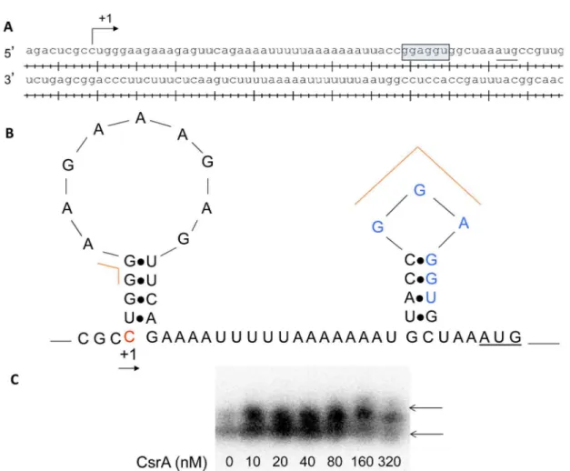
reduction in the half-life ofluxStranscript in the wild-type relative to theΔcsrAmutant (Fig 7B). CsrA is known to bind leader regions of target genes with high affinity for GGA sites. We predicted the potential binding sites for CsrA in theluxSleader region with GGA sites using Lasergene program suite (DNAstar, Madison, WI, USA), (Fig 8A). Folding of the leader RNA using RNAFold in the Vienna RNA website indicated that one of the GGA site is actually pres-ent within the ribosome binding site ofluxS(Fig 8B). Our results indicate that CsrA stabilizes luxSmessage stability at the entry into stationary phase. We also determined the interaction of CsrA withluxSleader region. Our results indicate that CsrA binds toluxSleader region and
Fig 6. Extracellular accumulation of AI-2 and expression ofluxS::lacZfusion in acsrAmutant.(A) AI-2 accumulation in the supernatant was assayed byVibrio harveyireporter assay in SM1005 (luxS::lacZ), SM1030 (ΔcsrA luxS::lacZ) and SM1031 (ΔcsrA/pcsrA luxS::lacZ) (B) Expression ofluxS::lacZwas monitored byβ-galactosidase assay in same sets of strains at indicted time points. Dotted lines indicate growth of corresponding bacterial strains. The graphs represent mean of three experiments. Representative data point used for calculation of significance is shown in asterisk.
doi:10.1371/journal.pone.0157532.g006
Fig 7. Effect ofcsrAon message stability ofluxSinE.coli.Total RNA was isolated from late logarithmic growth phase. (A) Message stability ofluxSoricd(housekeeping control) was evaluated before and following addition of rifampicin for 10 minutes. (B) Relative intensity of theluxSin the wild-type and the mutant as compared to intensity oficdbefore addition of rifampicin was set at 100%. This experiment was repeated two times.
influencesluxSmessage stability (Fig 8C). We also determined the effect ofhfqon theluxSfusion expression. We found that loss ofhfqreduced theluxSfusion expression significantly and com-plementation ofhfqin the mutant restoredluxSexpression to wild-type levels (S3 Fig).
Effect of expression of Lsr promoter,
lsrA
and
lsrk
in
uvrY
and
csrA
mutant
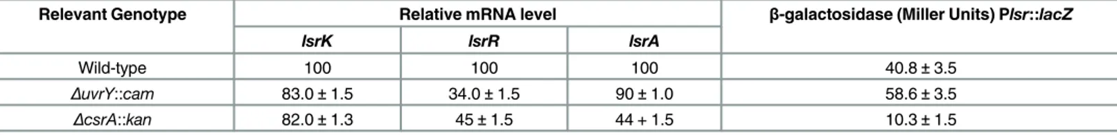
In order to determine whetheruvrYandcsrAplay a role in the regulation of uptake of AI-2 sig-nal, the expression of thelsrAorlsrKor the Lsr promoter was evaluated inuvrYandcsrAmutant. The Lsr promoter activity increased marginally inΔuvrYmutant and approximately four-fold reduced in thecsrAmutant at the entry of stationary phase (Fig 9). Expression of thelsrRwas reduced both in theuvrYandcsrAmutant (Table 4). The uptake of AI-2 was also evaluated in theuvrYandcsrAmutants and indicated an inverse relationship in thelsroperon expression.
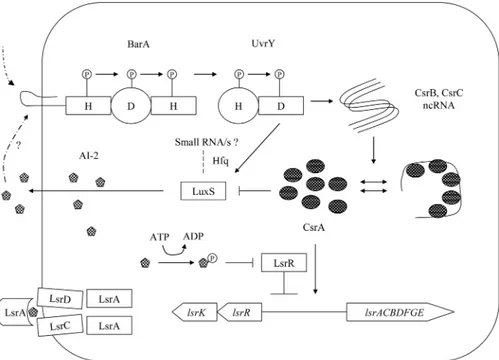
Predicted integrative model of AI-2 based cell-signaling and the BarA/
UvrY/CsrA pathway
We predicted a model integrating the BarA/UvrY/CsrA pathway with AI-2 based signaling (Fig 10). The BarA/UvrY/CsrA pathway controls both the synthesis and uptake of AI-2 via
Fig 8. Regulatory interactions of CsrA withluxSupstream region.A. Transcription start site and ribosome binding sites ofluxSwere indicated. B. Predicted binding sites of CsrA inluxSupstream regulatory region. C. CsrA-luxS mRNA regulatory interactions. 5’- end labeledluxSleader transcript was incubated with CsrA at concentration as shown below each lane. Positions of free and bound RNA are shown. The shift in the labeled probe was shown by an arrow.
transcriptional and posttranscriptional mode of regulation ofluxSandlsrtransporter. In this model both BarA, the sensor kinase and UvrY, the response regulator positively regulates the expression ofluxSand AI-2 accumulation. CsrA on the other hand, negatively regulatesluxS expression and AI-2 accumulation in the extracellular milieu. By contrast, the BarA/UvrY neg-atively controls Lsr promoter activity, whereas CsrA positively controls Lsr promoter activity. Such regulation suggests a balance between synthesis and uptake of AI-2 inE.coli. The repres-sion ofluxSis dependent on cAMP-CRP anduvrYinfluences this regulation. The role of small RNAs could be important for regulation ofluxSinE.coli.
Discussions
Quorum sensing is a bacterial population dependent cell-to-cell signaling mechanism mediated via synthesis, release and uptake of small freely diffusible molecules called autoinducers [47,
48]. Apart from the requirement of QS on cell-density, other environmental cues such as pH, nutrient depletion and stress are important for quorum sensing systems to synchronize coordi-nated response for adaptation and survival of bacteria [49]. InPseudomonas aeruginosa, the integration of metabolic cues with cell-cell signaling requires several regulators at the level of transcription and post transcription [50]. Metabolic adaptation through two-component regu-latory system is achieved by signal detection and response by appropriate changes in gene expression by the cognate response regulator.
The conserved pathways of BarA/UvrY/CsrA and its orthologs in theγ-subdivision of pro-teobacteria regulates virulence and secondary metabolism inE.coli,S.typhimurium,P. fluores-censandV.fischeri[51–58]. Our earlier work inE.colidemonstrated the pleiotropic role of BarA/UvrY/CsrA pathway in regulation of biofilm formation, motility and virulence gene expression [29–31]. Here we have shown the association of this regulatory pathway withluxS based AI-2 quorum sensing. Our data shows that multiple regulatory factors control the expression of LuxS including the BarA/UvrY/CsrA pathway, and global virulence regulator, crpand small regulatory RNAs. We found the interaction of UvrY withluxSpromoter is very
Fig 9. Effect oflsrpromoter activity inuvrYandcsrAmutant.SM1059 (wild type/p-lsr), SM1060 (ΔuvrY/p-lsr) and SM1061 (ΔcsrA/p-lsr) were tested for the expression of Lsr promoter activity. This experiment was repeated three times and mean of three experiments for each time points were plotted (p<0.05). Representative data point used for calculation of significance is shown in asterisk.
doi:10.1371/journal.pone.0157532.g009
Table 4. Effect of mutation ofuvrYandcsrAon expression of Lsr transporter.
Relevant Genotype Relative mRNA level β-galactosidase (Miller Units) Plsr::lacZ
lsrK lsrR lsrA
Wild-type 100 100 100 40.8±3.5
ΔuvrY::cam 83.0±1.5 34.0±1.5 90±1.0 58.6±3.5
ΔcsrA::kan 82.0±1.3 45±1.5 44 + 1.5 10.3±1.5
weakin vitroand that the interaction can be demonstratedin vivo. This suggests that there are other additional factors such as DNA bending proteins (HU, IHF, etc.) that might play a role in this regulation [59]. These findings indicate that the BarA/UvrY/CsrA pathway integrates with AI-2 based cell-cell signaling inEscherichia coliand as a consequence the regulatory path-way play a critical role in metabolism, persistence, virulence and other community associated microbial behaviors. The control of synthesis and uptake of AI-2 suggest regulatory crosstalk of metabolic pathways with cell-cell signaling. Our results are similar to the regulation inVibrio cholerawhere CsrA and three small RNA’s regulate quorum sensing [60]. VarS/VarA two-component regulatory system inVibrio choleraalso regulates quorum sensing [20]. Further-moreluxSinfluences biofilm formation in both AI-2 and AI-2 independent manner inE.coli [61]. Our results further are in consistence with the role of cAMP-CRP mediated repression of luxSin AI-2 synthesis [62]. Swarming, a flagella and cell-density dependent phenomenon, is also reduced inuvrYdelectation mutant and complementation ofuvrYin the mutant restored swarming in UPEC [31]. This further confirms the integration of the BarA/UvrY/CsrA path-way with AI-2 based quorum sensing signaling and biofilm formation.
This study indicates that the BarA-UvrY-CsrA pathway regulateE.colimetabolism, nutri-ent acquisition and cell-cell signaling and forms an underlying basis of bacterium-host signal-ing recognition and pathogenesis. Because bothluxSand the BarA/UvrY/CsrA pathway are conserved in several bacterial species, such regulation and integration might be investigated in clinically relevant pathogens. The integration of the BarA/UvrY/CsrA pathway and its homo-logs with cell-to-cell signaling in diverse pathogenic bacteria may be explored to determine if integration of metabolic pathway with cell-to cell signaling can be viewed as common themes in biological regulation. However, many strains ofE.colilack a functionallsrgene which might lead to accumulation of AI-2 in the extracellular environment and consequently cross species signaling might be more frequent in certain niche for coordinating changes in gene expression
Fig 10. Regulatory circuit of the BarA/UvrY/CsrA with AI-2 based cell-to-cell signaling.The BarA/UvrY TCS regulatesluxSexpression positively at transcriptional level whereas CsrA negatively regulatesluxS
post-transcriptionally. A possible role of quorum sensing regulatory RNA is indicated in the circuit.
[63]. Depending on the polymicrobial environment, the regulatory interactions of the pathway will influence accumulation of AI-2 in the extracellular environment and consequently AI-2 signaling can have varying role in the community structure and dynamics. Targeting two-com-ponent regulatory system that integrates with cell-to-cell signaling may influence both intra-species and interintra-species microbial communication in a niche-specific manner. These strategies may be effective for development of novel therapeutics particularly for infections that are diffi-cult to treat due to emergence of antibiotic resistance.
Supporting Information
S1 Fig. AI-2 accumulation inEscherichia coliMG1655 (harbouring wild typeluxSgene), luxSdeletion mutant and plasmid complemented strains ofluxSdeletion mutant.Note the wild type in this experiment is MG1655, which is not the same as SM1005, a merodiploid strain. Accumulation of AI-2 in the cell-free supernatant was assayed byVibrio harveyi reporter assay as described in the text. Dotted lines indicate growth of corresponding bacterial strains. The graphs represent mean of three experiments. Representative data point used for calculation of significance is shown in asterisk.
(TIF)
S2 Fig. Northern analysis ofluxSmessage inΔbarAorΔuvrYmutant.Relative pixel inten-sity of the signal to rRNA signal is expressed as numbers at the bottom. Deletion ofbarAor uvrYreduced expression ofluxSwhereas complementation of the mutant restored the expres-sion level ofluxSsimilar to the wild-type. This experiment was repeated twice.
(TIF)
S3 Fig. Effect ofhfqonluxS::lacZreporter activity.Mutation inhfqreduced reporter activity at the entry of stationary phase and complementation of Hfq in the mutant restored the expres-sion ofluxSfusion. This experiment was repeated two times. Representative data point used for calculation of significance is shown in asterisk.
(TIF)
S1 File. Data.
(XLSX)
Acknowledgments
We sincerely acknowledge B. Bassler for providing theV.harveyireporter strains. We thank T. Romeo, S. Adhya, D. J. Jin, B. Wanner, R. W. Simmons, T. Larson, T. Conaway for strains, plasmids and bacteriophages. Many thanks go to D. K. Chattoraj and members of late T. Cebu-la’s laboratory for their advice, guidance and support.
Author Contributions
Conceived and designed the experiments: AM CDH IRP SM. Performed the experiments: AM CDH IRP AC. Analyzed the data: AM CDH IRP SM. Wrote the paper: AM SM.
References
1. Waters CM, Bassler BL. Quorum sensing: cell-to-cell communication in bacteria. Annu Rev Cell Dev Biol. 2005; 21:319–46. doi:10.1146/annurev.cellbio.21.012704.131001PMID:16212498.
3. Yang Q, Defoirdt T. Quorum sensing positively regulates flagellar motility in pathogenic Vibrio harveyi. Environ Microbiol. 2014. doi:10.1111/1462-2920.12420PMID:24528485.
4. Zhu J, Miller MB, Vance RE, Dziejman M, Bassler BL, Mekalanos JJ. Quorum-sensing regulators con-trol virulence gene expression in Vibrio cholerae. Proc Natl Acad Sci U S A. 2002; 99(5):3129–34. doi: 10.1073/pnas.052694299PMID:11854465; PubMed Central PMCID: PMC122484.
5. Quinones B, Dulla G, Lindow SE. Quorum sensing regulates exopolysaccharide production, motility, and virulence in Pseudomonas syringae. Mol Plant Microbe Interact. 2005; 18(7):682–93. doi:10.1094/ MPMI-18-0682PMID:16042014.
6. Queck SY, Weitere M, Moreno AM, Rice SA, Kjelleberg S. The role of quorum sensing mediated devel-opmental traits in the resistance of Serratia marcescens biofilms against protozoan grazing. Environ Microbiol. 2006; 8(6):1017–25. doi:10.1111/j.1462-2920.2006.00993.xPMID:16689722.
7. Ray VA, Visick KL. LuxU connects quorum sensing to biofilm formation in Vibrio fischeri. Mol Microbiol. 2012; 86(4):954–70. doi:10.1111/mmi.12035PMID:23035866; PubMed Central PMCID:
PMC3566283.
8. Vidal JE, Ludewick HP, Kunkel RM, Zahner D, Klugman KP. The LuxS-dependent quorum-sensing system regulates early biofilm formation by Streptococcus pneumoniae strain D39. Infect Immun. 2011; 79(10):4050–60. doi:10.1128/IAI.05186-11PMID:21825061; PubMed Central PMCID: PMC3187250.
9. Bhardwaj AK, Vinothkumar K, Rajpara N. Bacterial quorum sensing inhibitors: attractive alternatives for control of infectious pathogens showing multiple drug resistance. Recent Pat Antiinfect Drug Discov. 2013; 8(1):68–83. PMID:23394143.
10. Walters M, Sperandio V. Quorum sensing in Escherichia coli and Salmonella. Int J Med Microbiol. 2006; 296(2–3):125–31. doi:10.1016/j.ijmm.2006.01.041PMID:16487745.
11. Ahmer BM. Cell-to-cell signalling in Escherichia coli and Salmonella enterica. Mol Microbiol. 2004; 52 (4):933–45. doi:10.1111/j.1365-2958.2004.04054.xPMID:15130116.
12. Xavier KB, Bassler BL. LuxS quorum sensing: more than just a numbers game. Curr Opin Microbiol. 2003; 6(2):191–7. PMID:12732311.
13. Santiago-Rodriguez TM, Patricio AR, Rivera JI, Coradin M, Gonzalez A, Tirado G, et al. luxS in bacteria isolated from 25- to 40-million-year-old amber. FEMS Microbiol Lett. 2014; 350(1):117–24. doi:10. 1111/1574-6968.12275PMID:24102660.
14. De Keersmaecker SC, Sonck K, Vanderleyden J. Let LuxS speak up in AI-2 signaling. Trends Micro-biol. 2006; 14(3):114–9. doi:10.1016/j.tim.2006.01.003PMID:16459080.
15. Taga ME, Semmelhack JL, Bassler BL. The LuxS-dependent autoinducer AI-2 controls the expression of an ABC transporter that functions in AI-2 uptake in Salmonella typhimurium. Mol Microbiol. 2001; 42 (3):777–93. PMID:11722742.
16. Xavier KB, Bassler BL. Regulation of uptake and processing of the quorum-sensing autoinducer AI-2 in Escherichia coli. J Bacteriol. 2005; 187(1):238–48. doi:10.1128/JB.187.1.238–248.2005PMID: 15601708; PubMed Central PMCID: PMC538819.
17. Dong YH, Zhang XF, Soo HM, Greenberg EP, Zhang LH. The two-component response regulator PprB modulates quorum-sensing signal production and global gene expression in Pseudomonas aerugi-nosa. Mol Microbiol. 2005; 56(5):1287–301. doi:10.1111/j.1365-2958.2005.04612.xPMID:15882421.
18. Fujii T, Ingham C, Nakayama J, Beerthuyzen M, Kunuki R, Molenaar D, et al. Two homologous Agr-like quorum-sensing systems cooperatively control adherence, cell morphology, and cell viability properties in Lactobacillus plantarum WCFS1. J Bacteriol. 2008; 190(23):7655–65. doi:10.1128/JB.01489-07 PMID:18805979; PubMed Central PMCID: PMC2583610.
19. Sperandio V, Torres AG, Kaper JB. Quorum sensing Escherichia coli regulators B and C (QseBC): a novel two-component regulatory system involved in the regulation of flagella and motility by quorum sensing in E. coli. Mol Microbiol. 2002; 43(3):809–21. PMID:11929534.
20. Tsou AM, Liu Z, Cai T, Zhu J. The VarS/VarA two-component system modulates the activity of the Vib-rio cholerae quorum-sensing transcriptional regulator HapR. Microbiology. 2011; 157(Pt 6):1620–8. doi:10.1099/mic.0.046235–0PMID:21393367; PubMed Central PMCID: PMC3167916.
21. Reading NC, Torres AG, Kendall MM, Hughes DT, Yamamoto K, Sperandio V. A novel two-component signaling system that activates transcription of an enterohemorrhagic Escherichia coli effector involved in remodeling of host actin. J Bacteriol. 2007; 189(6):2468–76. doi:10.1128/JB.01848-06PMID: 17220220; PubMed Central PMCID: PMC1899401.
22. Krell T, Lacal J, Busch A, Silva-Jimenez H, Guazzaroni ME, Ramos JL. Bacterial sensor kinases: diver-sity in the recognition of environmental signals. Annual review of microbiology. 2010; 64:539–59. Epub 2010/09/10. doi:10.1146/annurev.micro.112408.134054PMID:20825354.
24. Mizuno T. His-Asp phosphotransfer signal transduction. J Biochem. 1998; 123(4):555–63. PMID: 9538242.
25. Jung K, Fried L, Behr S, Heermann R. Histidine kinases and response regulators in networks. Curr Opin Microbiol. 2012; 15(2):118–24. doi:10.1016/j.mib.2011.11.009PMID:22172627.
26. Egger LA, Park H, Inouye M. Signal transduction via the histidyl-aspartyl phosphorelay. Genes Cells. 1997; 2(3):167–84. PMID:9189755.
27. Khorchid A, Ikura M. Bacterial histidine kinase as signal sensor and transducer. Int J Biochem Cell Biol. 2006; 38(3):307–12. doi:10.1016/j.biocel.2005.08.018PMID:16242988.
28. Mizuno T. Compilation of all genes encoding two-component phosphotransfer signal transducers in the genome of Escherichia coli. DNA Res. 1997; 4(2):161–8. PMID:9205844.
29. Herren CD, Mitra A, Palaniyandi SK, Coleman A, Elankumaran S, Mukhopadhyay S. The BarA-UvrY two-component system regulates virulence in avian pathogenic Escherichia coli O78:K80:H9. Infect Immun. 2006; 74(8):4900–9. doi:10.1128/IAI.00412-06PMID:16861679; PubMed Central PMCID: PMC1539585.
30. Palaniyandi S, Mitra A, Herren CD, Lockatell CV, Johnson DE, Zhu X, et al. BarA-UvrY two-component system regulates virulence of uropathogenic E. coli CFT073. PLoS One. 2012; 7(2):e31348. doi:10. 1371/journal.pone.0031348PMID:22363626; PubMed Central PMCID: PMC3283629.
31. Mitra A, Palaniyandi S, Herren CD, Zhu X, Mukhopadhyay S. Pleiotropic roles of uvrY on biofilm forma-tion, motility and virulence in uropathogenic Escherichia coli CFT073. PLoS One. 2013; 8(2):e55492. doi:10.1371/journal.pone.0055492PMID:23383333; PubMed Central PMCID: PMC3562240.
32. Sperandio V, Mellies JL, Nguyen W, Shin S, Kaper JB. Quorum sensing controls expression of the type III secretion gene transcription and protein secretion in enterohemorrhagic and enteropathogenic Escherichia coli. Proc Natl Acad Sci U S A. 1999; 96(26):15196–201. PMID:10611361; PubMed Cen-tral PMCID: PMC24796.
33. Antunes LC, Ferreira RB, Buckner MM, Finlay BB. Quorum sensing in bacterial virulence. Microbiology. 2010; 156(Pt 8):2271–82. doi:10.1099/mic.0.038794–0PMID:20488878.
34. Palaniyandi S, Mitra A, Herren CD, Zhu X, Mukhopadhyay S. LuxS contributes to virulence in avian pathogenic Escherichia coli O78:K80:H9. Vet Microbiol. 2013; 166(3–4):567–75. doi:10.1016/j.vetmic. 2013.07.009PMID:23958403.
35. Pernestig AK, Melefors O, Georgellis D. Identification of UvrY as the cognate response regulator for the BarA sensor kinase in Escherichia coli. J Biol Chem. 2001; 276(1):225–31. doi:10.1074/jbc.
M001550200PMID:11022030.
36. Gonzalez Barrios AF, Zuo R, Hashimoto Y, Yang L, Bentley WE, Wood TK. Autoinducer 2 controls bio-film formation in Escherichia coli through a novel motility quorum-sensing regulator (MqsR, B3022). J Bacteriol. 2006; 188(1):305–16. doi:10.1128/JB.188.1.305–316.2006PMID:16352847; PubMed Cen-tral PMCID: PMC1317603.
37. Sambrook J, Russell D. W. Molecular cloning: a laboratory manual. 3 ed. Cold Spring Harbor, NY: Cold Spring Harbor Laboratory Press; 2001.
38. Datsenko KA, Wanner BL. One-step inactivation of chromosomal genes in Escherichia coli K-12 using PCR products. Proc Natl Acad Sci U S A. 2000; 97(12):6640–5. doi:10.1073/pnas.120163297PMID: 10829079; PubMed Central PMCID: PMC18686.
39. Miller JH. A short course in bacterial genetics: a laboratory manual and handbook for Escherichia coli and related bacteria: Cold Spring Harbor Laboratory Press; 1992.
40. Podkovyrov SM, Larson TJ. A new vector-host system for construction of lacZ transcriptional fusions where only low-level gene expression is desirable. Gene. 1995; 156(1):151–2. PMID:7737510.
41. Simons RW, Houman F, Kleckner N. Improved single and multicopy lac-based cloning vectors for pro-tein and operon fusions. Gene. 1987; 53(1):85–96. PMID:3596251.
42. Altier C, Suyemoto M, Lawhon SD. Regulation of Salmonella enterica serovar typhimurium invasion genes by csrA. Infect Immun. 2000; 68(12):6790–7. PMID:11083797; PubMed Central PMCID: PMC97782.
43. Surette MG, Bassler BL. Quorum sensing in Escherichia coli and Salmonella typhimurium. Proc Natl Acad Sci U S A. 1998; 95(12):7046–50. PMID:9618536; PubMed Central PMCID: PMC22733.
44. Wade JT, Reppas NB, Church GM, Struhl K. Genomic analysis of LexA binding reveals the permissive nature of the Escherichia coli genome and identifies unconventional target sites. Genes Dev. 2005; 19 (21):2619–30. PMID:16264194.
46. Hardie KR, Cooksley C, Green AD, Winzer K. Autoinducer 2 activity in Escherichia coli culture superna-tants can be actively reduced despite maintenance of an active synthase, LuxS. Microbiology. 2003; 149(Pt 3):715–28. PMID:12634340.
47. Williams P, Winzer K, Chan WC, Camara M. Look who's talking: communication and quorum sensing in the bacterial world. Philos Trans R Soc Lond B Biol Sci. 2007; 362(1483):1119–34. doi:10.1098/rstb. 2007.2039PMID:17360280; PubMed Central PMCID: PMC2435577.
48. Bassler BL, Losick R. Bacterially speaking. Cell. 2006; 125(2):237–46. doi:10.1016/j.cell.2006.04.001 PMID:16630813.
49. Lee J, Wu J, Deng Y, Wang J, Wang C, Wang J, et al. A cell-cell communication signal integrates quo-rum sensing and stress response. Nat Chem Biol. 2013; 9(5):339–43. doi:10.1038/nchembio.1225 PMID:23542643.
50. Juhas M, Eberl L, Tummler B. Quorum sensing: the power of cooperation in the world of Pseudomonas. Environ Microbiol. 2005; 7(4):459–71. doi:10.1111/j.1462-2920.2005.00769.xPMID:15816912.
51. Suzuki K, Wang X, Weilbacher T, Pernestig AK, Melefors O, Georgellis D, et al. Regulatory circuitry of the CsrA/CsrB and BarA/UvrY systems of Escherichia coli. J Bacteriol. 2002; 184(18):5130–40. PMID: 12193630; PubMed Central PMCID: PMC135316.
52. Ahmer BM, van Reeuwijk J, Watson PR, Wallis TS, Heffron F. Salmonella SirA is a global regulator of genes mediating enteropathogenesis. Mol Microbiol. 1999; 31(3):971–82. PMID:10048039.
53. Teplitski M, Goodier RI, Ahmer BM. Pathways leading from BarA/SirA to motility and virulence gene expression in Salmonella. J Bacteriol. 2003; 185(24):7257–65. PMID:14645287; PubMed Central PMCID: PMC296259.
54. Laville J, Voisard C, Keel C, Maurhofer M, Defago G, Haas D. Global control in Pseudomonas fluores-cens mediating antibiotic synthesis and suppression of black root rot of tobacco. Proc Natl Acad Sci U S A. 1992; 89(5):1562–6. PMID:1311842; PubMed Central PMCID: PMC48492.
55. Gaffney TD, Lam ST, Ligon J, Gates K, Frazelle A, Di Maio J, et al. Global regulation of expression of antifungal factors by a Pseudomonas fluorescens biological control strain. Mol Plant Microbe Interact. 1994; 7(4):455–63. PMID:8075420.
56. Eriksson AR, Andersson RA, Pirhonen M, Palva ET. Two-component regulators involved in the global control of virulence in Erwinia carotovora subsp. carotovora. Mol Plant Microbe Interact. 1998; 11 (8):743–52. doi:10.1094/MPMI.1998.11.8.743PMID:9675890.
57. Lapouge K, Schubert M, Allain FH, Haas D. Gac/Rsm signal transduction pathway of gamma-proteo-bacteria: from RNA recognition to regulation of social behaviour. Mol Microbiol. 2008; 67(2):241–53. doi:10.1111/j.1365-2958.2007.06042.xPMID:18047567.
58. Jang J, Jung KT, Park J, Yoo CK, Rhie GE. The Vibrio cholerae VarS/VarA two-component system controls the expression of virulence proteins through ToxT regulation. Microbiology. 2011; 157(Pt 5):1466–73. doi:10.1099/mic.0.043737–0PMID:21330435.
59. Swinger KK, Rice PA. IHF and HU: flexible architects of bent DNA. Current opinion in structural biology. 2004; 14(1):28–35. doi:10.1016/j.sbi.2003.12.003PMID:15102446.
60. Lenz DH, Miller MB, Zhu J, Kulkarni RV, Bassler BL. CsrA and three redundant small RNAs regulate quorum sensing in Vibrio cholerae. Mol Microbiol. 2005; 58(4):1186–202. doi:10.1111/j.1365-2958. 2005.04902.xPMID:16262799.
61. Niu C, Robbins CM, Pittman KJ, Osborn j L, Stubblefield BA, Simmons RB, et al. LuxS influences Escherichia coli biofilm formation through autoinducer-2-dependent and autoinducer-2-independent modalities. FEMS Microbiol Ecol. 2013; 83(3):778–91. doi:10.1111/1574-6941.12034PMID: 23078586.
62. Wang L, Hashimoto Y, Tsao CY, Valdes JJ, Bentley WE. Cyclic AMP (cAMP) and cAMP receptor pro-tein influence both synthesis and uptake of extracellular autoinducer 2 in Escherichia coli. J Bacteriol. 2005; 187(6):2066–76. doi:10.1128/JB.187.6.2066–2076.2005PMID:15743955; PubMed Central PMCID: PMC1064054.