Mannitol Utilisation is Required for Protection of
Staphylococcus aureus
from Human Skin Antimicrobial
Fatty Acids
John G. Kenny1., Josephine Moran1., Stacey L. Kolar2, Alexander Ulanov3, Zhong Li3, Lindsey N. Shaw2, Elisabet Josefsson4, Malcolm J. Horsburgh1*
1Institute of Integrative Biology, University of Liverpool, Liverpool, Merseyside, United Kingdom,2Department of Cell Biology, Microbiology & Molecular Biology, University of South Florida, Tampa, Florida, United States of America,3Roy J. Carver Biotechnology Center, University of Illinois, Urbana-Champaign, Illinois, United States of America,4Department of Rheumatology and Inflammation Research, University of Gothenburg, Go¨teborg, Sweden
Abstract
Mannitol (Mtl) fermentation, with the subsequent production of acid, is a species signature ofStaphylococcus aureus, and discriminates it from most other members of the genus. Inactivation of the genemtlD,encoding Mtl-1-P dehydrogenase was found to markedly reduce survival in the presence of the antimicrobial fatty acid, linoleic acid. We demonstrate that the sugar alcohol has a potentiating action for this membrane-acting antimicrobial. Analysis of cellular metabolites revealed that, during exponential growth, themtlDmutant accumulated high levels of Mtl and Mtl-P. The latter metabolite was not detected in its isogenic parent strain or a deletion mutant of the entiremtlABFDoperon. In addition, themtlDmutant strain exhibited a decreased MIC for H2O2, however virulence was unaffected in a model of septic arthritis.
Citation:Kenny JG, Moran J, Kolar SL, Ulanov A, Li Z, et al. (2013) Mannitol Utilisation is Required for Protection ofStaphylococcus aureusfrom Human Skin Antimicrobial Fatty Acids. PLoS ONE 8(7): e67698. doi:10.1371/journal.pone.0067698
Editor:Michael Otto, National Institutes of Health, United States of America
ReceivedOctober 23, 2012;AcceptedMay 21, 2013;PublishedJuly 4, 2013
Copyright:ß2013 Kenny et al. This is an open-access article distributed under the terms of the Creative Commons Attribution License, which permits unrestricted use, distribution, and reproduction in any medium, provided the original author and source are credited.
Funding:This study was supported by BBSRC grant BB/D003563/1 (MJH) and in part AI090350 (LNS) from the National Institute of Allergies and Infectious Diseases. The funders had no role in study design, data collection and analysis, decision to publish, or preparation of the manuscript.
Competing Interests:MJH has funding from Unilever Plc to investigate skin survival mechanisms of skin staphylococci. This does not alter the authors’ adherence to all the PLOS ONE policies on sharing data and materials.
* E-mail: [email protected]
.These authors contributed equally to this work.
Introduction
S. aureusis a common skin and soft tissue pathogen capable of causing more severe infections including sepsis, osteomyelitis, and endocarditis [1]. The range of infections is due to a multitude of encoded virulence factors and nasopharyngeal carriage is frequent and a risk factor [2,3]. The spread of antibiotic-resistant strains and the emergence of community-acquired MRSA have increased the impact ofS. aureuson public health and it has necessitated the development of new therapeutics plus a better understanding of transmission and skin survival [4].
Several different barrier functions are proposed to retard the survival ofS. aureuson human skin, these include the antimicrobial peptides cathelicidin LL-37 and humanb-defensin 2, as well as dermicidin, psoriasin, RNase3 and RNase7. One focus for study of survival is the antimicrobial activity of long chain (typically C$16) unsaturated free fatty acids that generate the acid mantle on skin [5,6,7,8,9]. These antimicrobial fatty acids (AFAs) are components of the innate immune system that function on skin and in abscesses [9,10,11,12,13,14,15,16,17,18]. The amphipathic properties of AFAs are proposed to disrupt membrane function by altering permeability and fluidity and this is supported by transcriptional analyses of linoleic acid-treatedS. aureus[6]. Cells exposed to sub-inhibitory concentrations of linoleic acid respond by upregulating transcription of genes encoding capsule, peptidoglycan and carotenoid biosynthetic enzymes and pathways for stress resistance
[6]; glycolysis and fermentation pathway genes are concomitantly upregulated. In S. aureus protection against AFAs is afforded by reducing cell surface hydrophobicity [6,7,19] and the described transcriptional upregulation of cell surface components is proposed to mediate this effect [6]. The transcript encoding the cell surface protein SasF is upregulated.30 fold after addition of linoleic acid and inactivation of the gene decreases survival, but not via detectable changes to surface hydrophobicity [6]. In contrast, cell wall teichoic acid (WTA) and the iron-regulated surface protein IsdA increase survival from AFAs by decreasing surface hydrophobicity [7,19]. Inhibitory concentrations of AFAs cause leakage of proteins and inhibit respiration [20,21,22,23].
In this study, extended screening of S. aureus mutants with reduced survival from AFAs identified identical clones with defective mannitol (Mtl) metabolism. Since the capacity of staphylococci to ferment Mtl is most frequently associated with the pathogensS. aureus,S. saprophyticusandS. haemolyticus, we sought to determine the nature of the survival defect in the context of cellular resistance and virulence.
Materials and Methods
Mannitol broth contained: peptone 10 g l21
, Mtl 10 g l21 , beef extract 1 g l21
, NaCl 10 g l21
; fructose broth contained fructose in place of Mtl. Cultures were incubated at 37uC with shaking at 250 rpm and growth was monitored by measuring OD600. When included, antibiotics were added at the following concentrations: erythromycin, 5mg ml21
; lincomycin, 25mg ml21
; tetracycline, 5mg ml21
, chloramphenicol 5mg ml21
. Antibiotics were not included in comparative growth experiments.
Construction ofmtlMutants and Complementation Plasmids
Construction ofmtlDandmtlABFDallelic replacement mutants was performed using methods described previously [24]. Ampli-fication of mtlD for allelic replacement used upstream and downstream primer pairs, mtlD_BamHI
CGACGGATCC-GATGTTGATGGCAACACATC with mtlD_NotI
ATAACTGCGGCCGCCAGCACCAAAGTGAACTGC and
mtlD_KpnI CCGGTACCTAGCCGATGAAATAATTG with
mtlD_EcoRI ACATGAATTCAACTAATGACAAGGTTGC and for mtlABFD operon allelic replacement the primer pair mtlA
_-BamHI CGACGGATCCTAACTTCTGTATCTGTTTCTG
and mtlA_NotI ATAACTGCGGCCGCTCTCTTCAGTTTGT-GACATG. The downstream operon fragment was amplified using
mtlD_KpnI andmtlD_EcoRI. The tetracycline resistance gene (tet) was amplified from pDG1513 [25] followed by simultaneous cloning of tet disrupted alleles into pMUTIN4 [24,26] and the resultant plasmids pJK1 and pJK2 containing the mtlD-tet and
mtlABFD-tet inserts, respectively, were used to generate allelic replacement mutants in strain SH1000. Plasmids to complement the mtl mutations were made by ligating the mtlABFD operon, amplified using mtlA_SalI ACGCGTCGACC-GAACTTTCCCCCTTTCC and mtlD_BamHI ACGCG-GATCCGAACTACTACATTATTACTGATTG or mtlD_SalI with mtlD_BamHI. The amplicons and pSK5632 [27] were digested and ligated prior to directly transforming Liv1019 (RN4220mtlD::tet), selecting for acid production on Mtl salt agar containing 5mg ml21
chloramphenicol. The selected plasmid, pMJH71, was purified and used to transform Liv1023 (SH1000
mtlD::tet) and Liv1024 (SH1000mtlABFD::tet). Antimicrobial Fatty Acid Survival and MICs
An agar plate assay for AFA survival described previously [6] was used to measure comparative growth. Serial dilutions of the mutant strains were plated onto BHI agar containing millimolar concentrations of AFA, prior to viable counting. Minimum inhibitory concentrations (MIC) of AFAs were performed in 96 well plates using ethanol as a solvent.
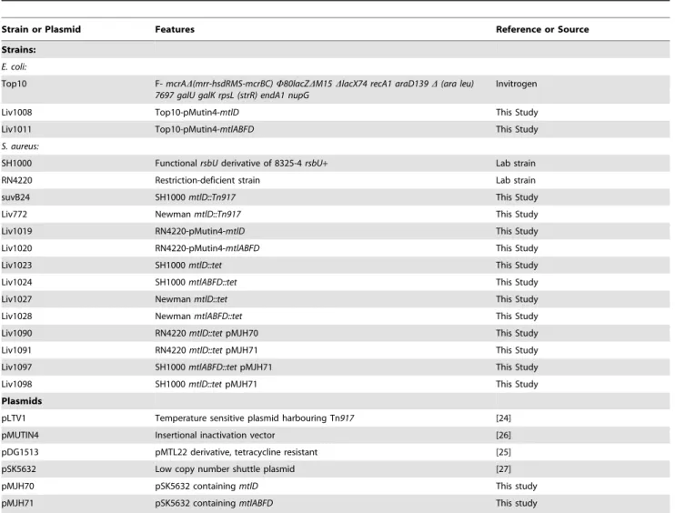
Table 1.Strains and plasmids used in the study.
Strain or Plasmid Features Reference or Source
Strains:
E. coli:
Top10 F-mcrAD(mrr-hsdRMS-mcrBC)W80lacZDM15DlacX74 recA1 araD139D(ara leu) 7697 galU galK rpsL (strR) endA1 nupG
Invitrogen
Liv1008 Top10-pMutin4-mtlD This Study
Liv1011 Top10-pMutin4-mtlABFD This Study
S. aureus:
SH1000 FunctionalrsbUderivative of 8325-4rsbU+ Lab strain
RN4220 Restriction-deficient strain Lab strain
suvB24 SH1000mtlD::Tn917 This Study
Liv772 NewmanmtlD::Tn917 This Study
Liv1019 RN4220-pMutin4-mtlD This Study
Liv1020 RN4220-pMutin4-mtlABFD This Study
Liv1023 SH1000mtlD::tet This Study
Liv1024 SH1000mtlABFD::tet This Study
Liv1027 NewmanmtlD::tet This Study
Liv1028 NewmanmtlABFD::tet This Study
Liv1090 RN4220mtlD::tetpMJH70 This Study
Liv1091 RN4220mtlD::tetpMJH71 This Study
Liv1097 SH1000mtlABFD::tetpMJH71 This Study
Liv1098 SH1000mtlD::tetpMJH71 This Study
Plasmids
pLTV1 Temperature sensitive plasmid harbouring Tn917 [24]
pMUTIN4 Insertional inactivation vector [26]
pDG1513 pMTL22 derivative, tetracycline resistant [25]
pSK5632 Low copy number shuttle plasmid [27]
pMJH70 pSK5632 containingmtlD This study
pMJH71 pSK5632 containingmtlABFD This study
Zeta potential and Hexadecane Partitioning
Zeta potential was determined using electrophoretic light scattering (ELS) in which the velocity of charged particles under the influence of an applied electric field is measured by monitoring the frequency shift of the scattered light from the particles. Culture (,800ml) was injected into a capillary cell and measured using a Zetasizer Nano (Malvern Instruments) with the detector positioned at a 17uscattering angle. The data were analysed and interpreted using the associated software. All charges were recorded as the mean of 5 consecutive measurements. Hexadecane partitioning was performed as previously described [6].
BioLog Phenotypic Arrays
BioLog phenotypic arrays were used to monitor growth of bacterial strains in 96 well plates under a wide range of conditions using redox levels within the growth media as a measure of bacterial growth [28]. Strains SH1000 or suvB24 were resus-pended from BHI plates to a transmittance of 81% using a BioLog turbidometer then added to the appropriate inoculation fluid for each assay plate. Comprehensive details of the growth factors tested using these assay plates PM1-PM10 can be found at http:// www.biolog.com/pdf/pm_lit/PM1-PM10.pdf. Following inocula-tion the array plates were incubated at 37uC and monitored for turbidity using the OmniLog plate reader at 30 min intervals over a 47 hour period. The assay was performed in triplicate and the mean values were used to compare growth.
Metabolite Analysis
Four hour cultures of strains SH1000, Liv1023 (SH1000
mtlD::tet) and Liv1024 (SH1000 mtlABFD::tet) were harvested by centrifugation and washed 3 times in PBS, before being resuspended in 3 ml of PBS. Cells were lysed using a bead-beater for three 1 min intervals at 4uC, with chilling between breakages. Cytoplasmic fractions were centrifuged, and metabolic reactions quenched via the addition of methanol. Samples were then dried down and derivatized as described previously [29,30] with the following modifications. Samples were incubated for 90 min at 50uC with 80ml of methoxyamine hydrochloride in pyridine (20 mg ml21) following a 60 min treatment at 50uC with 80ml MSTFA. Five ml of an internal standard (C31 fatty acid) was added prior to trimethylsilylation, and sample volumes of 1mL were injected with a split ratio of 7:1. The GC-MS system consisted of an Agilent 7890A (Agilent Inc, Palo Alto, CA, USA) gas chromatograph, an Agilent 5975C mass selective detector and Agilent 7683B autosampler. Gas chromatography was performed on a 60 m HP-5MS column with 0.25 mm inner diameter and 0.25mm film thickness (Agilent Inc, Palo Alto, CA, USA), and an injection temperature of 250uC. The interface was set to 250uC, and the ion source adjusted to 230uC. Helium carrier gas was set at a constant flow rate of 1.5 ml min21
. The temperature program was 5 min isothermal heating at 70uC, followed by an oven temperature increase of 5uC min21to 310uC, and a final 20 min at 310uC. The mass spectrometer was operated in positive electron impact mode (EI) at 69.9 eV ionization energy in a m/z 30– 800 scan range. The spectra of all chromatogram peaks were compared with electron impact mass spectrum libraries NIST08 (NIST, MD, USA), WILEY08 (Palisade Corporation, NY, USA), and a custom library. To allow comparison between sample sets, all data were normalized to internal standards in each chromato-gram, and the weights of each sample. The chromatograms and mass spectra were evaluated using the MSD ChemStation (Agilent, Palo Alto, CA, USA) and AMDIS (NIST, Gaithersburg, MD, USA) programs. The retention time and mass spectra were implemented within the AMDIS method formats. The resulting
data from triplicate samples (with less than 10% variability) was analyzed using a t-test. Samples with a p,0.05 and greater than 2-fold variation were then analyzed using the MetPA enrichment pathway analysis web application (http://metpa.metabolomics. ca/) [31].
Experimental Septic Arthritis
A previously described mouse model of septic arthritis was used to test the in vivo role of mtlD in virulence [32,33]. Seven week female NMRI mice were obtained from Charles River Labora-tories (Sulzfeld, Germany) and maintained in the animal facility of the Department of Rheumatology and Inflammation Research, University of Go¨teborg, Sweden. All mice were maintained according to the local ethic board animal husbandry standards. The mice were housed 10 to a cage under standard conditions of temperature and light and were fed standard laboratory chow and waterad libitum. Mice were inoculated in the tail vein with 0.2 ml of bacterial suspension cultured and bacteria in kidney abscesses were enumerated after 14 days as described previously [6]. Presented data represent CFU per kidney pair.
Results
Identification of amtlDAFA Survival Mutant
A screen of S. aureus Tn917 library transposants identified multiple clones with greatly reduced survival on BHI agar containing 1 mM linoleic acid (C18:2D9D12), in addition to those mutants described previously [6]. DNA sequence determination by arbitrary-primed PCR [6] revealed these clones were identical, with Tn917inserted inmtlDat nucleotide position 317/1107. The
mtlABFD operon encodes the Mtl-specific phosphotransferase system (PTS) trasnsporter (MtlAB) and the operon transcriptional repressor (MtlF); Mtl-1-P 5-dehydrogenase, encoded by mtlD, catalyses the conversion of Mtl-1-P to fructose-6-P (Figure 1). The
mtlD mutant suvB24 (SH1000 mtlD::Tn917) selected for further study showed clearly reduced survival on linoleic acid agar compared to its isogenic parent strain (Figure 2). Transduction of suvB24 into S. aureus Newman (Liv772; Table 1) identified a proportionately similar reduction in linoleic acid survival in this distinct strain background (data not shown).
Figure 1. Mannitol uptake pathway inS. aureus. ThemtlABFD
operon encodes the Mtl-specific PTS (MtlAB) and the operon transcriptional repressor (MtlF); Mtl-1-P 5-dehydrogenase, encoded by
mtlD, catalyses the conversion of Mtl-1-P to fructose-6-P which enters into the Embden-Meyerhoff and hexosemonophosphate glycolytic pathways.
Culture Phenotypes ofmtlMutants
To investigate the role of the mtlD gene product in host cell physiology and to help explain the mechanism for reduced linoleic acid agar survival, growth of the suvB24 mutant was compared with its isogenic parental strain using a Biolog phenotype array (Biolog Inc. California, USA). Comparative growth arrays in the presence of various carbon, nitrogen, phophorous and sulphur compounds and a variety of amino acids, peptide nitrogen sources, osmolytes and pH ranges [28] identified that reduced Mtl metabolism was the only significantly altered phenotype (data not shown).
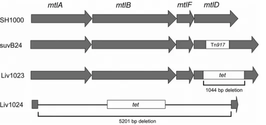
To confirm the role of the Mtl PTS operon in S. aureus cell survival, allelic replacement mutants were generated for mtlD, Liv1023 (SH1000 mtlD::tet) and for the entire mtlABFD operon, Liv1024 (SH1000 mtlABFD::tet) (Figure 3), using methods de-scribed previously [34,35,36]. Two complementation vectors were also generated by cloning themtlDgene and themtlABFDoperon into the low copy shuttle vector pSK5632, producing plasmids pMJH70 and pMJH71, respectively. Cloning of the mtlABFD
operon was achieved by transforming ligation products into strain Liv1021 (RN4220mtlD::tet) selecting for fermentation on mannitol salt agar (MSA), since cloning of the operon inE. coliTOP10 was not successful, potentially due to toxicity. Complementation with
mtlDalone did not restore Mtl fermentation on MSA due to the absence of a promoter for this distal gene; consequently complementation experiments were performed using pMJH71. Culture of Liv1023 (SH1000 mtlD::tet) and Liv1024 (SH1000
mtlABFD::tet) on MSA at 37uC demonstrated the inability of these mutants to ferment Mtl to produce acid (Figure 4). Weak growth was observed for Liv1023 on MSA agar in contrast to Liv1024, which grew similarly to the wild-type SH1000 strain. Metabolism
was restored in the complemented strains Liv1097 (SH1000
mtlABFD::tet pMJH71) and LIV1098 (SH1000mtlD::tet pMJH71) (Figure 4). Transduction of the mtlD and mtlABFD inactivations into S. aureus Newman (Liv1027 and Liv1028, respectively) confirmed the absence of Mtl fermentation in both mutants (data not shown).
Comparative growth assays of the allelic replacement mutants on linoleic acid agar confirmed that Liv1023 (SH1000 mtlD::tet) had an AFA growth defect similar to suvB24 (SH1000
mtlD::Tn917) with greater than 3-log reduction in survival (Figure 5). Similarly reduced levels of survival were observed following growth on agar supplemented with millimolar concen-trations of oleic acid (C18:1D9) or sapienic acid (C16:1D6) (data not shown) demonstrating that inactivation of mtlD caused reduced survival to multiple AFAs. Allelic replacement of the mtlABFD
operon did not impair survival from AFAs, in contrast to inactivation ofmtlDalone. Proportionately reduced AFA survival was observed with anmtlDbut not anmtlABFDinactivation inS. aureusNewman (Liv1027 and Liv1028, respectively; Table 1) (data not shown). Reduced survival of the mtlD mutant was fully complemented with the entire mtlABFD operon present on pMJH71 using strain Liv1098 (SH1000 mtlD::tet pMJH71) (Figure 2). The reduced survival of Liv1023 (SH1000 mtlD::tet) on linoleic acid agar was supported with a significantly reduced linoleic acid MIC (0.4560.02 mM) (p,0.004) in BHI medium, compared to SH1000 (0.960.04 mM), Liv1024 (0.6960.02 mM) and Liv1098 (0.8560.03 mM).
Strain Liv1023 (SH1000mtlD::tet) exhibited a profound growth defect when cultured in broth containing Mtl as the carbohydrate source (peptone 10 g l21
, Mtl 10 g l21
, beef extract 1 g l21 , NaCl 10 g l21
) (Figure 6A). Substituting the sugar alcohol Mtl for the Figure 2. Comparative survival ofS. aureusstrains.Growth of dilutions from overnight cultures on BHI agar in the presence and absence of 1 mM linoleic acid. SuvB24 (SH1000mtlD::Tn917) and Liv1023 (SH1000mtlD::tet) displayed.500-fold reduced survival on linoleic acid relative to wild type (SH1000), Liv1024 (SH1000mtlABFD::tet) and the complemented mutant strain Liv1098 (SH1000mtlD::tetpMJH71).
sugars fructose or glucose restored normal growth, demonstrating the Mtl-specific defect (data not shown). S. aureus accumulates intracellular Mtl following incubation in the presence of glucose. To test if this accumulation affected survival from AFAs, the relative survival of exponential cells (OD600= 1) of SH1000 incubated in PBS containing 1% (w/v) glucose was determined after growth on 1 mM linoleic acid agar. No clear difference in survival of the strains was observed.
All strains grew equally well at 37uC in BHI broth (data not shown), however a pronounced reduction in growth rate was observed for strain Liv1023 (SH1000mtlD::tet) when cultured in BHI broth at 25uC (Figure 6B). This defect was specific to inactivation ofmtlDbut not for deletion of the complete operon. Starvation survival with limiting glucose was not impaired inmtl
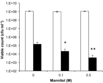
mutant strains [35,37]. Growth ofS. aureusSH1000 was tested in the absence or presence of mannitol (0.1 M, 0.5 M), with or without 1 mM linoleic acid to test for synergy. Mannitol was shown to have similar properties as ethanol [13], by acting synergistically with linoleic acid as evident by the reduced viable count with increasing mannitol concentration (Figure 7).
Analysis of Cellular Metabolites
A comparative metabolomics analysis was undertaken to identify the intracellular metabolites of exponentially growing cells of strains SH1000, Liv1023 (SH1000 mtlD::tet) and Liv1024 (SH1000
mtlABFD::tet). This revealed that inactivation ofmtlDresulted in an accumulation of Mtl and Mtl-P, the latter being undetectable in both SH1000 and Liv1024 (Table 2 and supplementary table 1). The total relative levels of Mtl species were over 20-fold greater in Liv1023 (SH1000mtlD::tet) than SH1000. The near absence of Mtl in strain Liv1024 supports data that the MtlAB PTS transporter is the main portal for Mtl uptake [38]. Inactivation ofmtlDandmtlABFDresulted in the absence of cellular Sorbitol-6-P (Table 2). Further clear differences in metabolite levels were evident in strain Liv1023 (mtlD::tet) relative to SH1000 and Liv1024 (Table S1).
Figure 3. Schematic representation of themtlABFDlocus.Position of the transposon insertion and allelic replacements created during this study.
doi:10.1371/journal.pone.0067698.g003
Figure 4. Mtl fermentation capability ofS. aureus strains.Mtl fermentation is revealed by acid formation and colour change of the pH indicator to yellow. Liv1023 (SH1000 mtlD::tet) and Liv1024 (SH1000
mtlABFD::tet) do not ferment Mtl and this capability was restored by complementation with the entire locus in strains Liv1098 (SH1000
mtlD::tetpMJH71) and Liv1097 (SH1000 mtlABFD::tetpMJH71). Weak growth of Liv1023 was observed.
doi:10.1371/journal.pone.0067698.g004
Figure 5. Survival on linoleic acid agar.Comparative survival of strains on BHI agar supplemented with 1 mM linoleic acid. Strains SH1000 (open circles), Liv1023 (SH1000 mtlD::tet) (filled squares), Liv1024 (SH1000mtlABFD::tet) (open triangles) and Liv1098 (SH1000
mtlD::tetpMJH71) were diluted in PBS and equivalent volumes were plated onto the agar. SE from triplicate experiments is shown with error bars inside symbols.
Resistance and Cell Surface Properties ofmtlMutants A range of antimicrobial agents were tested to determine if the observed reduced resistance of Liv1023 (SH1000 mtlD::tet) extended beyond AFAs. Growth and MICs were comparable between Liv1023 (SH1000mtlD::tet) and SH1000 in the presence of a range of concentrations of NaCl, lauroyl sarcosine, SDS, dichlorophenyl and the human cathelicidin LL37 (Sigma). Liv1023 (SH1000mtlD::tet) was observed to exhibit a lower MIC for H2O2 (1 mM) compared to SH1000 (4 mM) and Liv1024 (SH1000mtlABFD::tet) (4 mM).
The hydrophobicity and zeta potential of all of the strains was similar when tested using either hexadecane partitioning or measured using a zetasizer (Malvern, UK), respectively (data not shown). The levels of carotenoid in cell membranes were similar
between SH1000 and themtlmutants, as judged by spectropho-tometric analysis of methanol-extracted cells from overnight and 2 day-old cultures (data not shown).
Virulence ofmtlDMutant
The decreasedin vitroAFA survival and reduced H2O2MIC of themtlDmutant prompted testing of its virulence compared to the isogenic parent strain using a previously described model of experimental septic arthritis (Figure 8). This model was tested to determine whether inactivation of the mtlABFD locus affected virulence, since its contribution to metabolismin vivois unknown and the model generates abscesses where AFAs accumulate [14]. This revealed that SH1000mtlDdid not have reduced virulence, at least under the conditions studied [6,32,33].
Figure 6. Growth phenotype ofmtlDinactivatedS. aureus.(A) Culture of strains in broth containing Mtl at 37uC. Liv1023 (SH1000
mtlD::tet) (
N
) had a significantly reduced growth rate (P,0.01, Student’s t-test) compared to wild-type (SH1000) (&), Liv1024 (SH1000mtlABFD::tet) (m), strains Liv1098 (SH1000mtlD::tetpMJH71) (%) and
Liv1097 (SH1000mtlABFD::tetpMJH71) (#). Calculated doubling times between 2 h and 3 h of growth: SH1000 = 0.33, Liv1023 (SH1000
mtlD::tet) = 1.7. Error bars indicate 1 SEM (n = 3). (B) Culture of strains in BHI broth at 25uC. Liv1023 (SH1000mtlD::tet) (
N
) has a significantly reduced growth rate (P,0.001, Student’s t-test) compared to wild-type (SH1000) (&), Liv1024 (SH1000 mtlABFD::tet) (m), strains Liv1098 (SH1000 mtlD::tet pMJH71) (%) and Liv1097 (SH1000 mtlABFD::tetpMJH71) (#). Calculated doubling times between 5 h and 13 h of growth: SH1000 = 2.46, Liv1023 = 3.13. Representative dataset from triplicate assay.
doi:10.1371/journal.pone.0067698.g006
Figure 7. Growth ofS. aureusin the presence of mannitol and linoleic acid.Bacteria were cultured on BHI agar containing either no or added mannitol (0.1 M, 0.5 M) in the presence (black bars) or absence (white bars) of 1 mM linoleic acid. Differences in viable cells recovered in the presence of mannitol were significantly reduced compared to the absence of mannitol (P = 0.03 and P = 0.001 for 0.1 M and 0.5 M, respectively).
doi:10.1371/journal.pone.0067698.g007
Table 2.Sugar alcohols present inS. aureusstrains.
Relative mean concentration
Metabolite SH1000 Liv1023 Liv1024
Arabitol 116.1 (3.9) 107.5 (19.7) 51.4 (7.9) Mannitol 417.6 (29.5) 1351.4 (82.5) 5.9 (0.9) Mannitol-P ND 8161.3 (119.7 ND Ribitol 240 (22.3) 214.8 (17.6) 272.2 (27.1) Sorbitol-6-P 149.4 (18.2) ND ND
Discussion
The intrinsic importance ofS. aureuscarriage and transmission in relation to disease and its hypothesized link with virulence [39] requires that determinants are identified and characterised that promote survival in its primary niche and during its transient residence on human skin. From the study of gene mutantsS. aureus
defence from AFAs is achieved via a variety of surface components (IsdA, WTA, SasF) and regulation of peptidoglycan biosynthesis (VraRS, VraE), where a reduction in hydrophobicity to minimize access of the AFA to the membrane explains the contribution of several of these components to survival [6,7,19]. In addition, the arginine deiminase pathway increases survival [6], where its various contributions to metabolic versatility and its potential to modify local pH could explain its role.
Determining that an Mtl-1-P-dehydrogenase mutant, but not an
mtlABFD transport operon mutant, has greatly reduced survival from AFAs implicates the accumulation of Mtl-1-P as being the causative factor. As the most abundant natural hexitol, Mtl is a carbon source for staphylococci and the inducible oxidation of Mtl-1-P generates fructose-6-P for entry into the Embden-Meyerhoff and hexosemonophosphate glycolytic pathways [38,40]. All strains of S. aureus accumulate Mtl, despite not all being capable of using it for metabolism during aerobic growth. In
S. aureusthe cellular accumulation of Mtl was identified in resting cells when incubated in glucose or cultured in media without added carbohydrate [38]. Mtl accumulation was proposed to enhance metabolic versatility inS. aureus,however its mechanistic role is incompletely understood [41]. Following stress, such as after exposure to AFAs, utilisation of the pathway for Mtl conversion to fructose-6-P would regenerate NADH, thereby alleviating the pressure upon regenerating reactions downstream of pyruvate. In our previous studies [6], exposure of S. aureus to linoleic acid caused downregulated transcription of the mtlABFDlocus, which suggests, either that reduced levels of intracellular Mtl is a preferred metabolic state following exposure to AFAs, or that lower amounts of Mtl-1-P arising from metabolism (concomitantly regenerating NADH) limited induction of the operon. A potential explanation for the reduced AFA survival of themtlDmutant is its reduced adaptive capacity due to an inability to metabolise Mtl. The near wild-type AFA survival of themtlABFDoperon mutant
argues against this Mtl metabolism hypothesis, however, unless there is an alternative metabolic reserve. 3-phosphoglycerate could serve as just such an alternative metabolic source and substrate for regenerating NADH, and of note there is 3-fold reduced 3-PGA in themtlDmutant.
Metabolite analysis of theS. aureus mtlDmutant, when compared to themtlABFDtransport mutant and the parental strain, revealed that 14% of the total Mtl that accumulated intracellularly was not phosphorylated. Since the EIIMtl mannitol transporter (encoded bymtlA) phosphorylates the imported Mtl and since the Mtl-1-P-dehydrogenase activity is ablated in the mtlD mutant, the conversion of Mtl-1-P to Mtl in themtlDmutant is likely to arise from phosphotransferase reactions as described by Saier and Newman [42]. Alternatively, an undescribed phosphatase activity might account for the presence of Mtl. InLactobacillus plantaruma hypothetical phosphatase activity of EIIMtl was proposed to explain the apearance of Mtl in engineered strains [43]. In the study of Mtl overproducing strains ofL. lactisa Mtl-1-phosphatase activity was proposed to explain the presence of unphosphorylated Mtl, wheremtlAwas absent, and thus an EIIMtl activity, could not be present. Analysis of the metabolites of growing cells of theS. aureus mtlDmutant cultured in BHI broth, when compared to the
mtlABFD transport mutant and the parental strain, revealed further differences aside from sugar alcohol content (Table S1). These metabolite changes e.g. aminoadipic acid, 3-phosphoglyc-erate, hydroxypentanoic acid, heptanoic acid and tetradecanoyl-glycerol, do not indicate a clearly defined mechanistic explanation for decreased AFA MIC.
Growth of the mtlD mutant was strongly retarded in media containing mannitol, highlighting the deleterious effects resulting from the likely unrestricted accumulation of Mtl/Mtl-1-P. A direct link between the intracellular accumulation of Mtl/Mtl-1-P and reduced resistance to AFAs inS. aureuscurrently lacks an evidence-based mechanism. However, several features of themtlD pheno-type could result from a membrane-associated effect. Alcohol has a well-described potentiating mechanism with respect to AFAs and their membrane activity [13], since it is capable of solubilising membrane lipids due to its polarity and lipophilicity. Intracellu-larly accumulated sugar alcohol, Mtl, might act simiIntracellu-larly to potentiate AFA action, since it was demonstrated in this study that Mtl acted synergistically with linoleic acid when added externally Figure 8. Virulence ofmtlDin a murine infection.(A) Effect of WT SH1000 or Liv1023 (SH1000mtlD::tet) on percentage change in weight of infected mice. There were no significant differences using Dunn’s test. (B) Effect of mutations ofmtlDon cfu ofS. aureus SH1000 in kidneys of infected mice. There were no significant differences using the Mann Whitney Test.
in BHI agar. Two further phenotypes point towards a membrane-specific alteration in the mtlD mutant; the reduced growth rate that was oberved for the mtlDmutant at 25uC and the reduced MIC for H2O2 which did not result from differences in catalase specific activity (data not shown). A perturbation in peroxide permeability at the membrane is consistent with the reduced MICs observed and might arise via Mtl potentiating the linoleic acid by virtue of the polarity and lipophilicity of alcohols affecting diffusion across the membrane, but this was not investigated further. No differences were observed between the staphylox-anthin levels in methanol extracts of any of the strains, which might be expected if the intracellular accumulation of Mtl altered membrane fluidity (data not shown) [44]. Mtl is frequently included in membrane preparations as a stabilising entity, either through direct effects or via osmotic stabilisation. The expression of a bacterial mtlD in Saccharomyces cerevisiae was sufficient to generate mannitol which was proposed to act as an osmolyte and was sufficient to rescue the phenotypes of a glycerol deficient mutant, producing an increased resistance to high salt and H2O2 [45]. Mtl is also proposed to function as an osmoprotectant in cells of petunia as well as improving cold tolerance [46].
The observed phenotype of reduced survival in the presence of AFAs did not translate to a reduction in virulence in a murine arthritis model or reduced MIC levels to a range of other membrane-acting agents. Thus, the changes to cellular physiology in themtlDmutant are discrete, at least in this model of infection tested and other disease models, such as skin survival, remain to be tested.
Supporting Information
Table S1 Metabolites present in S. aureus strains. Metabolites were identified using GC-MS analysis of cytoplasmic fractions from exponential growth phase cells. 131 unique metabolites were compared and chromatograms and mass spectra were evaluated as described previously [8,32] using the MSD ChemStation (Agilent, Palo Alto, CA, USA) and AMDIS (NIST, Gaithersburg, MD, USA) programs. The resulting data from triplicate samples (with less than 10% variability) were analyzed using a t-test. Samples with greater than 2-fold variation (p,0.05) were analyzed using the MetPA enrichment pathway analysis web application (http://metpa.metabolomics.ca/) [45]. ND, not de-tectable.
(XLSX)
Acknowledgments
We thank Ing-Marie Jonsson (Gothenburg) for technical help with virulence studies.
Author Contributions
Conceived and designed the experiments: JGK JM ZL EJ LNS MJH. Performed the experiments: JGK JM SLK AU EJ MJH. Analyzed the data: JGK JM SLK AU EJ MJH. Contributed reagents/materials/analysis tools: ZL LNS EJ MJH. Wrote the paper: JM MJH.
References
1. Lowy FD (1998)Staphylococcus aureusinfection. N Engl J Med 339: 520–532. 2. Foster TJ (2005) Immune evasion by staphylococci. Nat Rev Microbiol 3: 948–
58.
3. Gorwitz RJ, Kruszon-Moran D, McAllister SK, McQuillan G, McDougal LK, et al (2008) Changes in the prevalence of nasal colonization withStaphylococcus aureusin the United States, 2001–2004. J Infect Dis 197: 1226–34.
4. Deleo FR, Otto M, Kreiswirth BN, Chambers HF (2010) Community-associated methicillin-resistantStaphylococcus aureus. Lancet 375: 1557–1568.
5. Kelsey JA, Bayles KW, Shafii B, McGuire MA (2006) Fatty acids and monoacylglycerols inhibit growth ofStaphylococcus aureusLipids 41: 951–961. 6. Kenny JG, Ward D, Josefsson E, Jonsson IM, Hinds J, et al. (2009) The
Staphylococcus aureusresponse to unsaturated long chain free fatty acids: survival mechanisms and virulence implications. PLoS ONE 4: e4344.
7. Kohler T, Weidenmaier C, Peschel A (2009) Wall teichoic acid protects
Staphylococcus aureusagainst antimicrobial fatty acids from human skin. J Bacteriol 191: 4482–4484.
8. Campbell IM, Crozier DN, Pawagi AB, Buivids IA (1983) In vitro response of
Staphylococcus aureusfrom cystic fibrosis patients to combinations of linoleic and oleic acids added to nutrient medium. J Clin Microbiol 18: 408–415. 9. Zheng CJ, Yoo JS, Lee TG, Cho HY, Kim YH, et al. (2005) Fatty acid synthesis
is a target for antibacterial activity of unsaturated fatty acids. FEBS Lett 579: 5157–5162.
10. Ansari MN, Nicolaides N, Fu HC (1970) Fatty acid composition of the living layer and stratum corneum lipids of human sole skin epidermis. Lipids 5: 838– 845.
11. Bergsson G, JArnfinnsson J, Steingrı´msson O, Thormar H (2001) Killing of Gram-positive cocci by fatty acids and monoglycerides. APMIS. 109: 670–678. 12. Do TQ, Moshkani S, Castillo P, Anunta S, Pogosyan A, et al (2008) Lipids including cholesteryl linoleate and cholesteryl arachidonate contribute to the inherent antibacterial activity of human nasal fluid. J Immunol 181: 4177–4187. 13. Drake DR, Brogden KA, Dawson DV, Wertz PW (2008) Thematic review series: skin lipids. Antimicrobial lipids at the skin surface. J Lipid Res. 49: 4–11. 14. Dye ES, Kapral FA (1981) Characterization of a bactericidal lipid developing
within staphylococcal abscesses. Infect Immun 32: 98–104.
15. Engler HD, Kapral FA (1992) The production of a bactericidal monoglyceride in staphylococcal abscesses. J Med Microbiol. 37: 238–244.
16. Nieman C (1954) Influence of trace amounts of fatty acids on the growth of microorganisms. Bacteriol Rev 18: 147–161.
17. Pappas A, Anthonavage M, Gordon JS (2002) Metabolic fate and selective utilization of major fatty acids in human sebaceous gland. J Invest Dermatol 118: 164–171.
18. Won SR, Hong MJ, Kim YM, Li CY, Kim JW, et al. (2007). Oleic acid: an efficient inhibitor of glucosyltransferase. FEBS Lett 581: 4999–5002.
19. Clarke SR, Mohamed R, Bian L, Routh AF, Kokai-Kun JF, et al. (2007) The
Staphylococcus aureussurface protein IsdA mediates resistance to innate defenses of human skin. Cell Host Microbe 1: 199–212.
20. Galbraith H, Miller TB (1973) Effect of long chain fatty acids on bacterial respiration and amino acid uptake. J Appl Bacteriol 36: 659–675.
21. Galbraith H, Miller TB (1973) Effect of metal cations and pH on the antibacterial activity and uptake of long-chain fatty acids. J Appl Bacteriol 36: 635–646.
22. Galbraith H, Miller TB (1973) Physicochemical effects of long-chain fatty acids on bacterial cells and their protoplasts. J Appl Bacteriol 36: 647–658. 23. Greenway DL, Dyke KG (1979) Mechanism of the inhibitory action of linoleic
acid on the growth ofStaphylococcus aureus. J Gen Microbiol 115: 233–245. 24. Horsburgh MJ, Wiltshire MD, Crossley H, Ingham E, Foster SJ (2004) PheP, a
putative amino acid permease ofStaphylococcus aureus, contributes to survival in vivo and during starvation. Infect Immun 72: 3073–3076.
25. Guerot-Fleury AM, Shazand K, Frandsen N, Stragier P (1995) Antibiotic resistance cassettes forBacillus subtilis. Gene 167: 335–336.
26. Vagner V, Dervyn E, Ehrlich ED (1998) A vector for systematic inactivation in
Bacillus subtilis. Microbiology 144: 3097–3104.
27. Grkovic S, Brown MH, Hardie KM, Firth N, Skurry RA (2003) Stable low-copy-numberStaphylococcus aureusshuttle vectors. Microbiology 149: 785–794. 28. Bochner BR, Gadzinski P, Panomitros E (2001) Phenotype microarrays for
high-throughput phenotypic testing and assay of gene function. Gen Res 11: 1246– 1255.
29. Dawe AL, Van Voorhies WA, Lau TA, Ulanov AV, Li Z (2009) Major impacts on the primary metabolism of the plant pathogenCryphonectria parasiticaby the virulence-attenuating virus CHV1-EP713. Microbiology 155: 3913–3921. 30. Roessner U, Wagner C, Kopka J, Trethewey RN, Willmitzer L (2000) Technical
advance: simultaneous analysis of metabolites in potato tuber by gas chromatography-mass spectrometry. Plant J 23: 131–42.
31. Xia J, Wishart DS (2010) MetPA: a web-based metabolomics tool for pathway analysis and visualization. Bioinformatics 26: 2342–2344.
32. Bremell T, Lange S, Yacoub A, Ryden C, Tarkowski A (1991) Experimental
Staphylococcus aureusarthritis in mice. Infect Immun 59: 2615–2623.
33. Josefsson E, Kubica M, Mydel P, Potempa J, Tarkowski A (2008) In vivo sortase A and clumping factor A mRNA expression duringStaphylococcus aureusinfection. Microb Pathog 44: 103–110.
34. Horsburgh MJ, Aish JL, White IJ, Shaw L, Lithgow JK, et al. (2002) SigmaB modulates virulence determinant expression and stress resistance: characteriza-tion of a funccharacteriza-tional rsbU strain derived from Staphylococcus aureus 8325–4. J Bacteriol 184: 5457–5467.
36. Horsburgh MJ, Thackray PD, Moir A (2001) Transcriptional responses during outgrowth ofBacillus subtilisendospores. Microbiology 147: 2933–2941. 37. Watson SP, Clements MO, Foster SJ (1998) Characterization of the
starvation-survival response ofStaphylococcus aureus. J Bacteriol 180: 1750–1758. 38. Edwards KG, Blumenthal HJ, Khan M, Slodki ME (1981) Intracellular
mannitol, a product of glucose metabolism in staphylococci. J Bacteriol 146: 1020–1029.
39. Massey RC, Horsburgh MJ, Lina G, Ho¨o¨k M, Recker M (2006) The evolution and maintenance of virulence inStaphylococcus aureus: a role for host-to-host transmission? Nat Rev Microbiol 4: 953–8.
40. Watanabe S, Hamano M, Kakeshita H, Bunai K, Tojo S, et al. (2003) Mannitol-1-phosphate dehydrogenase (MtlD) is required for mannitol and glucitol assimilation in Bacillus subtilis: possible cooperation of mtl andgut operons. J Bacteriol 185: 4816–4824.
41. Solomon PS, Waters ODC, Oliver RP (2007) Decoding the mannitol enigma in filamentous fungi. Trends Microbiol 15: 257–252.
42. Saier MH Jr, Newman MJ (1976) Direct transfer of the phosphoryl moiety of mannitol 1-phosphate to [14C]mannitol catalyzed by the enzyme II complexes
of the phosphoenolpyruvate: mannitol phosphotransferase systems inSpirochaeta aurantiaandSalmonella typhimurium. J Biol Chem 251: 3834–7.
43. Wisselink HW, Moers AP, Mars AE, Hoefnagel MH, de Vos WM, et al. (2005) Overproduction of heterologous mannitol 1-phosphatase: a key factor for engineering mannitol production byLactococcus lactis. Appl Environ Microbiol 71: 1507–14.
44. Mishra NN, Liu GY, Yeaman MR, Nast CC, Proctor RA (2011) Carotenoid-related alteration of cell membrane fluidity impacts Staphylococcus aureus
susceptibility to host defense peptides. Antimicrob Agents Chemother 55: 526–31.
45. Chaturvedi V, Bartiss A, Wong B (1997) Expression of bacterial mtlD in
Saccharomyces cerevisiae results in mannitol synthesis and protects a glycerol-defective mutant from high-salt and oxidative stress. J Bacteriol 179: 157–62. 46. Chiang Y, Stushnoff C, McSay AE (2005) Overexpression of