Rapid Pathogen-Induced Apoptosis: A Mechanism Used
by Dendritic Cells to Limit Intracellular Replication of
Legionella pneumophila
Catarina V. Nogueira1,2, Tullia Lindsten3, Amanda M. Jamieson4, Christopher L. Case1, Sunny Shin1, Craig B. Thompson3, Craig R. Roy1*
1Section of Microbial Pathogenesis, Yale University School of Medicine, New Haven, Connecticut, United States of America,2Graduate Program in Areas of Basic and Applied Biology, Instituto de Cieˆncias Biomedicas Dr. Abel Salazar, Universidade do Porto, Porto, Portugal,3Department of Cancer Biology, Abramson Family Cancer Research Institute, University of Pennsylvania, Philadelphia, Pennsylvania, United States of America,4Department of Immunobiology, Howard Hughes Medical Institute, Yale University School of Medicine, New Haven, Connecticut, United States of America
Abstract
Dendritic cells (DCs) are specialized phagocytes that internalize exogenous antigens and microbes at peripheral sites, and then migrate to lymphatic organs to display foreign peptides to naı¨ve T cells. There are several examples where DCs have been shown to be more efficient at restricting the intracellular replication of pathogens compared to macrophages, a property that could prevent DCs from enhancing pathogen dissemination. To understand DC responses to pathogens, we investigated the mechanisms by which mouse DCs are able to restrict replication of the intracellular pathogenLegionella pneumophila. We show that both DCs and macrophages have the ability to interfere with L. pneumophila replication
through a cell death pathway mediated by caspase-1 and Naip5.L. pneumophilathat avoided Naip5-dependent responses,
however, showed robust replication in macrophages but remained unable to replicate in DCs. Apoptotic cell death
mediated by caspase-3 was found to occur much earlier in DCs following infection by L. pneumophila compared to
macrophages infected similarly. Eliminating the pro-apoptotic proteins Bax and Bak or overproducing the anti-apoptotic protein Bcl-2 were both found to restoreL. pneumophilareplication in DCs. Thus, DCs have a microbial response pathway that rapidly activates apoptosis to limit pathogen replication.
Citation:Nogueira CV, Lindsten T, Jamieson AM, Case CL, Shin S, et al. (2009) Rapid Pathogen-Induced Apoptosis: A Mechanism Used by Dendritic Cells to Limit Intracellular Replication ofLegionella pneumophila. PLoS Pathog 5(6): e1000478. doi:10.1371/journal.ppat.1000478
Editor:Brad T. Cookson, University of Washington, United States of America ReceivedNovember 20, 2008;AcceptedMay 18, 2009;PublishedJune 12, 2009
Copyright:ß2009 Nogueira et al. This is an open-access article distributed under the terms of the Creative Commons Attribution License, which permits unrestricted use, distribution, and reproduction in any medium, provided the original author and source are credited.
Funding:Supported by Fundac¸a˜o para a Cieˆncia e a Tecnologia (BD/11758/2003) (C.N.); NIH Ruth L. Kirchenstein National Research Service Award and the Irvington Institute Fellowship Program of the Cancer Research Institute (S.S.), National Science Foundation Graduate Research Fellowship (C.L.C.), and NIH grant R01-AI048770 (C.R.R.). The funders had no role in study design, data collection and analysis, decision to publish, or preparation of the manuscript.
Competing Interests:The authors have declared that no competing interests exist. * E-mail: [email protected]
Introduction
Macrophages and dendritic cells (DCs) are the sentinels of the innate immune system. They are key in sensing infection and activating downstream antimicrobial responses [1,2]. These profes-sional phagocytes are activated following stimulation of pattern-recognition receptors, such as transmembrane Toll-like receptors (TLRs) and cytoplasmic nucleotide-binding domain and leucine-rich repeat containing receptors (NLRs) by pathogen-associated molecular patterns (PAMPs). Signaling through these receptors induces the expression and secretion of proinflammatory cytokines, chemokines and other antimicrobial defense molecules [3–7].
Bacterial pathogens that are able to infect and establish residence within macrophages and DCs provide a unique challenge to the innate immune system, as many pathogens have evolved virulence factors that subvert the cellular processes of these cells. One such pathogen isLegionella pneumophila, the etiological agent of the severe pneumonia known as Legionnaires’ disease [8,9].L. pneumophilais able to infect alveolar macrophages and modulate transport of the phagosome in which it resides to avoid fusion with endosomes and lysosomes [10].L. pneumophilahas the ability to recruit vesicles in transit between the endoplasmic reticulum (ER) and Golgi
apparatus and use these vesicles to remodel the L. pneumophila -containing vacuole (LCV) to create a unique ER-derived vacuole that supports intracellular replication [10–17]. Modulation of intracellular transport of the LCV requires a functional type IV secretion system (TFSS) encoded by thedotandicmgenes, which translocates bacterial effectors directly into the host cytosol [18–21]. Many of the translocated effector proteins engage host factors involved in vesicular transport and assist in LCV transport [22–29].
L. pneumophila mutants defective in the Dot/Icm system do not replicate intracellularly, as they are unable to modulate intracellular transport and occupy a more conventional phagosome that undergoes rapid endocytic maturation [19,30].
into the host cell cytosol [38–40]. Naip5 in conjunction with the NLR protein Ipaf activates caspase-1, which limitsL. pneumophila
replication in macrophages by inducing a pro-inflammatory cell death pathway known as pyroptosis [37–40].
Naip5 control of caspase-1 activation does not seem to be the only cellular mechanism used by innate immune cells to control L.
pneumophilareplication. In DCs infected withL. pneumophila, although phagosomes containing bacteria are able to mature into ER-derived organelles, bacterial replication is limited [41]. DCs are still able to process and present L. pneumophila antigens on MHC class II molecules, andde novosynthesis ofL. pneumophilaproteins inside DCs is critical for maximal stimulation of CD4+
T cells. This indicates that restriction of L. pneumophila replication could be important to the ability of DCs to present bacterial antigens to T cells and direct subsequent adaptive immune responses [41]. Interestingly, DCs are able to limit the intracellular replication of several other pathogens that are capable of replicating in macrophages, such as Listeria monocytogenes, Mycobacterium tuberculosisand Salmonella entericaSerovar Typhimurium [42–45].
Thus, it appears that there are inherent differences between DCs and macrophages with respect to their abilities to restrict replication of intracellular pathogens. We show here that one of these differences involves the ability of DCs to rapidly activate a cell intrinsic apoptotic cell death pathway in response to the intracellular pathogenL. pneumophila.
Results
Canonical pathogen surveillance pathways are not required for restriction ofL. pneumophilareplication by DCs
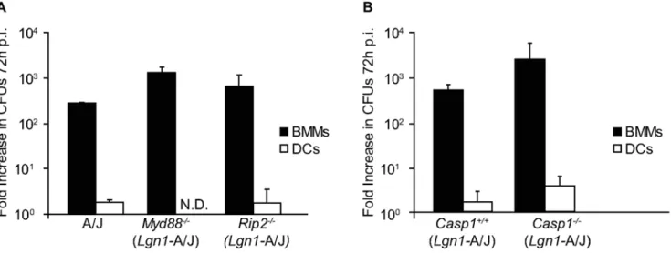
Signaling through TLRs in macrophages results in enhanced phagocytosis and phagosome fusion with lysosomes [46]. Thus, innate immune recognition ofL. pneumophilacould activate cellular processes that control bacterial replication in DCs. Cells deficient in the adapters MyD88 or Rip2 were used to interfere with the TLR and Nod signaling pathways respectively, to determine whetherL. pneumophilareplication in DCs is restricted by activation of signaling pathways controlled by innate immune receptors. Replication ofL. pneumophilawas not detected in DCs derived from A/J mice, which are defective for Naip5 signaling, or from A/J-derived mice deficient in either MyD88 or Rip2 (Figure 1A). By contrast, exponential replication of L. pneumophilaoccurred in the macrophages derived from these mice (Figure 1A).L. pneumophilaintracellular replication was not observed in DCs derived from mice deficient in both MyD88
Author Summary
The immune system is designed to identify microbes that enter the body and elicit responses that prevent the replication and dissemination of these organisms. Dendritic cells play an important role in regulating host immunity to pathogens. Their phagocytic capacity enables DCs to internalize and destroy most microbes, and the ability of DCs to migrate to specialized lymphoid organs is important for inducing antigen-specific immunity. Here, we analyzed
interactions between DCs and Legionella pneumophila, a
bacterial pathogen that can subvert phagocytic host cell functions to create a vacuole that permits intracellular replication. We found thatL. pneumophilainfection rapidly induced DCs to commit cell death through apoptosis. Rapid apoptosis was not observed after infection of macrophages, which are the phagocytic cells that supportL. pneumophila
replication in the lungs of infected animals. Using cells derived from knockout mice, we found that DCs deficient in the proteins Bax and Bak, which are essential for induction of the apoptosis pathway, were unable to restrict the intracel-lular replication ofL. pneumophila. Likewise, overproduction of Bcl-2, which is a negative regulator of apoptosis, resulted in DCs that were permissive forL. pneumophilareplication. These data indicate DCs have the ability to rapidly undergo apoptosis when infected with a microbe capable of replicating intracellularly, and this response effectively prevents pathogen replication. We hypothesize that this response may be designed to interfere with the migration of infected DCs through the lymphatic system, which would prevent DCs from serving as a ‘‘Trojan Horse’’ that transports pathogenic microbes from peripheral sites to central organs.
Figure 1. Restriction ofL. pneumophilareplication in DCs does not require signaling by MyD88, Rip2 or caspase-1.(A) BMMs (closed bars) and DCs (open bars) from A/J, Myd882/2 and Rip22/2 mice were infected with L. pneumophila WT for 72 h. Intracellular replication is determined by dividingL. pneumophilaCFUs recovered at 72 h by the CFUs recovered at 1 h post infection. (B) BMMs (closed bars) and DCs (open bars) derived fromCasp12/2or theCasp1+/+littermate mice were infected withL. pneumophilaWT. Intracellular replication is determined by dividing
L. pneumophilaCFUs recovered at 72 h by the CFUs recovered at 1 h post infection. Cells were homozygous for the permissiveLgn1allele from the A/ J mouse as indicated. Data are the mean6SD from three independent wells. N.D. = not detectable.
and Trif (data not shown), indicating that the lack of both of these TLR adaptor proteins did not restore L. pneumophila intracellular replication in DCs. Thus, DC restriction ofL. pneumophilareplication does not require TLR signaling through MyD88 and Trif or Nod1/2 signaling through Rip2.
Mouse macrophages restrict L. pneumophila replication by inducing a cell death pathway controlled by Naip5 and caspase-1 [37]. Mouse macrophages become permissive forL. pneumophila
replication if they are homozygous for the permissiveNaip5gene encoded in the A/J mouse or if caspase-1 is absent [35–37]. Intracellular replication of L. pneumophila was examined in DCs derived from Naip5-deficient mice to determine if the Naip5 protein produced by A/J-derived DCs retained an activity sufficient to restrict replication. The Naip52/2 DCs did not
support replication of L. pneumophila (Figure S1). It remained possible that proteins other than Naip5 might activate a caspase-1-dependent pathway that prevented L. pneumophila replication in DCs from A/J mice. To test this possibility, L. pneumophila
replication was measured in DCs derived from caspase-1-deficient mice homozygous for the A/J Naip5 allele (Casp12/2). L. pneumophila replication was not detected in Casp12/2 DCs,
whereas, L. pneumophila replication was similar in BMMs from these same Casp12/2 and caspase-1-sufficient mice (Casp1+/+ ) (Figure 1B). Thus, Naip5 and caspase-1 are not required for DC restriction ofL. pneumophilareplication.
DC apoptosis occurs rapidly afterL. pneumophila infection
Although caspase-1-mediated cell death was not required for DCs to restrict the replication ofL. pneumophila, it remained possible that another cell death pathway could be important for this process. Thus, we examined whether apoptosis occurred uponL. pneumophila
infection of DCs. TdT-mediated dUTP-biotin nick end-labeling (TUNEL) analysis was performed on DCs infected for 6 hours with either wild type (WT)L. pneumophilaor the isogenicDdotAstrain that has a nonfunctional Dot/Icm secretion system. Examination of DCs that had internalized WTL. pneumophilarevealed that 37% were TUNEL positive (Figure 2A and 2B, top panel). Only 1% of DCs containing theDdotAstrain were TUNEL positive (Figure 2A and 2B, top panel). The majority of DCs were TUNEL positive following induction of apoptosis with staurosporine (staur), a broad-spectrum protein kinase inhibitor (Figure 2B, bottom panel). Similar results were obtained usingCasp12/2DCs (Figure S2), indicating
that the absence of caspase-1 did not prevent apoptosis in DCs infected withL. pneumophila.
Macrophages and DCs were infected with WTL. pneumophilato compare the kinetics of apoptosis. At 1-hour post infection, infected DCs became TUNEL positive, whereas, TUNEL-positive macro-phages were not apparent until12-hours post infection (Figure 2C). In addition to using TUNEL staining, the kinetics of apoptosis was determined by measuring caspase-3/7 activity in DCs and macrophages afterL. pneumophilainfection. At 4-hours post infection there was a significant Dot/Icm-dependent increase in caspase-3/7 activity in DC extracts, but not in corresponding macrophage extracts (Figure S3). A significant increase in Caspase-3/7 activity was not observed for macrophages until 11-hours post infection (Figure S3). Thus, apoptosis in DCs was induced byL. pneumophila
with faster kinetics than in similarly infected macrophages.
Caspase-3-mediated effector responses are induced byL. pneumophilaafter DC infection
Caspase-3 mediates many of the downstream effector responses in the apoptotic cell death pathway, including fragmentation of
DNA in the nucleus [47]. Caspase-3-deficient mice (Casp32/2) were
used to determine whether DNA fragmentation induced afterL. pneumophilainfection of DCs was due to induction of the apoptotic cell death pathway. TUNEL analysis performed on DCs derived fromCasp32/2andCasp3+/+
mice 6 hours after infection withL. pneumophilarevealed that 57% of the infectedCasp3+/+
DCs were TUNEL positive, whereas only 9.5% of Casp32/2 DCs infected
with WTL. pneumophilawere TUNEL positive (Figure 3A, left panel and 3B). BothCasp32/2andCasp3+/+DCs infected with the
DdotA
strain showed minimal TUNEL staining (Figure 3A, right panel and 3B). Thus, L. pneumophila infection of DCs rapidly activates downstream components of the apoptotic cell death pathway.
Caspase-3 is involved in DC restriction ofL. pneumophila replication
To determine whether activation of the apoptotic cell death pathway was important for DC restriction of L. pneumophila
replication, DCs from A/J-derivedCasp32/2 and Casp3+/+mice infected with WTL. pneumophila were examined by fluorescence microscopy. The efficiency of L. pneumophila internalization determined 2 hours after infection was equivalent for Casp32/2
and Casp3+/+
DCs (Figure 4A, top panel). When DCs were examined 10 hours after infection, there was a significant increase in the percentage of infected Casp32/2 DCs that contained vacuoles supporting L. pneumophila replication (R.V.) (19%) compared to Casp3+/+
DCs (6%) (Figure 4A, bottom panel). Representative images in Figure 4A show that the number ofL. pneumophilain vacuoles that supported replication was higher in
Casp32/2DCs, and that most of the infectedCasp3+/+DCs had condensed or fragmented nuclei. These data were corroborated by determining colony-forming units (CFUs) over time. There was roughly a 10-fold increase inL. pneumophilaCFUs 72 hours after
Casp32/2DCs were infected with WTL. pneumophilacompared to
a slight decrease in CFUs recovered from Casp3+/+
DCs at 72 hours (Figure 4C). DCs eliminated theDdotAstrain with equal efficiency. Macrophages derived from these mice were infected in parallel. The infectedCasp3+/+ macrophages had normal nuclei (Figure 4B) and supported L. pneumophila replication to similar levels as theCasp32/2 macrophages (Figure 4B and 4D). These
data indicate that caspase-3 plays a role in restrictingL. pneumophila
replication in DCs, but not macrophages.
Cell death mediated by Bax and Bak restrictsL. pneumophilareplication in DCs
Bax and Bak play a central role in regulating apoptosis. When activated by members of the BH3-only protein family, Bax and Bak create a channel in the membrane of mitochondria that releases cytochrome c. This results in activation of the apoptosome and the subsequent activation of effector caspases, such as caspase-3 [48–51]. DCs derived from C57BL/6 (B6) and from mice deficient in Bak (Bak2/2) or both Bax and Bak (Bax2/2Bak2/2) were analyzed to
determine if Bax and Bak have a role in cell death induced byL. pneumophila. TUNEL analysis demonstrated that WTL. pneumophila
induced equivalent levels of cell death in DCs derived from B6 and
Bax2/2Bak2/2 mice (Figure 5A), suggesting that the
Naip5-dependent pathway of cell death remained functional in DCs. AL. pneumophilastrain containing an in-frame deletion of theflaA gene encoding flagellin was used to bypass Naip5-mediated cell death [38– 40]. A dramatic reduction in cell death was observed forBax2/2 Bak2/2 DCs infected with L. pneumophila DflaA (Figure 5A).
induces DC cell death by a Bax/Bak-dependent pathway and a Naip5-dependent pathway.
Replication of WT L. pneumophila was not detected in either
Bak2/2 orBax2/2Bak2/2 DCs (Figure 5B), which is consistent with the Naip5-mediated pathway being operational in these cells.
L. pneumophila DflaA replicated to similar levels as WT L. pneumophilain DCs derived fromCasp32/2mice homozygous for the A/J Naip5 allele (Figure S4), indicating that eliminating flagellin does not significantly enhance the capacity of L. pneumophilato replicate in DCs with a genetic defect in the Naip5 signaling pathway. DCs from Bax2/2Bak2/2 mice supported replication of L. pneumophila DflaA, whereas, replication of L.
pneumophilaDflaAwas not detected in DCs from control B6 mice (Figure 5B). Limited replication of theDflaAstrain was observed in
Bak2/2 DCs; however, replication was not as robust as that
observed in the Bax2/2Bak2/2 DCs (Figure 5B). Single cell
analysis revealed that the efficiency of infection was equivalent in B6,Bak2/2andBax2/2Bak2/2DCs (Figure 5C, top panel). Large
vacuoles harboring replicating bacteria were abundant inBax2/2 Bak2/2 DCs infected for 10-hours with L. pneumophila DflaA
(Figure 5C, bottom panel), whereas, vacuoles containing replicat-ingL. pneumophilaDflaAwere rare in the B6 andBak2/2DCs.
The development of vacuoles containing replicatingL. pneumo-philaDflaAwas evaluated in DCs derived from B6,Casp32/2and
Figure 2.L. pneumophilainfection of DCs induces nuclear DNA fragmentation.(A) Fluorescence micrographs show TUNEL staining (green) of DCs from A/J mice infected for 6 h with eitherL. pneumophilaWT (top left panel) orDdotA(top right panel). Total DNA was stained with DAPI (blue) and bacteria are red. Non-infected DCs (bottom left panel) or DCs treated with staurosporine for 5 h (bottom right panel) were used as negative and positive controls, respectively. (B) Quantification of the percentage of infected cells that were TUNEL positive (top graph); Quantification of the percentage of total cells that were TUNEL positive in the non-infected DCs and staurosporine-treated DCs (bottom graph). (C) Displayed are the percentage ofL. pneumophilaWT infected DCs (closed bars) or BMMs (open bars) that were TUNEL positive at 1, 2, 4, 6 and 12 h after infection. Data are represented by the mean6SD of 500 cells counted per each coverslip in triplicate. ** p,0.01. Bar = 10mm.
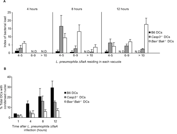
Bax2/2Bak2/2 mice. Vacuoles containing replicatingL. pneumo-philaDflaAwere detectable in bothCasp32/2, andBax2/2Bak2/2
DCs at 8-hours post infection (Figure 6A). Large vacuoles containing.10L. pneumophilaDflaAwere frequent in theBax2/2 Bak2/2 DCs at 12-hours post infection, but were found less
frequently in theCasp32/2DCs (Figure 6A). AlthoughCasp32/2
DCs exhibited enhanced resistance to cell death induced by L. pneumophilaDflaA, they were not as resistant to cell death as the
Bax2/2Bak2/2 DCs (Figure 6B), which likely explains why the
Bax2/2Bak2/2DCs were slightly more permissive for replication ofL. pneumophilaDflaAat 12-hours post infection compared to the
Casp32/2DCs. These data indicateL. pneumophilaactivation of the
intrinsic cell death pathway in DCs is sufficient to limit intracellular replication.
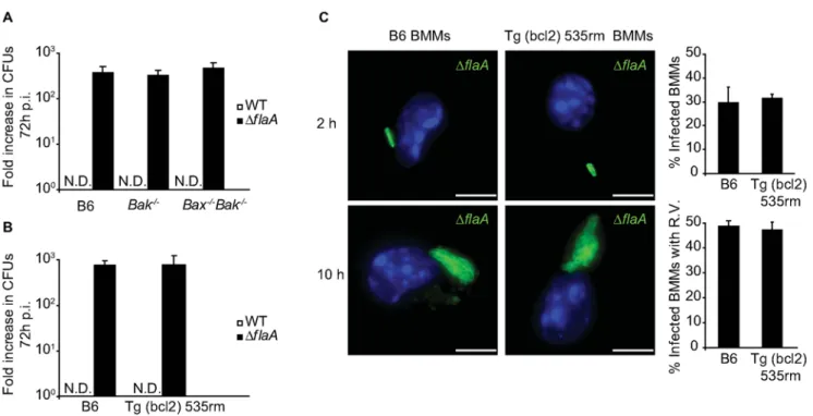
Bcl-2 overproduction antagonizes restriction ofL. pneumophilareplication by DCs
Bcl-2 is a pro-survival protein that regulates apoptosis [52,53]. Overexpression of pro-survival proteins such as those from the Bcl-2 family can block mitochondria membrane permeabilization and prevent apoptosis [54–56]. DCs from transgenic mice expressing humanBCL2under the control of the CD68 promoter (Tg(bcl2) 535rm) (Jamieson & Medhzitov, unpublished data) were used to determine whether overproduction of Bcl-2 could interfere with the ability of DCs to restrictL. pneumophilareplication. Immunoblot analysis confirmed that both macrophages and DCs derived from Tg(bcl2) 535rm mice produced human Bcl-2, and that overpro-duction of Bcl-2 did not affect the levels of Bax and Bak in these cells (Figure 7A). Replication of WT L. pneumophila was not observed in Tg(bcl2) 535rm DCs, presumably because these cells produce a functional Naip5 protein (Figure 7B). Replication of the
DflaAstrain was observed in the Tg(bcl2) 535rm DCs, but not in control DCs from B6 mice (Figure 7B). Single cell analysis
confirmed replication of theDflaAstrain in Tg(bcl2) 535rm DCs (Figure 7C). At 10-hours post infection, 21% of theDflaA-infected Tg(bcl2) 535rm DCs had large vacuoles containing replicatingL. pneumophila, and most of the infected Tg(bcl2) 535rm DCs were devoid of apoptotic features, such as condensed and fragmented nuclei, that were observed in infected control DCs derived from B6 mice (Figure 7C). TUNEL staining confirmed that the Tg(bcl2) 535rm DCs were more resistant to apoptosis after infection byL. pneumophilaDflaAcompared to control B6 DCs (Figure 7D). Thus, Bcl-2 overproduction limited DC apoptosis in response to L. pneumophilaand resulted in enhanced intracellular replication.
DCs have a unique ability to efficiently restrictL. pneumophilareplication by apoptosis
Macrophages derived fromBax2/2Bak2/2and Tg(bcl2) 535rm
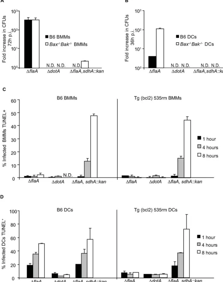
mice were used to determine whether rapid induction of programmed cell death as a mechanism to restrictL. pneumophila
replication was an exclusive property displayed by DCs. Replication of WT L. pneumophila was restricted by the Bax2/
2Bak2/2 macrophages and Tg(bcl2) 535rm macrophages as
efficiently as control B6 macrophages (Figure 8A and 8B). When the DflaA strain was used to bypass Naip5-mediated growth restriction, bacterial replication was not enhanced in theBax2/2 Bak2/2macrophages or Tg(bcl2) 535rm macrophages compared
to control B6 macrophages (Figure 8A and 8B). Single cell analysis confirmed these growth curve results, and showed that Bax and Bak function was not required for Naip5-mediated growth restriction of WT L. pneumophila and had no measurable effect on limiting the growth of the DflaA strain in macrophages (Figure 8C).
Previous studies have shown that macrophages infected with a
L. pneumophila mutant deficient in the effector protein SdhA undergo rapid cell death by an unknown pathway [57]. This
Figure 3. Caspase-3 is required for nuclear DNA fragmentation followingL. pneumophilainfection.(A) Fluorescence micrographs show TUNEL staining (green) ofCasp3+/+
andCasp32/2DCs infected for 6 h with eitherL. pneumophilaWT (left panel) orDdotA(right panel). Total DNA
was stained with DAPI (blue) and bacteria are red. (B) The graph shows percentage of infectedCasp3+/+(closed bars) andCasp32/2DCs (open bars)
that were TUNEL positive. Cells were homozygous for the permissiveLgn1allele from the A/J mouse as indicated. Data are represented by the mean6SD of 500 cells counted per each coverslip in triplicate. ** p,0.01. Bar = 10mm.
observation suggests that one possible reason DCs die quickly after
L. pneumophila infection is because a proposed anti-apoptotic activity mediated by the translocated SdhA protein might not be effective at preventing cell death in DCs. This would explain why the phenotype of DCs infected by L. pneumophila capable of translocating the SdhA protein appears to be similar to the phenotype of macrophages infected by an sdhA mutant. If this hypothesis is correct, then perturbing cell death pathways activated by Bax and Bak should restore replication of an sdhA
mutant in macrophages, and the elimination of SdhA should not affect replication ofL. pneumophilain DCs deficient in Bax and Bak signaling. To test this hypothesis we inactivated sdhA in the L.
pneumophilaDflaAstrain to generateL. pneumophilaDflaA,sdhA::kan. Elimination of Bax and Bak did not restore replication of L. pneumophilaDflaA,sdhA::kanin macrophages (Figure 9A) and theL. pneumophilaDflaA,sdhA::kanstrain was unable to replicate inBax2/
2Bak2/2DCs (Figure 9B). After infection byL. pneumophilaDflaA, sdhA::kan, cell death levels measured by TUNEL staining were similar in Tg(bcl2) 535rm macrophages and control B6 macro-phages (Figure 9C). TheL. pneumophilaDflaA,sdhA::kanstrain also induced cell death in DCs derived from Tg(bcl2) 535rm mice (Figure 9D). Thus, theL. pneumophila sdhAmutant phenotype was similar in both macrophages and DCs, which indicates that SdhA is necessary to prevent L. pneumophila from killing both
macro-Figure 4. Caspase-3 is required for the restriction ofL. pneumophilareplication in DCs but not macrophages.(A)Casp3+/+andCasp32/2
DCs were infected withL. pneumophilaWT (green) and fixed at either 2 or 10 h after infection. Total DNA was stained with DAPI (blue). On the right are graphical representations of the percentage of infectedCasp3+/+andCasp32/2DCs at 2 h post infection and the percentage of infected DCs with
vacuoles containing replicatingL. pneumophilaat 10 h post infection. (B) Fluorescence micrographs ofCasp3+/+
andCasp32/2BMMs (blue) that were
fixed at either 2 or 10 h after infection withL. pneumophilaWT (green); On the right are graphical representations of the percentage of infected BMMs at 2 h post infection and the percentage of infected BMMs with vacuoles containing replicatingL. pneumophilaat 10 h post infection. Data represent the mean6SD of 500 cells counted per coverslip in triplicate. R.V. = vacuoles containing replicating bacteria. ** p,0.01. Bar = 10mm. (C) DCs or (D) BMMs fromCasp3+/+
(triangles) orCasp32/2mice (squares) were infected with eitherL. pneumophilaWT (closed symbols) orDdotA(open symbols) and intracellular bacterial replication was measured over a period of 72 h. The fold increase in replication was determined by dividingL. pneumophila
CFUs recovered at the indicated time point by theL. pneumophilaCFUs recovered at 1 h post infection. Cells were homozygous for the permissive
phages and DCs by a pathway that does not require Bax and Bak function.
Discussion
Two cell death pathways were found to restrictL. pneumophila
replication in DCs. The first pathway was described previously in macrophages and involved activation of Naip5 by a process requiring L. pneumophila flagellin [37–40]. It had been shown clearly that stimulation of Naip5 byL. pneumophilaflagellin results in the activation of caspase-1 [37–40], which is a critical mediator
of pyroptosis. Recent data indicate that Naip5 activation of caspase-1 also results in the activation of caspase-7 [58], and that Naip5-dependent activation of caspase-7 is important for restric-tion ofL. pneumophila replication in mouse macrophages. Many details of the Naip5 signaling pathway remain to be determined, including the full repertoire of proteins required for Naip5-mediated cell death and all the cell types capable of restricting the replication of L. pneumophila by this pathway. Our data help to answer some of these questions by showing that components of the Naip5 pathway required for flagellin sensing and downstream effector responses are functioning in DCs. Additionally, the
Figure 5.BaxandBakare required forL. pneumophilagrowth restriction in DCs.(A) The graph shows percentage of B6 (open bars) and
Bax2/2Bak2/2DCs (closed bars) infected withL. pneumophilaWT,DdotAorDflaAthat were TUNEL positive at 6 h post infection. Data represent the
mean6SD of 300 cells counted per coverslip in triplicate. ** p,0.01. (B) B6,Bak2/2and Bax2/2Bak2/2were infected with eitherL. pneumophilaWT
(white bars) orDflaA(black bars) for 36 h. Intracellular replication was determined by dividingL. pneumophilaCFUs recovered at 36 h by the CFUs recovered at 1 h post infection. Data for each time point are the average of values obtained from three independent wells. ** p,0.01. (C) Fluorescence micrographs of B6,Bak2/2andBax2/2Bak2/2DCs that were infected withL. pneumophilaDflaAand fixed at either 2 h or 10 h post
infection. DCs were stained with an antibody specific for MHC II (red), DAPI (blue) and an anti-L. pneumophilaantibody (green). On the right are graphical representations of the percentage of B6,Bak2/2andBax2/2Bak2/2DCs infected at 2 h and the percentage of infected B6;Bak2/2and
Bax2/2Bak2/2DCs with vacuoles containing replicating bacteria at 10 h post infection. Data represent the mean
6SD of 500 cells counted per coverslip in triplicate. All cells had a dominantLgn1allele producing a functional Naip5 protein. R.V. = vacuoles containing replicating bacteria. N.D. = not detectable. ** p,0.01. Bar = 10mm.
observation that overproduction of Bcl-2 or elimination of Bax and Bak did not affect restriction of WTL. pneumophila replication in macrophages and DCs with a functional Naip5 protein provides evidence that this pathway is not functionally dependent on the mitochondrial pathway of apoptosis. Thus, both macrophages and
DCs have the capacity to undergo Naip5-dependent pyroptosis. In addition to restricting pathogen replication, activation of caspase-1 during this response generates bioactive IL-1b and IL-18 to stimulate additional antimicrobial responses and promote the recruitment of other immune cells [59–62]. This suggests that pyroptosis is a general innate immune response mediated by both macrophages and DCs to initiate early pro-inflammatory events at the site of microbial infection.
A second cell death pathway, which involved Bax and Bak regulation of caspase-3 activation, was found to efficiently restrict
L. pneumophilareplication in DCs. When the pyroptosis pathway was inactivated, either by using DCs with a defectiveNaip5allele or by using L. pneumophila that had the gene encoding flagellin deleted, the cell death pathway regulated by Bax and Bak was as efficient as the pyroptosis pathway at restricting replication ofL. pneumophila. A similar number of replicating L. pneumophila were contained in vacuoles in DCs deficient in Bax and Bak at 10-hours post infection (Figure 5C) when compared to macrophages
Figure 6. Enhanced replication ofL. pneumophilain DCs correlates with reduced apoptosis.(A) The graph shows the percentage of infected B6 (black bars),Casp32/2 (gray bars) orBax2/2Bak2/2DCs (white bars) that form vacuoles containing 4–5, 6–9 or.10 replicatingL. pneumophilaDflaAat 4, 8 and 12 h post infection. The index of bacterial load was calculated by dividing the percentage of vacuoles containing the indicated number of bacteria at the time points given by the percentage of infected DCs at 2 h p.i. and multiplying this number by 100. (B) The graph shows the percentage of total B6 (black bars),Casp32/2(gray bars) andBax2/2Bak2/2DCs (white bars) that had condensed nuclei at 1, 4, 8 and
12 hours after L. pneumophilaDflaAinfection. All cells had a dominant Lgn1allele producing a functional Naip5 protein. Data represent the mean6SD of 300 cells counted per coverslip in triplicate.
doi:10.1371/journal.ppat.1000478.g006
Table 1.Caspase-3/7 activity 6 h post-infection in relative fluorescence units.
L. pneumophila B6 DCs6SD Bak2/2DCs6SD
Bax2/2Bak2/2
DCs6SD non-infected 1913361950 134336680 100006624
DflaA 3246761358 123336404 85676321 DdotA 1763361357 106666404 843361001
(Figure 8C). Additionally, the number ofL. pneumophilarecovered from DCs deficient in caspase-3 was similar after 24-hours of infection when compared to macrophages (Figure 4). Because the addition of bacteria stimulates the maturation of DCs in culture, and mature DCs become non-phagocytic,L. pneumophila replica-tion in cultured DCs was not amplified by reinfecreplica-tion. This explains why replication subsided after L. pneumophila exited infected DCs at 24-hours post infection, but continued over a 72-hour period in macrophages (Figure 4). Thus, rapid activation of the intrinsic cell death pathway appears to be the primary mechanism by which DCs from permissive strains of mice restrict the intracellular replication ofL. pneumophila.
L. pneumophilawas capable of replication in DCs deficient in caspase-3; however, DCs deficient in both Bax and Bak were more permissive. This suggests that deletion of Bax and Bak more acutely blocks the apoptotic pathway, perhaps because other effector caspases can compensate for caspase-3 deficiency.
Consistent with this explanation,Bax2/2Bak2/2mice have severe developmental defects and most die perinatally, whereas,Casp32/2
mice are viable and have fewer developmental defects [63–65]. Accordingly, L. pneumophila infection induced the mitochondrial pathway of apoptosis inCasp32/2DCs, but the absence of caspase-3 was sufficient to delay cell death for a long enough period of time that vacuoles containing replicatingL. pneumophilawere detected. By contrast, apoptosis was not induced uponL. pneumophilainfection of
Bax2/2Bak2/2DCs and in the absence of cell deathL. pneumophila
was able to replicate for a longer period of time as was indicated by an increase in the number of large vacuoles containing over 10 bacteria. These data also suggest that cell death, as opposed to another activity mediated specifically by caspase-3, was sufficient to restrictL. pneumophilareplication.
The finding that overproduction of Bcl-2 resulted in enhanced bacterial replication in DCs supports the hypothesis that the mitochondrial pathway of apoptosis is important for restriction of
Figure 7. L. pneumophilareplication occurs in DCs overexpressing Bcl-2.(A) Immunoblot analysis of total human Bcl-2, Bax and Bak expression in Tg (bcl2) 535rm BMMs and DCs or the respective B6 littermates. Blots were reprobed for actin as a loading control. (B) B6 and Tg (bcl2) 535rm DCs were infected with eitherL. pneumophilaWT (white bars) orDflaA(black bars) for 36 h. Intracellular replication was determined by dividing the CFUs recovered at 36 h by the CFUs recovered at 1 h after infection. Data for each time point are the average of values obtained from three independent wells. * p,0.05. N.D. = not detectable. (C) Fluorescence micrographs of B6 or Tg (bcl2) 535rm DCs that were infected withL. pneumophilaDflaAand fixed at either 2 h or 10 h post infection. DCs were stained with an antibody specific for MHC II (red), DAPI (blue) and an
anti-L. pneumophilaantibody (green). On the right are graphical representations of the percentage of infected B6 or Tg (bcl2) 535rm DCs at 2 h post infection and the percentage of infected DCs with vacuoles containing replicatingL. pneumophilaDflaAat 10 h post infection. Data represent the mean6SD representative of 500 cells counted per coverslip in triplicate. R.V. = vacuoles containing replicating bacteria. ** p,0.01. Bar = 10mm. (D) The graph shows the percentage of B6 (open bars) and Tg (bcl2) 535rm DCs (closed bars) infected withL. pneumophilaWT,DdotAorDflaAthat were TUNEL positive at 6 h post infection. All cells had a dominant Lgn1allele producing a functional Naip5 protein. Data are represented by the mean6SD of 300 cells per coverslip in triplicate. ** p,0.01.
L. pneumophila replication in DCs. Bcl-2 functions as a negative regulator of Bax and Bak function, preventing their activation and insertion into the mitochondrial membrane [66,67]. Thus, the observation that Bcl-2 overproduction phenocopies a deficiency in Bax and Bak indicates thatL. pneumophilainfection of DCs triggers a cell-autonomous response that activates the mitochondrial pathway of apoptosis, leading to restriction of intracellular bacterial proliferation.
Previous studies in macrophages and macrophage-like cells have demonstrated that L. pneumophila is capable of activating the mitochondrial pathway of apoptosis [68–71]; however, our data indicate that the timing of this response is different in DCs compared to macrophages. In macrophages the response is slower, and morphological signs of apoptosis were typically not observed in cells until the late stages of infection after robust bacterial replication had occurred. Host cell apoptosis induced by L. pneumophilain both macrophages and DCs required a functional Dot/Icm secretion system, but not bacterial replication. This suggests that apoptosis is activated in response to either direct activities of bacterial effector proteins translocated by the Dot/Icm system or by host cell disturbances that are caused by the cumulative actions of multiple effector proteins.
The balance of pro-apoptotic to anti-apoptotic factors is important in the regulation of the mitochondrial pathway of apoptosis. Microbial infection affects this balance both by triggering the activation of pro-apoptotic factors and by inducing expression of anti-apoptotic proteins [49,72–74]. For many non-pathogenic bacteria, these two events are balanced and apoptosis is prevented. The added stress on cells infected with pathogenic
microbes, however, will typically result in apoptosis unless the pathogen has the ability to alter the function of proteins involved in regulating cell death [75,76]. Thus, differences in the expression of Bcl-2 family members or in the functioning of effector proteins could account for the faster kinetics of apoptosis in DCs compared to macrophages followingL. pneumophilainfection.
Two effector proteins translocated into host cells by the L. pneumophilaDot/Icm system have been implicated in preventing cell death. The effector protein SidF appears to interfere with the function of pro-apoptotic 2 family members BNIP3 and Bcl-Rambo [77]. Although macrophages infected with asidFmutant show increased apoptosis 14-hours after infection, this increase in apoptosis does not impact bacterial replication greatly [77]. By contrast, the effector SdhA is required to prevent macrophage cell death during infection by a mechanism that is not understood, and the cell death induced by ansdhAmutant greatly reduces bacterial replication in macrophages [57]. We found that thesdhAmutant induced cell death in both macrophages and DCs, and that this cell death pathway was not inhibited by Bcl-2 over-expression or elimination of Bax and Bak. Additionally, intracellular growth of the sdhA mutant was not restored in macrophages deficient in caspase-3 (data not shown). Thus, both macrophages and DCs are equally susceptible to cell death induced by thesdhAmutant, and the cell death pathway triggered by the sdhA mutant does not require several of the central components of the apoptosis pathway. These data are consistent with there being an intrinsic difference between macrophages and DCs with respect to their ability to activate the mitochondrial cell death pathway in response toL. pneumophila.
Figure 8. Interfering with Bax and Bak function does not enhanceL. pneumophilareplication in macrophages.(A) B6,Bak2/2andBax2/2 Bak2/2BMMs or (B) B6 and Tg (bcl2) 535rm BMMs were infected with eitherL. pneumophilaWT (white bars) orDflaA(black bars) for 72 hours. Intracellular replication is determined by dividing theL. pneumophilaCFUs recovered at 72 h by the CFUs recovered 1 h after infection. Data are the average of values obtained from three independent wells. N.D. = not detectable. (C) Fluorescence micrographs of B6 and Tg (bcl2) 535rm BMMs that were infected withL. pneumophilaDflaAand fixed either at 2 h or 10 h post infection. BMMs were stained with DAPI (blue) and an anti-L. pneumophila
antibody (green). On the right are graphical representations of the percentage of B6 or Tg (bcl2) 535rm BMMs infected at 2 h and the percentage of infected BMMs with vacuoles containing replicatingL. pneumophilaat 10 h post infection. Data represent the mean6SD of 500 cells counted per coverslip in triplicate. All cells had a dominantLgn1 allele producing a functional Naip5 protein. R.V. = vacuoles containing replicating bacteria. Bar = 10mm.
Figure 9.L. pneumophila sdhAmutants induce rapid cell death in macrophages and DCs by a pathway that does not require Bax and Bak.(A) Intracellular replication ofL. pneumophilaDflaA,DdotAandDflaA,sdhA::kanwas measured in B6 (black bars) andBax2/2Bak2/2BMMs (white
In addition to L. pneumophila, there are many other reports demonstrating that DCs are able to restrict the replication of pathogens capable of growing within macrophages [42–45]. DCs are very proficient at migrating from peripheral tissues to the host lymphatic system following exposure to maturation stimuli, such as encounters with microbes. Because of this property, it has been suggested that DCs can function as a ‘‘Trojan Horse’’ capable of systemic dissemination of pathogens internalized at peripheral sites of infection [42,44,78]. Here we show that rapid cell death is one mechanism DCs use to avoid being subverted by an intracellular pathogen. In addition to preventing pathogen replication and dissemination, apoptotic DCs harboring intracellular pathogens would become substrates for phagocytosis by neighboring DCs and macrophages, and most mechanisms used by intracellular pathogens to subvert host cellular function would be ineffective as long as the pathogen were residing in an apoptotic cell. Thus, apoptotic bodies containing pathogens would be degraded in lysosomes, resulting in the release of pathogen-derived molecules that could stimulate innate immune receptors and trigger adaptive responses by being presented on the cell surface in association with host MHC proteins. Based on these data, we hypothesize that rapid pathogen-induced apoptosis by DCs is an important innate immune response to intracellular pathogens.
Materials and Methods
Bacterial cultures
L. pneumophilaserogroup 1 strain, Lp01 [18], an isogenicdotA
mutant strain (DdotA), and a flagellin-deficient mutant strain (DflaA) [79] were cultured on charcoal yeast extract agar (CYE) [80] for 2 days prior to use in experiments. TheDflaA, sdhA::kanstrain was cultured on CYE with 10mg/mL kanamycin. The plasmid pAM239 was used to produce DSred or GFP in theL. pneumophila
strains indicated [81]. For experiments utilizing bacteria express-ing DSred or GFP, L. pneumophila was grown on plates supplemented with chloramphenicol (6.25mg/ml), and DSred or GFP expression was induced after infection by adding IPTG (0.2 mM) to the tissue culture medium.
Mice
A/J and C57BL/6 (B6) mice were purchased from Jackson Laboratories.Caspase-12/2(Casp12/2),Caspase-32/2 (Casp32/2), Myd882/2,Rip22/2(Ripk22/2;Rick2/2),Bak2/2,Bax2/2Bak2/2
and Naip52/2 mice have been described [59,63,65,82–84]. Myd882/2Trif2/2mice homozygous for the B6 Lgn1allele were
provided by R. Medzhitov. Myd882/2 and Rip22/2 mice were
crossed with A/J mice to generate progeny homozygous for the A/ JLgn1allele as described previously [31].Casp12/2andCasp32/2
mice homozygous for the permissive A/J Lgn1 allele were backcrossed to the A/J background for 4 and 5 generations respectively. Transgenic C57BL/6 mice over expressing human BCL2 under the control of the CD68 promoter (Tg(bcl2) 535rm) (Jamieson & Medhzitov, unpublished data), were kindly provided by R. Medzhitov. All animals were maintained in accordance with the guidelines of the Yale Institutional Animal Use and Care Committee.
Macrophage and dendritic cell cultures
Bone-marrow derived macrophages (BMMs) were prepared as described previously with some modifications [85]. Briefly, bone marrow was collected from the femurs and tibiae of mice. Cells were plated on non-tissue culture-treated dishes and incubated at 37uC in RPMI-1640 containing 20% heat-inactivated fetal bovine serum (FBS), 30% macrophage colony-stimulating factor (M-CSF)-conditioned medium, and 1% penicillin-streptomycin. On day 7, cells were harvested and resuspended in RPMI 1640 containing 10% FBS and 15% M-CSF-conditioned medium. Cells were then plated in 24-well tissue culture-treated plates and incubated at 37uC. Bone marrow derived-DCs (BMDCs) were prepared as described in Lutzet al. [86]. Modifications were as follows. Cells were plated on non-tissue culture-treated dishes and incubated at 37uC in RPMI-1640 supplemented with 10% heat-inactivated FBS, 50mM 2-mercaptoethanol, 1% penincillin-streptomycin and 1% GM-CSF (DC medium). Cells were harvested and used on day 10.
Intracellular replication assays
Intracellular replication of L. pneumophila in BMMs was measured as described previously [79] and modified slightly for DCs.L. pneumophilawas added to DCs at a multiplicity of infection (MOI) of 20. The plates were centrifuged at 150 g for 5 minutes (min) and then incubated at 37uC for 30 min. Cells were removed from the wells and DCs were positively selected on magnetic columns using anti-CD11c-coated magnetic beads (Miltenyi Biotech). To remove extracellular bacteria, DCs were washed 36 with PBS containing 2 mM EDTA and 0.5% BSA while
bound to the column. DCs were eluted and 26105 DCs were added to individual wells in 48-well plates. Adherent and non-adherent DCs were taken from individual wells and lysed with sterile H2O at the indicated times after infection, and these fractions were pooled with the culture supernatants. Dilutions from the pooled fractions were plated on CYE agar to determine bacterial CFUs. Data are the mean CFUs recovered from three independent wells6SD. Bacterial replication was calculated by determining the fold increase in CFUs.
Single cell immunofluorescence assays to measureL. pneumophilauptake and formation of vacuoles containing replicating bacteria (RV)
L. pneumophilauptake and intracellular growth in bothCasp32/2
and Casp3+/+
DCs was performed as previously described [41]. Intracellular replication in B6,Bak2/2, andBax2/2Bak2/2DCs was performed following the same protocol described previously with some modifications to the immunofluorescence staining [41]. Briefly, after permeabilization for 15 min at room temperature (R.T.) in RPMI containing 0.05% saponin, coverslips were incubated for 1 h at R.T. in permeabilization solution containing anti-MHC II I-Ab+d+q
, I-Ed+k
antibody (TIB 120; American Type Culture Collection (ATCC), Rockville, MD). Coverslips were washed 36in RPMI containing 0.05% saponin. Coverslips were incubated 45 min at R.T. with Alexa Fluor 568- conjugated goat anti-rat (Invitrogen-Molecular Probes) in permeabilization solu-tion and then washed 36with PBS. Coverslips were mounted on
Bak2/2DCs (white bars) at 36 h after infection. The fold increase in intracellular replication was determined by dividingL. pneumophilaCFUs recovered
at 36 h by theL. pneumophilaCFUs recovered at 1 h post infection. Data are the mean6SD from three independent wells. N.D. = not detectable. (C) The graph shows the percentage of B6 and Tg (bcl2) 535rm BMMs or (D) DCs infected withL. pneumophilaDflaA,DdotAorDflaA,sdhA::kanthat were TUNEL positive at 1 h (black bars), 4 h (gray bars) and 8 h (white bars) after infection. All cells had a dominantLgn1allele producing a functional Naip5 protein. Data are represented by the mean6SD of 300 cells counted per each coverslip in triplicate.
slides and examined by fluorescence microscopy. TIB 120 staining of MHC II was used to identify DCs. Assays to measure uptake and formation of vacuoles containing replicatingL. pneumophilain BMMs were conducted similarly [14]. Data are represented by the mean number of cells observed in three independent coverslips.
TUNEL staining
DCs previously selected by CD11c magnetic beads were infected withL. pneumophilaat an MOI of 25 or treated for 5 h with staurosporine (1mg/ml) and assayed for nuclear DNA fragmentation by TUNEL with thein situcell death detection kit (Roche). Samples were then analyzed by fluorescence microscopy and all data points represent the average number of TUNEL positive cells6SD obtained from three independent coverslips.
Immunoblotting
BMMs and DCs were directly lysed in SDS-PAGE sample buffer. Lysates were separated by SDS-PAGE, and proteins were transferred (Wet Transfer Cell; Bio-Rad) at 100 V for 1 h to Immobilon P membranes (Millipore) in transfer buffer (50 mM Tris, 40 mM glycine, and 10% methanol). Membranes were blocked for 1 h at 25uC in Tris-buffered saline (TBS), 5% nonfat dry milk, and 0.1% Tween-20. Membranes were incubated with primary antibody overnight at 5uC and incubated with horseradish peroxidase-conjugated secondary antibody 1 h at R.T. Rabbit anti-human Bcl-2, rabbit anti-Bax, and rabbit anti-Bak (Cell Signaling Technology) were used. Western Lightning Chemiluminescence Reagent Plus (Perkin Elmer) was used for antibody detection.
Caspase-3/7 activity
Macrophages and DCs were plated in 96 well plates at a concentration of 56104 cells/well. Cells were infected with L. pneumophilaat an MOI of 50, incubated at 37uC for 1, 2, 4, 6 and 11 hours and then frozen at220uC to lyse the cells. Caspase-3/7 activity was measured using the Apo-One Homogeneous caspase-3/7 kit (Promega). Relative fluorescence units (RFU) measured at each time point is proportional to the amount of caspase-3/7 activity. All data points represent the average values6SD obtained from three wells assayed independently.
Gene ID numbers
MyD88: 17874; Rip2: 192656; Caspase-1: 12362; Caspase-3: 12367; Bax: 12028; Bak: 12018; Human Bcl-2: 596; Naip5: 17951.
Protein ID numbers
MyD88: P22366; Rip2: P58801; Caspase-1: P29452; Caspase-3: P70677; Bax: Q07813; Bak: O08734; human Bcl-2: P10415; Naip5: Q8CGT2.
Supporting Information
Figure S1 Naip5-deficient DCs restrictL. pneumophilareplication. Quantification of the percentage ofL. pneumophila WT or DdotA
infected B6 (black bars) and Naip52/2 DCs (white bars) with
vacuoles containing replicating bacteria at 10 h post-infection. Data represent the mean6SD of 500 cells counted per coverslip in triplicate. R.V. = vacuoles containing replicating bacteria. Found at: doi:10.1371/journal.ppat.1000478.s001 (0.46 MB EPS)
Figure S2 Nuclear fragmentation in DCs induced by L. pneumophila is caspase-1-independent. Quantification of the per-centage of B6 (closed bars) andCasp12/2DCs (open bars) infected
with eitherL. pneumophilaWT orDdotAthat are TUNEL positive 6 h after infection. Data are represented by the mean6SD of 300 cells counted per each coverslip in triplicate.
Found at: doi:10.1371/journal.ppat.1000478.s002 (0.46 MB EPS)
Figure S3 L. pneumophila-induced activation of caspase-3/7 occurs faster in DCs compared to macropahges. (A) DCs and (B) BMMs were infected with eitherL. pneumophila WT orDdotAfor 1 h, 2 h, 4 h, 6 h and 11 h as indicated. Caspase-3/7 activity is indicated as relative fluorescence units (RFU) measured at each time point. Data are expressed as mean6SD obtained from 3 independent wells. * p,0.05. **p,0.01.
Found at: doi:10.1371/journal.ppat.1000478.s003 (0.49 MB EPS)
Figure S4 L. pneumophilaWT andDflaAreplicate to similar levels in caspase-3-deficient DCs homozygous for the A/J Lgn1 allele. Intracellular replication ofL. pneumophilaWT,DflaAorDdotAwas compared in Casp32/2 DCs at 36 h after infection. The fold
increase in intracellular replication was determined by dividingL. pneumophilaCFUs recovered at 36 h by the CFUs recovered at 1 h post infection. Data are the mean6SD from three independent wells. N.D. = not detectable.
Found at: doi:10.1371/journal.ppat.1000478.s004 (0.46 MB EPS)
Acknowledgments
We are grateful to Dr. Shizuo Akira for permission to useMyd882/2, and Trif2/2mice, to Dr Richard Flavell for permission to useCasp12/2and
Casp32/2mice and to Dr. Russell Vance for permission to useNaip52/2
mice; Annie Neild, Jonathan Kagan, Igor Brodsky, Anja Lu¨hrmann and Kristina Archer for critical manuscript review; Dr. Salome´ Gomes, Dr. Rui Appelberg, Dr Manuel Santos and the Roy lab for helpful discussions and assistance.
Author Contributions
Conceived and designed the experiments: CVN TL AMJ CRR. Performed the experiments: CVN TL SS. Analyzed the data: CVN TL CRR. Contributed reagents/materials/analysis tools: CVN TL AMJ CLC SS CBT. Wrote the paper: CVN TL CRR.
References
1. Steinman RM (1991) The dendritic cell system and its role in immunogenicity. Annu Rev Immunol 9: 271–296.
2. Taylor PR, Martinez-Pomares L, Stacey M, Lin HH, Brown GD, et al. (2005) Macrophage receptors and immune recognition. Annu Rev Immunol 23: 901–944. 3. Akira S, Takeda K (2004) Functions of toll-like receptors: lessons from KO mice.
C R Biol 327: 581–589.
4. Flo TH, Smith KD, Sato S, Rodriguez DJ, Holmes MA, et al. (2004) Lipocalin 2 mediates an innate immune response to bacterial infection by sequestrating iron. Nature 432: 917–921.
5. Janeway CA Jr, Medzhitov R (2002) Innate immune recognition. Annu Rev Immunol 20: 197–216.
6. Miao EA, Andersen-Nissen E, Warren SE, Aderem A (2007) TLR5 and Ipaf: dual sensors of bacterial flagellin in the innate immune system. Semin Immunopathol 29: 275–288.
7. Ishii KJ, Koyama S, Nakagawa A, Coban C, Akira S (2008) Host innate immune receptors and beyond: making sense of microbial infections. Cell Host Microbe 3: 352–363.
8. Fraser DW, Tsai TR, Orenstein W, Parkin WE, Beecham HJ, et al. (1977) Legionnaires’ disease: description of an epidemic of pneumonia. N Engl J Med 297: 1189–1197.
9. McDade JE, Shepard CC, Fraser DW, Tsai TR, Redus MA, et al. (1977) Legionnaires’ disease: isolation of a bacterium and demonstration of its role in other respiratory disease. N Engl J Med 297: 1197–1203.
10. Roy CR, Tilney LG (2002) The road less traveled: transport ofLegionellato the endoplasmic reticulum. J Cell Biol 158: 415–419.
12. Horwitz MA (1983) Formation of a novel phagosome by the Legionnaires’ disease bacterium (Legionella pneumophila) in human monocytes. J Exp Med 158: 1319–1331.
13. Horwitz MA, Silverstein SC (1980) Legionnaires’ disease bacterium (Legionella pneumophila) multiples intracellularly in human monocytes. J Clin Invest 66: 441–450.
14. Kagan JC, Roy CR (2002)Legionellaphagosomes intercept vesicular traffic from endoplasmic reticulum exit sites. Nat Cell Biol 4: 945–954.
15. Tilney LG, Harb OS, Connelly PS, Robinson CG, Roy CR (2001) How the parasitic bacteriumLegionella pneumophilamodifies its phagosome and transforms it into rough ER: implications for conversion of plasma membrane to the ER membrane. J Cell Sci 114: 4637–4650.
16. Robinson CG, Roy CR (2006) Attachment and fusion of endoplasmic reticulum with vacuoles containingLegionella pneumophila. Cell Microbiol 8: 793–805. 17. Swanson MS, Isberg RR (1995) Association ofLegionella pneumophilawith the
macrophage endoplasmic reticulum. Infect Immun 63: 3609–3620. 18. Berger KH, Isberg RR (1993) Two distinct defects in intracellular growth
complemented by a single genetic locus inLegionella pneumophila. Mol Microbiol 7: 7–19.
19. Marra A, Blander SJ, Horwitz MA, Shuman HA (1992) Identification of a
Legionella pneumophilalocus required for intracellular multiplication in human macrophages. Proc Natl Acad Sci U S A 89: 9607–9611.
20. Segal G, Purcell M, Shuman HA (1998) Host cell killing and bacterial conjugation require overlapping sets of genes within a 22-kb region of the
Legionella pneumophilagenome. Proc Natl Acad Sci U S A 95: 1669–1674. 21. Vogel JP, Andrews HL, Wong SK, Isberg RR (1998) Conjugative transfer by the
virulence system ofLegionella pneumophila. Science 279: 873–876.
22. Chen J, de Felipe KS, Clarke M, Lu H, Anderson OR, et al. (2004)Legionella
effectors that promote nonlytic release from protozoa. Science 303: 1358–1361. 23. Conover GM, Derre I, Vogel JP, Isberg RR (2003) TheLegionella pneumophila
LidA protein: a translocated substrate of the Dot/Icm system associated with maintenance of bacterial integrity. Mol Microbiol 48: 305–321.
24. Luo ZQ, Isberg RR (2004) Multiple substrates of theLegionella pneumophilaDot/ Icm system identified by interbacterial protein transfer. Proc Natl Acad Sci U S A 101: 841–846.
25. Ingmundson A, Delprato A, Lambright DG, Roy CR (2007) Legionella pneumophila proteins that regulate Rab1 membrane cycling. Nature 450: 365–369.
26. Nagai H, Kagan JC, Zhu X, Kahn RA, Roy CR (2002) A bacterial guanine nucleotide exchange factor activates ARF onLegionellaphagosomes. Science 295: 679–682.
27. Machner MP, Isberg RR (2006) Targeting of host Rab GTPase function by the intravacuolar pathogenLegionella pneumophila. Dev Cell 11: 47–56.
28. Machner MP, Isberg RR (2007) A bifunctional bacterial protein links GDI displacement to Rab1 activation. Science 318: 974–977.
29. Murata T, Delprato A, Ingmundson A, Toomre DK, Lambright DG, et al. (2006) TheLegionella pneumophila effector protein DrrA is a Rab1 guanine nucleotide-exchange factor. Nat Cell Biol 8: 971–977.
30. Horwitz MA (1987) Characterization of avirulent mutantLegionella pneumophila
that survive but do not multiply within human monocytes. J Exp Med 166: 1310–1328.
31. Archer KA, Roy CR (2006) MyD88-dependent responses involving toll-like receptor 2 are important for protection and clearance ofLegionella pneumophilain a mouse model of Legionnaires’ disease. Infect Immun 74: 3325–3333. 32. Hawn TR, Smith KD, Aderem A, Skerrett SJ (2006) Myeloid differentiation
primary response gene (88)- and toll-like receptor 2-deficient mice are susceptible to infection with aerosolizedLegionella pneumophila. J Infect Dis 193: 1693–1702. 33. Sporri R, Joller N, Albers U, Hilbi H, Oxenius A (2006) MyD88-dependent IFN-cproduction by NK cells is key for control ofLegionella pneumophilainfection. J Immunol 176: 6162–6171.
34. Derre I, Isberg RR (2004) Macrophages from mice with the restrictiveLgn1
allele exhibit multifactorial resistance toLegionella pneumophila. Infect Immun 72: 6221–6229.
35. Diez E, Lee SH, Gauthier S, Yaraghi Z, Tremblay M, et al. (2003)Birc1eis the gene within theLgn1locus associated with resistance toLegionella pneumophila. Nat Genet 33: 55–60.
36. Wright EK, Goodart SA, Growney JD, Hadinoto V, Endrizzi MG, et al. (2003)
Naip5affects host susceptibility to the intracellular pathogenLegionella pneumophila. Curr Biol 13: 27–36.
37. Zamboni DS, Kobayashi KS, Kohlsdorf T, Ogura Y, Long EM, et al. (2006) The Birc1e cytosolic pattern-recognition receptor contributes to the detection and control ofLegionella pneumophilainfection. Nat Immunol 7: 318–325. 38. Amer A, Franchi L, Kanneganti TD, Body-Malapel M, Ozoren N, et al. (2006)
Regulation ofLegionellaphagosome maturation and infection through flagellin and host Ipaf. J Biol Chem 281: 35217–35223.
39. Molofsky AB, Byrne BG, Whitfield NN, Madigan CA, Fuse ET, et al. (2006) Cytosolic recognition of flagellin by mouse macrophages restricts Legionella pneumophilainfection. J Exp Med 203: 1093–1104.
40. Ren T, Zamboni DS, Roy CR, Dietrich WF, Vance RE (2006) Flagellin-deficientLegionellamutants evade caspase-1- andNaip5-mediated macrophage immunity. PLoS Pathog 2: e18. doi:10.1371/journal.ppat.0020018. 41. Neild AL, Roy CR (2003)Legionellareveal dendritic cell functions that facilitate
selection of antigens for MHC class II presentation. Immunity 18: 813–823.
42. Herrmann JL, Lagrange PH (2005) Dendritic cells andMycobacterium tuberculosis: which is the Trojan horse? Pathol Biol (Paris) 53: 35–40.
43. Niedergang F, Sirard JC, Blanc CT, Kraehenbuhl JP (2000) Entry and survival of Salmonella typhimurium in dendritic cells and presentation of recombinant antigens do not require macrophage-specific virulence factors. Proc Natl Acad Sci U S A 97: 14650–14655.
44. Pron B, Boumaila C, Jaubert F, Berche P, Milon G, et al. (2001) Dendritic cells are early cellular targets ofListeria monocytogenesafter intestinal delivery and are involved in bacterial spread in the host. Cell Microbiol 3: 331–340. 45. Tailleux L, Schwartz O, Herrmann JL, Pivert E, Jackson M, et al. (2003)
DC-SIGN is the majorMycobacterium tuberculosisreceptor on human dendritic cells. J Exp Med 197: 121–127.
46. Blander JM, Medzhitov R (2004) Regulation of phagosome maturation by signals from toll-like receptors. Science 304: 1014–1018.
47. Reed JC (1997) Cytochrome c: can’t live with it–can’t live without it. Cell 91: 559–562.
48. Adams JM (2003) Ways of dying: multiple pathways to apoptosis. Genes Dev 17: 2481–2495.
49. Byrne GI, Ojcius DM (2004)Chlamydiaand apoptosis: life and death decisions of an intracellular pathogen. Nat Rev Microbiol 2: 802–808.
50. Gross A, McDonnell JM, Korsmeyer SJ (1999) BCL-2 family members and the mitochondria in apoptosis. Genes Dev 13: 1899–1911.
51. Korsmeyer SJ, Wei MC, Saito M, Weiler S, Oh KJ, et al. (2000) Pro-apoptotic cascade activates BID, which oligomerizes BAK or BAX into pores that result in the release of cytochrome c. Cell Death Differ 7: 1166–1173.
52. Siegel RM (2006) Caspases at the crossroads of immune-cell life and death. Nat Rev Immunol 6: 308–317.
53. Strasser A (2005) The role of BH3-only proteins in the immune system. Nat Rev Immunol 5: 189–200.
54. Bouillet P, Purton JF, Godfrey DI, Zhang LC, Coultas L, et al. (2002) BH3-only Bcl-2 family member Bim is required for apoptosis of autoreactive thymocytes. Nature 415: 922–926.
55. Cory S, Adams JM (2002) The Bcl2 family: regulators of the cellular life-or-death switch. Nat Rev Cancer 2: 647–656.
56. Keeble JA, Gilmore AP (2007) Apoptosis commitment–translating survival signals into decisions on mitochondria. Cell Res 17: 976–984.
57. Laguna RK, Creasey EA, Li Z, Valtz N, Isberg RR (2006) A Legionella pneumophila-translocated substrate that is required for growth within macrophag-es and protection from host cell death. Proc Natl Acad Sci U S A 103: 18745–18750.
58. Akhter A, Gavrilin MA, Frantz L, Washington S, Ditty C, et al. (2009) Caspase-7 activation by the Nlrc4/Ipaf inflammasome restricts Legionella pneumophila
infection. PLoS Pathog 5: e1000361. doi:10.1371/journal.ppat.1000361. 59. Kuida K, Lippke JA, Ku G, Harding MW, Livingston DJ, et al. (1995) Altered
cytokine export and apoptosis in mice deficient in interleukin-1bconverting enzyme. Science 267: 2000–2003.
60. Mariathasan S, Monack DM (2007) Inflammasome adaptors and sensors: intracellular regulators of infection and inflammation. Nat Rev Immunol 7: 31–40.
61. Martinon F, Burns K, Tschopp J (2002) The inflammasome: a molecular platform triggering activation of inflammatory caspases and processing of
proIL-b. Mol Cell 10: 417–426.
62. Thornberry NA, Molineaux SM (1995) Interleukin-1bconverting enzyme: a novel cysteine protease required for IL-1b production and implicated in programmed cell death. Protein Sci 4: 3–12.
63. Kuida K, Zheng TS, Na S, Kuan C, Yang D, et al. (1996) Decreased apoptosis in the brain and premature lethality in CPP32-deficient mice. Nature 384: 368–372.
64. Lakhani SA, Masud A, Kuida K, Porter GA Jr, Booth CJ, et al. (2006) Caspases 3 and 7: key mediators of mitochondrial events of apoptosis. Science 311: 847–851.
65. Lindsten T, Ross AJ, King A, Zong WX, Rathmell JC, et al. (2000) The combined functions of proapoptotic Bcl-2 family members bak and bax are essential for normal development of multiple tissues. Mol Cell 6: 1389–1399. 66. Cheng EH, Wei MC, Weiler S, Flavell RA, Mak TW, et al. (2001) 2,
BCL-X(L) sequester BH3 domain-only molecules preventing BAX- and BAK-mediated mitochondrial apoptosis. Mol Cell 8: 705–711.
67. Willis SN, Fletcher JI, Kaufmann T, van Delft MF, Chen L, et al. (2007) Apoptosis initiated when BH3 ligands engage multiple Bcl-2 homologs, not Bax or Bak. Science 315: 856–859.
68. Abu-Zant A, Santic M, Molmeret M, Jones S, Helbig J, et al. (2005) Incomplete activation of macrophage apoptosis during intracellular replication ofLegionella pneumophila. Infect Immun 73: 5339–5349.
69. Gao LY, Abu Kwaik Y (1999) Activation of caspase 3 during Legionella pneumophila-induced apoptosis. Infect Immun 67: 4886–4894.
70. Hagele S, Hacker J, Brand BC (1998) Legionella pneumophila kills human phagocytes but not protozoan host cells by inducing apoptotic cell death. FEMS Microbiol Lett 169: 51–58.
71. Molmeret M, Zink SD, Han L, Abu-Zant A, Asari R, et al. (2004) Activation of caspase-3 by the Dot/Icm virulence system is essential for arrested biogenesis of theLegionella-containing phagosome. Cell Microbiol 6: 33–48.
73. Everett H, McFadden G (1999) Apoptosis: an innate immune response to virus infection. Trends Microbiol 7: 160–165.
74. Faherty CS, Maurelli AT (2008) Staying alive: bacterial inhibition of apoptosis during infection. Trends Microbiol 16: 173–180.
75. Philpott DJ, Girardin SE, Sansonetti PJ (2001) Innate immune responses of epithelial cells following infection with bacterial pathogens. Curr Opin Immunol 13: 410–416.
76. Sansonetti PJ (2004) War and peace at mucosal surfaces. Nat Rev Immunol 4: 953–964.
77. Banga S, Gao P, Shen X, Fiscus V, Zong WX, et al. (2007)Legionella pneumophila
inhibits macrophage apoptosis by targeting pro-death members of the Bcl2 protein family. Proc Natl Acad Sci U S A 104: 5121–5126.
78. Moll H, Flohe S, Rollinghoff M (1995) Dendritic cells inLeishmania major -immune mice harbor persistent parasites and mediate an antigen-specific T cell immune response. Eur J Immunol 25: 693–699.
79. Zuckman DM, Hung JB, Roy CR (1999) Pore-forming activity is not sufficient forLegionella pneumophilaphagosome trafficking and intracellular growth. Mol Microbiol 32: 990–1001.
80. Feeley JC, Gibson RJ, Gorman GW, Langford NC, Rasheed JK, et al. (1979) Charcoal-yeast extract agar: primary isolation medium forLegionella pneumophila. J Clin Microbiol 10: 437–441.
81. Coers J, Monahan C, Roy CR (1999) Modulation of phagosome biogenesis by
Legionella pneumophilacreates an organelle permissive for intracellular growth. Nat Cell Biol 1: 451–453.
82. Adachi O, Kawai T, Takeda K, Matsumoto M, Tsutsui H, et al. (1998) Targeted disruption of the MyD88gene results in loss of IL-1- and IL-18-mediated function. Immunity 9: 143–150.
83. Susa M, Ticac B, Rukavina T, Doric M, Marre R (1998)Legionella pneumophila
infection in intratracheally inoculated T cell-depleted or -nondepleted A/J mice. J Immunol 160: 316–321.
84. Lightfield KL, Persson J, Brubaker SW, Witte CE, von Moltke J, et al. (2008) Critical function for Naip5 in inflammasome activation by a conserved carboxy-terminal domain of flagellin. Nat Immunol 9: 1171–1178.
85. Celada A, Gray PW, Rinderknecht E, Schreiber RD (1984) Evidence for a gamma-interferon receptor that regulates macrophage tumoricidal activity. J Exp Med 160: 55–74.