ORI GI N AL ART I CLE
Abstract:
Background: Malaria is the most important parasitic infection of man. Microscopy remains gold standard in malaria diagnosis. Management of malaria requires rapid detection of parasite in human blood. Hence there is a need to develop another diagnostic method with less limitation, which will address this issue. Aim and Objectives: To find a low cost reliable and accurate method for malaria detection on peripheral smear.
Material and Methods: A prospective study of 40 cases was done. Two thin smears were prepared for each case; one was stained with Leishman stain and other with new methylene blue stain and examined under oil immersion. The smears were examined individually by two pathologists and results were prepared. Different parasitic morphologic forms were looked for. Parasitemia percentage was calculated. We also compared number of fields required to diagnose with both stains in positive cases. Results: In this study we found that 25 (83.3%) cases were detected in less than 50 fields using New Methylene blue stain against 18 (60.0%) cases with Leishman stain. We also found 100% sensitivity and specificity for New Methylene blue stain, whereas Leishman stain showed 90% sensitivity and specificity of 85%. Conclusion: The detection of malaria parasite was considerably easy with New Methylene Blue stain and required less time in comparison with Leishman stain.
Keywords: Malaria, New Methylene Blue Stain, Leishman stain, Parasitemia
New Methylene Blue Stain for Malaria Detection on Thin Smears
1 1* 1 1
Himanshu D. Mulay , Teena D. Murthy , Savitri M. Nerune , Amrutha M. R
1
Department of Pathology, B.L.D.E.U'S Shri. B. M. Patil Medical College, Hospital & Research Centre, Vijayapura-586103 (Karnataka) India
Introduction:
management of severe disease [2]. Microscopy requires technical expertise of the microscopist and is time consuming. To overcome the limitations of conventional microscopy many alternative rapid diagnostic non microscopic methods such as demonstration of acridine orange stained parasites in capillary centrifuged blood, rapid immuno-chromatographic assays and the sensitive molecular techniques like DNA hybridization and polymerase chain reaction have been introduced. These methods also have their own limitations and should be used as complementary method to conventional microscopy [6]. Management of malaria requires rapid detection of presence of parasite in human blood and early institutionali-zation of antimalarial drugs. According to WHO, all cases of suspected malaria should be confirmed by microscopy or rapid diagnostic tests before administering treatment [2]. Hence there is a need to develop another diagnostic method with less limitation, which will address this issue.
Material and Methods:
A prospective study of 40 cases including 30 cases positive for malaria and 10 cases negative for malaria diagnosed by Leishman stained smears, was done in haematopathology section of Department of Pathology, BLDE University's, Shri B. M. Patil Medical College, Vijayapura, Karnataka. All universal precautions were taken while preparing the smears. Two thin smears were prepared for each case; one was stained with
Leishman stain and other with new methylene blue stain and examined under oil immersion. The samples were divided into 3 groups based on the percentage of parasitemia. The smears by both the stains were examined individually by two pathologists and results were prepared. Smears were examined for different parasitic morphologic forms like trophozoites, schizonts and gametocytes. Also hemozoin pigment character was observed in the smears. Parasitemia percentage was calculated. The smears were scanned carefully and minimum of 200 oil immersion fields were examined before labeling a slide as 'Negative for Malaria'. If only occasional infected red cells are seen then 'less than 1%' value was given. One negative smear makes diagnosis of malaria unlikely but in clinically suspected cases; smears were repeated every 6-12 hours for 48 hours to rule out malaria. Also presence of malarial pigment in circulating neutrophils and monocytes was looked for.
Results:
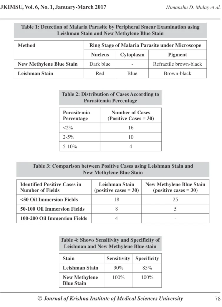
Malaria parasite can be detected by peripheral smear examination using Leishman stain and new methylene blue stain as in Table 1.
The percentage of parasitemia was calculated by calculating number of infected red blood cells per 1000 red cells and expressed as percentage (Table 2).
Parasitemia Percentage
Number of Cases (Positive Cases = 30)
<2% 16
2-5% 10
5-10% 4
Table 2: Distribution of Cases According to Parasitemia Percentage
Identified Positive Cases in Number of Fields
Leishman Stain (positive cases = 30)
New Methylene Blue Stain (positive cases = 30)
<50 Oil Immersion Fields 18 25
50-100 Oil Immersion Fields 8 5
100-200 Oil Immersion Fields 4
-Table 3: Comparison between Positive Cases using Leishman Stain and New Methylene Blue Stain
Stain Sensitivity Specificity
Leishman Stain 90% 85%
New Methylene Blue Stain
100% 100% Table 4: Shows Sensitivity and Specificity of
Leishman and New Methylene Blue stain
Method Ring Stage of Malaria Parasite under Microscope
Nucleus Cytoplasm Pigment
New Methylene Blue Stain Dark blue - Refractile brown-black
Leishman Stain Red Blue Brown-black
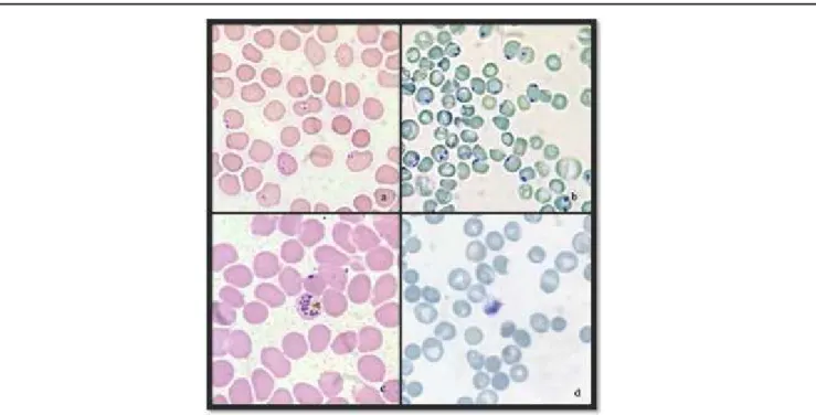
Fig. 1: a) Plasmodium Falciparum showing Two Rings on Leishman Stain b) Plasmodium Falciparum showing Two Chromatin Dots on New Methylene Blue Stain c) Schizonts of Plasmodium Vivax on Leishman Stain d) Schizonts of Plasmodium Vivax Showing Pigment and Nuclear Chromatin Dots on New Methylene Blue Stain
Discussion:
This is a novel and one of its kind studies as here we have compared the number of oil immersion fields required for making the diagnosis. It is recommended that at least 200 oil immersion fields should be examined before labeling a peripheral smear negative for malaria. In a laboratory in the resource poor endemic zone, this has become cumbersome for pathologists to screen multiple cases stained with Leishman stain. Thick smears have a higher sensitivity of detection of 5-10 parasites/µl but due to red cell lysis, morphology and species identification is difficult, for which a second thin smear stained with Leishman stain needs to be done. Thick smear method is tedious with multiple steps. Thin smears stained with Leishman stain are used for species identification and have a sensitivity of 200 parasites/ µl. But if the single thin smear prepared, is stained with new methylene blue stain the sensitivity is maintained along with the morphology. This helps to reduce the turnaround time, reduces false negative and false positive cases and the sensitivity of the test is also maintained.
The identification of parasite was easier with new methylene blue stain, as only the various forms of parasites, reticulocytes and leukocytes take the dark blue stain against the background of pale blue to pale green color, giving high contrast to parasites. There were no artifacts seen as in Leishman stain which usually results in confusion, like the stain precipitates and red cell artifacts leading to increase in false positive and false negative test results. Also new methylene blue stain allowed species identification as it preserves the morphology unlike thick smears. The number of chromatin dots and nuclear features were better appreciated in new methylene
blue stain than Leishman stain. It was observed that hemozoin pigment was better appreciated with new methylene blue stain than Leishman stain (Fig. 1 and 2).
In this study we found that 25 (83.3%) cases were detected in less than 50 fields using new methylene blue stain against 18 (60.0%) cases with Leishman stain. (Table 3) We found 100% sensitivity and specificity for new methylene blue stain, whereas Leishman stain showed 90% sensitivity and specificity of 85% because of false positive and false negative results due to artifacts (Table 4).
Lema et al. (1999) [6] found 98% sensitivity and 100% specificity for field stain which has new methylene blue as a component and 92% sensitivity and 85% specificity for Giemsa stain which is also a Romanowsky stain like Leishman stain. They also found that Giemsa staining is time consuming and is more suitable for batch staining rather than individual samples whereas Fields staining is quick and can be utilized for individual as well as batch staining. Occasional washing off of the blood film was the disadvantage with fields stain.
standard Giemsa stain. Occasional washing off of the blood films was noted with Modified Giemsa stain.
Mohapatra et al. (2014) [8] found that new methylene blue stain (in 90% cases) detected Gametocytes of Plasmodium vivax better than Giemsa stain (in 83% cases). They also detected gametocyte count 2-3 times higher in new methylene blue stained smears than Giemsa stained smears. Hemozoin pigment was better appreciated with new methylene blue stain. Like our study they also found that average parasitemia was nearly equal by both the stains, suggesting that both stains are equally efficient in detecting the asexual forms of parasite.
Conclusion:
The detection of malaria parasite was considerably easy with New Methylene Blue stain and required less time due to good contrast properties of the stain. The pigment character was also better appreciated by new methylene stain in comparison with Leishman stain.
Thus, in present study it was found that the peripheral thin smears with New Methylene blue stain is better and easier method for detection of malaria parasite than with the Leishman stain. Hence it can be used in place of Leishman stain in suspected cases of malaria.
References
1. Gera M, Chauhan A. Detection of malaria parasite using different microscopic methods and a comparison among them. IJSR 2015; 4(3):1635-8.
2. Chakrabortya K, Chattopadhyayb A, Chakrabarti A, Acharyad T, Dasguptae AK. A combined algorithm for malaria detection from thick smear blood slides. J
Health Med Informat 2015: 06.
3. Bhandari P, Raghuveer C, Rajeev A, Bhandari P. Comparative study of peripheral blood smear, quantitative buffy coat and modified centrifuged blood smear in malaria diagnosis. Indian J Pathol Microbiol
2008; 51(1):108-12
4. Khan SA, Anwar M, Hussain S, Qureshi AH, Ahmad M, Afzal S. Comparison of Optimal Malarial Test with Light Microscopy for the diagnosis of Malaria. J Pak
Med Assoc 2004; 54(8):404-7.
5. Sathpathi S, Mohanty A, Satpathi P, Mishra S, Behera P, Patel G et al. Comparing Leishman and Giemsa staining for the assessment of peripheral blood smear preparations in a malaria-endemic region in India.
Malar J 2014; 13:512.
6. Lema OE, Carter JY, Nagelkerke N, Wangai MW, Kitenge P, Gikunda SM et al. Comparison of five methods of malaria detection in the outpatient setting.
Am J Trop Med Hyg 1999; 60(2):177-82.
7. Iqbal J, Hira P, Al-Ali F, Khalid N, Sher A. Modified giemsa staining for rapid diagnosis of malaria infection. Med Princ Pract 2003; 12(3):156-9.
8. Mohapatra S, Sharma D, Gupta K, Deb M, Gaind R. A simple and rapid staining method for detection of hemozoin pigment by methylene blue stain. Trop
Parasitol 2014; 4(1): 59-62.
*
Author for Correspondence: Dr. Teena D. Murthy, Department of Pathology, B.L.D.E.U'S Shri. B. M. Patil