179
J
ournal of Epilepsy and ClinicalNeurophysiology
J Epilepsy Clin Neurophysiol 2007; 13(4):179-182
* Neurologista pediátrica, Pós-Graduanda do Departmento de Neurologia e Neurocirurgia da Universidade Federal de São Paulo – UNIFESP, São Paulo, SP, Brasil.
** Professora Doutora, Coordenadora do Programa de Cirurgia de Epilepsia Pediátrica do Hospital São Paulo da UNIFESP. Received Sept. 28, 2007; accepted Oct. 26, 2007.
Sleep Spindles: Validated Concepts and Breakthroughs
Mônica Jaques Spinosa*, Eliana Garzon**
Unidade de Pesquisa e Tratamento de Epilepsia do Hospital São Paulo da Universidade Federal de São Paulo – UNIPETE/UNIFESP
RESUMO
Fusos de sono: validando e quebrando conceitos
Introdução: Fusos de sono, marco da sincronização do sono no estágio 2 do sono não-REM, são ondas rítmicas e monomórficas, entre 10 e 14 Hz, com amplitude máxima no vértex (Cz), e irradiação bilateral para áreas centrais (C3 e C4). Objetivo: Neste artigo apresentamos uma revisão sobre fusos de sono abran-gendo conceitos bem estabelecidos de morfologia, mecanismos de geração, características maturacionais, padrões anormais e aspectos patológicos, uma vez que o conhecimento se faz necessário para a identificação de padrões normais e patológicos. Conclusões: Fusos centro-parietal e frontal são padrões fisiológiocs nor-mais do sono fase 2 enquanto fusos extremos é um padrão patológico encontrado em crianças abaixo de 5 anos com retardo mental. Fronteiras da pesquisas quanto à consolidação da memória, potenciação sináptica e plasticidade cerebral também foram revisados.
Unitermos: fusos de sono, sono não-REM, EEG.
ABSTRACT
Introduction: Sleep spindles, the hallmark of sleep synchronization in stage 2 non-REM sleep, are rhythmic and monomorphic waves, between 10 and 14 Hz, with a maximum amplitude in the vertex (Cz), with bi-lateral irradiation to central regions (C3 and C4). Objective: In this article we present an overview of sleep spindles ranging from well established concepts of morphology, generation mechanisms, maturational features, abnormal patterns and pathological aspects since the knowledge is important to identify the normal and abnormal patterns. Conclusions: Centro-parietal and frontal spindle are normal pattern and extreme spindle is abnormal, mostly found in children mentally retarded up to 5 years-old. In this review research boundaries of memory consolidation, synaptic potentiation and brain plasticity were also presented.
Key words: sleep spindles, non-REM sleep, EEG.
INTRODUCTION
Sleep spindles, the hallmark of sleep synchronization in stage 2 non-REM sleep, are rhythmic oscillations between 10 to 14 Hz. In this article we present an overview of sleep spindles ranging from well established concepts of morphology, generation mechanisms, maturational features, abnormal patterns and pathological aspects to research boundaries of memory consolidation, synaptic potentiation and brain plasticity.
GENERAL FEATURES AND GENERATION MECHANISMS
180 this sleep stage, they also occur during slow wave sleep, scattered throughout the tracing and frequently associated with sharp and slow transients comprising K complexes. Sleep spindles show maximum amplitude in the vertex (Cz), with bilateral irradiation to central regions (C3 and C4). Nonetheless, topography of sleep spindles, as well as their frequency, vary as sleep deepens. This feature motivated Gibbs and Gibbs1 to describe 3 types of spindles found in the normal subject. Fourteen Hz spindles project in central regions with a maximum at the vertex, 12 Hz spindles occur in a more profound light sleep and have a frontal maximum, while 10 Hz spindles occur in a moderately deep sleep and are more generalized, involving greater areas of both cerebral hemispheres. It is important to note that these variations may be encountered simultaneously in the same individual.
The hypothesis of a sleep spindle generator among the thalamic nuclei dates back to 1942. Thalamic reticular thalamic nucleus (RE) is a thin sheet of GABAergic neurons located at the lateral aspect of the thalamus that is separated from the remaining thalamic nuclei by the external medullary lamina. As a potential candidate for sleep spindle generator, RE had the unique quality of possessing ample connections with virtually all thalamic nuclei. Spindle oscillations in thalamic nuclei were abolished by the disconnection from RE, using cats in experimental studies.2 The hypothesis of RE as the generator of sleep spindles was reinforced by the observation that this nucleus was capable of producing focal spindles even when disconnected from the thalamus and the cerebral cortex.3 Sleep spindles are generated by rhythmic discharges at the RE that lead to 100-150 ms hyerpolarization at thalamocortical relay cells. The intrinsic property of low calcium threshold conductance of corticothalamic relay cells produces a rebound depolarization when hyper-polarization reaches -85 mV. These post-inhibitory bursts reach the neocortex as synchronous excitatory post-synaptic potentials of rhythmic nature that are registered at the scalp EEG as sleep spindles.4 It should be noted that this merely represents a simplification of the basic route of spindle generation. However, it has been observed that the mechanisms involved in the synchro-nization process that precede and are essential to the appearance of spindles during non-REM sleep depend on an intricate network of thalamocorticothalamic circuits and fundamentally on the withdrawal of cholinergic inputs originated at the brainstem reticular formation and basal forebrain that dictate desynchronization during the waking state and desynchronized REM sleep.4
MATURATIONAL FEATURES
Sleep spindles undergo great maturational changes in the first 2 years of life. Bursts of irregular, low-voltage,
short-lasting, rhythmic activity within the 12-14 Hz frequency range projected over fronto-central regions can be recognized in full term infants of 4 to 8 weeks.5
Sleep spindles usually emerge as scarce, low voltage, irregular spindles that grow in number, amplitude and regularity in 1 to 2 weeks after their appearance.5 It must be noted that the maturation of sleep spindles is a dynamic and individual process that follows guidelines but has great interindividual differences that must not be mistaken for abnormalities. Most EEG readers tend to consider the absence of sleep spindles in a routine EEG with sure documentation of sufficient stage 2 sleep abnormal after 3 months of age.
Another typical trait of infants in the first semester is the long duration of spindles, which can last up to 10 seconds (Figure 1). Duration of spindles gradually decreases in the second half of the first year to reach 1-2 seconds that will remain mostly unchanged throughout childhood. In the first year of life spindles tend to be asynchronous, frequently alternating between cerebral hemispheres, with maximum amplitude at C3 or C4 electrodes and less often at the vertex (Cz). The asynchrony of spindles is equally balanced between both hemispheres throughout the EEG recording, with an overall symmetry in number of spindles per hemisphere. Interhemispheric synchrony is achieved around 2 years of age.
In 1961, Fois6 described the sharp spindles of infancy, a normal pattern of sleep spindles that is peculiar to infants up to 2 years of age. These are asynchronous, medium voltage 12-17 Hz spindles, with peak frequency at 14 Hz, projected over centro-parietal regions with sharp wave morphology, generally of the negative component.
If superficially analyzed, it may seem that sleep spindles do not suffer major changes after the second year of life. However, evidence supports a reduction of sleep spindles abundance, frequency, amplitude and duration in the elderly,7,8 proving that spindles maturation is a continuous dynamic process that extends into adulthood and old-age. It is worth mentioning that this reduction has been shown to affect the frequency range below 14Hz but not the higher frequencies of spindles from 14 to 15 Hz.7 Since melatonin is known to enhance spindle activity,9 it has been speculated that the decline in sleep spindles observed in the elderly may be attributed to the decrease in circulating melatonin during night-time observed in this age-group.10 No correlation was found between sleep spindle decline and cognitive impairment; however, the decrease in spindle amplitude was associated with brain atrophy, suggesting that there is a still obscure etiology behind the reduction of amplitude of sleep spindles in the elderly population.8
181
ABNORMAL PATTERNS AND PATHOLOGICAL ASPECTS
Gibbs and Gibbs11 reviewing their records of children with mental retardation in search of an electroencephalo-graphical pattern correlated with cognitive deficits, des-cribed an abnormal pattern of sleep spindles. Extreme spindles, so named in reference to their high amplitude (ranging from 200 to 400 µV), were not associated with epilepsy but with mental retardation. This pattern consists of continuous or nearly continuous 8-15 Hz spindles, frequently of sharp morphology, observed in non-REM sleep and often remaining during wakefulness, maximal in centro-parietal regions but with a much wider distri-bution than that observed in normal spindles (Figure 2).
Extreme spindles are mostly found in children below 5 years of age and have not been observed in those above 12. The authors found this abnormal spindle in 1/3000 healthy children and 17% of 300 mentally retarded children.
In 1967, Niedermeyer and Capute12 described the fast and spikey spindle variant, originally encountered in 4 children ranging from 3 to 13 years-old with organic brain diseases (cerebral palsy, hydrocephalus and porencephaly, epilepsy, progressive cerebellar degeneration). These ab-normal spindles occur in childhood and adolescence, have a negative sharp component, range in frequency from 16-20 Hz, may be asynchronous and have variable dis-tributions with a common accentuation on central areas.
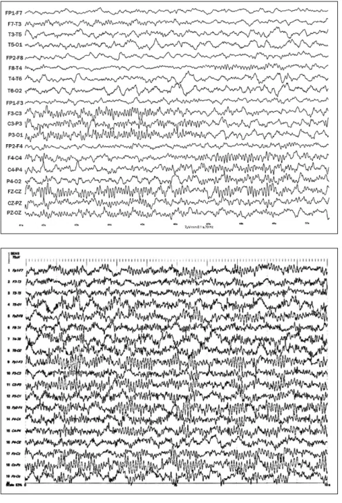
Figure 1. Centro-parietal sleep spindle – BCMF, female, 4 months old. Sleep stage 2. Observe sleep spindle, asynchronous, lasting 7 seconds and first recorded at left centro-parietal region and midline and then evolving to right centro-parietal region.
Figure 2. Extreme spindle. GS, male, 2yrs old. West Syndrome as antecedent. Sleep stage 2. Observe continuous and high amplitude bilateral frontal sleep spindle with a quite diffuse spreading.
182 Apart from these two peculiar abnormal spindle variants, sleep spindles have been reported to be absent or reduced in patients with mental retardation,13 cerebral palsy14 and phenylcetonuria.15
RESEARCH BOUNDARIES
The physiological role of sleep spindles remains an unresolved yet intriguing topic. The function of sleep spindles as sleep promoters and maintainers has long been postulated. Over the last few years, attention has been drawn towards research on topics concerning sleep and its role in cognition. Sleep spindles are reduced in mental retardation13,15 and also in various forms of dementia.16 In children, it has been shown that sleep deprivation is related to lower cognitive performance including abstract thinking and verbal creativity.17 Schabus et al.18 studied 12 adult healthy subjects performing declarative memory tests (using word-pair tasks) and showed that increased spindle activity is related to greater scores in the recall tests, documenting an association between sleep spindles and memory consolidation. The importance of sleep spindles in synaptic regulation and learning has been acknowledged even in daytime naps, on this occasion testing single hand motor learning, establishing that there is a positive correlation between higher performance and the amount of sleep spindles observed in central regions contralateral to the hand used in motor tasks.19 This resonant body of evidence suggests that sleep spindles are possibly a mechanism of brain plasticity. However, different types of learning show specific consolidation mechanisms during sleep, suggesting that synaptic consolidation and brain plasticity is far from being a unitary process that involves solely sleep spindles.
REFERENCES
1. Gibbs FA, Gibbs EL. Atlas of Electroencephalography. Cambridge: Addison-Wesley; 1962. v. 1. p. 94.
2. Steriade M, Deschenes M, Domich L, Mulle C. Abolition of spindle oscillations in thalamic neurons disconnected from nucleus reticularis thalami. J Neurophysiol. 1985; 54:1473-97.
3. Steriade M, Domich L, Oakson G, Deschenes M. The deafferented reticular thalamic nucleus generates spindle rhythmicity. J Neurophysiol. 1987; 57:260-73.
4. Steriade M, Gloor P, Llinás RR, Lopes da Silva FH, Mesulam MM. Basic mechanisms of cerebral rhythmic activities. Elecro-encephalogr Clin Neurophysiol. 1990; 76:481-508.
5. Tanguay PE, Ornitz EM, Kaplan A, Bozzo ES. Evolution of sleep spindles in childhood. Electroencephalogr Clin Neurophysiol. 1975; 38:175-81.
6. Fois A. The electroencephalogram of the normal child. Springfield: Thomas; 1961. p. 124.
7. Landolt HP, Dijk DJ, Achermann P, Borbély AA. Effect of age on the sleep EEG: slow-wave activity and spindle frequency activity in young and middle-aged men. Brain Res. 1996; 738:205-12. 8. Guazzelli M, Feinberg I, Aminoff M, Fein G, Floyd TC, Maggini C.
Sleep spindles in normal elderly: comparison with young adult patterns and relation to nocturnal awakening, cognitive function and brain atrophy. Electroencephalogr Clin Neurophysiol. 1986; 63:526-39.
9. Dijk DJ, Roth C, Landolt HP, Werth E, Aeppli M, Achermann P, Borbély AA. Melatonin effect on daytime sleep in men: suppression of EEG low frequency activity and enhancement of spindle frequency activity. Neurosci Lett. 1995; 201:13-6.
10. Van Coevorden A, Mockel J, Laurent E, Kerkhofs M, L’ Hermite-Balériaux M, Decoster C, Nève P, Van Cauter E. Neuroendocrine rhythms and sleep in aging men. Am J Physiol. 1991; 260:651-61. 11. Gibbs EL, Gibbs FA. Extreme spindles: correlation of
electro-encephalographic sleep pattern with mental retardation. Science. 1962; 138:1106-7.
12. Niedermeyer E, Capute AJ. A fast and spikey spindle variant in children with organic brain disease. Electroencephalogr Clin Neurophysiol. 1967; 23:67-73.
13. Shibagaki M, Kiyono S, Watanabe K. Nocturnal sleep in severely mentally retarded children: abnormal EEG patterns in sleep cycle. Electroencephalogr Clin Neurophysiol. 1980; 49:337-44. 14. Shibagaki M, Kiyono S, Takeuchi T. Nocturnal sleep in mentally
retarded infants with cerebral palsy. Electroencephalogr Clin Neurophysiol. 1985; 61:465-71.
15. Fois A, Rosenberg C, Gibbs FA. The electroencephalogram in phenylpyruvic oligophrenia. EEG Clin Neurophysiol. 1955; 7:569-72.
16. Prinz PN, Peskind ER, Vitaliano PP, Raskind MA, Eisdorfer C, Zemcuznikov N, Gerber CJ. Changes in the sleep and waking EEGs in nondemented and demented patients. J Am Geriatr Soc. 1982; 30:86-93.
17. Randazzo AC, Muehlbach MJ, Schweitzer PK, Walsh JK. Cognitive function following acute sleep restriction in children ages 10-14. Sleep. 1998; 21:861-8.
18. Schabus M, Gruber G, Parapatics S, Sauter C, Klösch G, Anderer P, Klimesch W, Saletu B, Zeitlhofer J. Sleep spindles and their significance for declarative memory consolidation. Sleep. 2004; 27:1479-85.
19. Nishida M, Walker MP. Daytime naps, motor memory consolidation and regionally specific sleep spindles. PLos ONE. 2007; 2:e341.
Endereço para correspondência:
Eliana Garzon
Departamento de Neurologia e Neurocirurgia – UNIFESP Rua Napoleão de Barros 715, 13° andar – Vila Clementino CEP 04024-002, São Paulo, SP, Brasil