ISSN 2278- 4136 ZDB-Number: 2668735-5
IC Journal No: 8192
Volume 1 Issue 5
Online Available at www.phytojournal.com
Journal of Pharmacognosy and Phytochemistry
Vol. 1 No. 5 2013 www.phytojournal.com Page | 112
Pharmacognostic Studies on Indian Madder
(
Rubia cordifolia
L.)
Devi Priya M.1 and E.A. Siril1*
1. Department of Botany, University of Kerala, Kariavattom, Trivandrum 695 581, India [E-mail: easiril@yahoo.com]
‘Indian Madder’ (Rubia cordifolia L.) has wide range of pharmacological properties. In the present study, an attempt was made to identify the pharmacognostic features of various parts of R. cordifolia to differentiate it from adulterants. The organoleptic characters, proximate analysis, physio-chemical behavior of powders were recorded. The stomatal index, palisade ratio and ash value were shown to be characteristic under standard conditions. Optical activity of various R. cordifolia extracts under visible and UV light (254 nm and 366 nm) were recorded. Phytochemical studies revealed the presence of anthraquinone and other metabolites in different plant parts. Powder analysis showed trace red colour in root powder under most of the standard test conditions. Phytochemical and pharmacognostic records evolved in the present study can be used for framing standard parameters for the proper identification of raw materials of R. cordifolia.
Keyword:Rubia cordifolia, Phytochemical, Powder, Extract
1. Introduction
Rubia cordifolia L. is a climbing or scrambling herb, with red rhizomatous base and roots. It is an essential raw drug for the traditional herbal formulations such as aswagandharistam, gulguluthikthkarishtam, jaatyaadi ghrita, madhookasavam, majishthaadi taila, useerasavam etc. It is a member of Rubiaceae family, distributed in hilly tracts of India up to 3750m. The plant (Fig. 1A) is commonly known as ‘Indian Madder’ and sold under the trade name ‘manjistha’. The plant has other vernacular names such as manjit in Hindi, chitravalli in Kannada, ceevalli in Tamil, manchatti and manjatti in Malayalam and manderti in Telengu[1].
Fig 1A: Rubiacordifolia Habit
The mature roots are chiefly valued plant part of
Vol. 1 No. 5 2013 www.phytojournal.com Page | 113
preparations based on `manjistha` are prescribed for treatment of major burns, bone fractures and dysentery. Manjistha is considered as tonic, antitussive and useful in chronic low fevers. Decoction from roots is prescribed to cure jaundice, paralytic affections, urinary troubles, amenorrhea and to the mother after delivery for cleansing and shrinking of the uterus.[2] The root decoction is effective to regulates menstruation cycles.. It is proved that various root extracts of
R. cordifolia are has astringent, themogenic, febrifuge, antidysenteric, antihelmintic, galacto-purifier, ophthalmic, and rejuvenating effect.[3,4] It is used to cure tuberculosis and intestinal ulcer. In modern pharmacopeia, R. cordifolia is reported to be active against a diversified panel of cancer cell lines, such as P388, L1210, L5178Y, B16 melanoma, Lewis lung carcinoma, and sarcoma-180[5].
The macroscopic and microscopic description of a medicinal plant is the first step towards identification and determination of purity. However, such an attempt has not been made on
R. cordifolia so far. In the present communication we devoted our effort to document the pharmacognostic features of the entire plant to identify the plant in its crude form.
2. Materials and Methods
The plants were collected from the natural habitat, Ellappara, (Latitude 9º36’, 49.89” N, Longitude 77º00’, 6.59”E Elevation 1158m)), Idukki Distt, Kerala and was positively identified and confirmed by the herbarium in the Department of Botany, University of Kerala. Morphological studies were performed by using simple, binocular, light microscope. For micro-characterization, free hand sections of about 10-20 µm thickness of stem, root, petiole and leaves were made and stained with aqueous safranine (0.5%) solution. After washing, the stained sections were mounted on clean micro slides and examined. The preparation was further observed in an image analyzer (Olympus BX 51) and the anatomical peculiarities were photo documented. As a part of quantitative microscopy, stomatal number, stomatal index and vein islet number
were determined by using fresh leaves of the plant.6
Shade dried, coarsely powdered raw drug was used for powder analysis, which includes the identification of organoleptic characters, determination of physicochemical characters, behaviour of the powder with different chemical reagents/solvents according to methods described in Indian pharmacopeia[7] and fluorescence as suggested by Chase and Pratt[8,9].
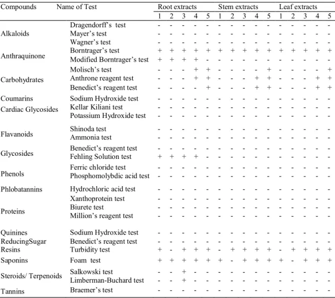
Preliminary phytochemical studies were carried out using 10 g powdered material and subjecting it to successive extraction in a Soxhlet apparatus with 150 ml solvents viz., hexane, chloroform, acetone, methanol, and water. The extraction was continued until the solvent became colourless. The extracts were collected; concentrated using a rotary evaporator and the obtained extracts were air dried. Presence of various phytoconstituents
viz., alkaloids (Dragendorff’s test, Mayer’s test, Wagner’s test), anthraquinones (Borntrager’s test, Modified Borntrager’s test), carbohydrates (Anthrone reagent test, Benedict’s reagent test, Molish's test), coumarins (Sodium hydroxide test), cardiac glycosides (Kellar kiliani test, Potassium Hydroxide test), flavonoids (Shinoda test, Ammonia test), glycosides (Benedict’s reagent test, Fehling solution test), phenols (Ferric chloride test, Phosphomolybdic acid test), phlobatannins (Hydrochloric acid test), protein (Biurete test, Millon's test, Xanthoprotein test), quinines (Sodium hydroxide test), reducing sugar (Benedict’s test), resins (Turbidity test), saponins (Foam test), steroids and terpenoids (Lieberman burchard test, Salkowski test) and tannins (Braemer’s test) were tested. 10, 11
3. Result and Discussion 3.1 Pharmacognostic Features
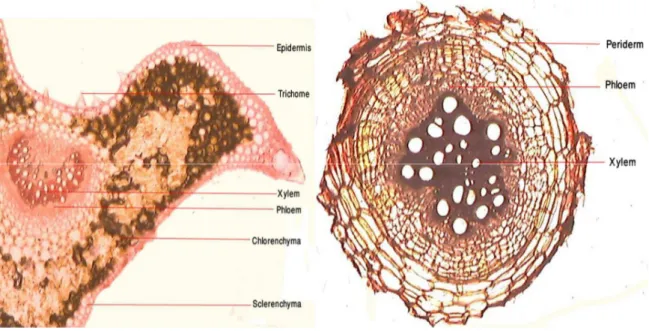
a. Stem: is quadrangular, divaricately branched,
Vol. 1 No. 5 2013 www.phytojournal.com Page | 114
and is composed of sieve tubes and phloem parenchyma. Cambium ring is represented in two layers. Secondary xylem forms a continuous cylinder, which is made of vessels, tracheids, fibres and xylem parenchyma. In secondary
xylem, vessels are large and uniformly arranged. Uniseriate medullary rays are also present. In older stem, large central parenchymatous pith is present.
Fig. 1B: C.S. of Stem Fig. 1C: T.S. of Leaf
b. Leaves: are 3.8-9 X 1.6-3.5 cm long, arranged
in a whorl of four, cordate-ovate to ovate-lanceolate, 3-9 palmately veined, upper surface mostly glabrous and rough. Lower leaves are larger than the upper, and all are scabrous above. Leaf base is slightly cordate. The margins are with minute white prickles. Leaf section showed single layered epidermis, covered with cuticle and possesses pyramidal hairs. Palisade cells are single layered and compactly packed and the spongy cells are multilayered and loosely arranged. In the lower portion of the midrib 2-4 layers made of collenchymatous cells. Vascular bundles are definite in number, conjoint, collateral and closed (Fig. 1C).
c. Petiole: is 5-10 cm long with sharp, recurved prickles. Stipules are completely absent or modified into leaves. The T.S. of the petiole showed a 'V' shaped prominent median groove. It had a single layered epidermis provided with pyramidal hairs. Below the epidermis at the lobes and base, 3-4 layers of sclerenchymatous cells were present. The cortical cells were made
of thin walled chlorenchyma. Vascular bundle is 'C'- shaped (Fig. 1D).
d. Root: Rubia cordifolia has long, cylindrical,
Vol. 1 No. 5 2013 www.phytojournal.com Page | 115 Fig. 1D: T.S. of Petiole Fig. 1E: C.S. of Root
3.2 Quantitative Microscopy
Leaf peelings from both abaxial and adaxial side showed paracytic (rubiaceous) type of stomata where the stoma is surrounded by two subsidiary cells, the long axis of which are parallel to the stoma. The palisade ratio (1.78), stomatal index (16.67) and vein-islet number (1.48) were calculated as a part of quantitative microscopy (Table 1).
Table 1: Quantitative microscopic examinations of R. cordifolia leaf peelings
Determinations Range Palisade ratio 1.78 Stomatal index 16.67 Vein-islet number 1.48
3.3 Powder Analysis
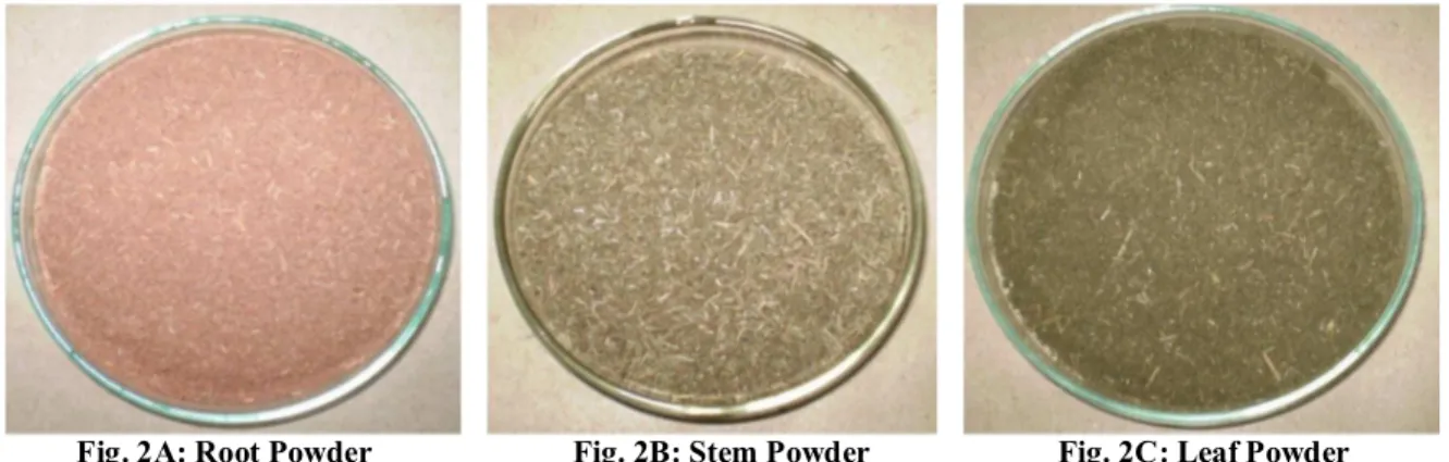
Powder Microscopy: Organoleptic
(morphological) characters refers to evaluation of drugs by colour, odour, taste, size, shape and consistence (touch and texture). These were identified by using the crude powder samples. The root powder was smooth, moderately fine
with coral pink colour (Table: 2).The stem and leaf powders were somewhat grayish. Stem powder was moderately coarse and fibrous. All the samples were with mouldy smell. The root samples were acrid and bitter in taste.
Table 2: Organoleptic characters of root, stem and leaf powders of R. cordifolia
Character Root Stem Leaf
Colour Coral Pink Gray Ivy Green Odour Mouldy Mouldy Mouldy Size Moderately
fine
Moderately coarse
Fine
Taste Acrid and bitter
Bitter Acrid
Texture Smooth Fibrous Smooth
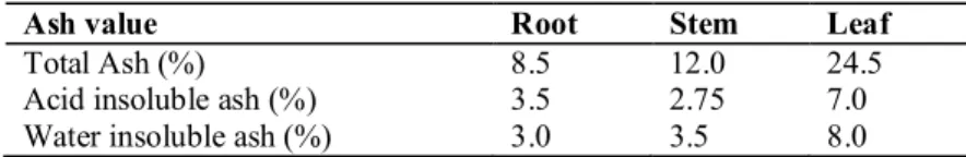
3.4 Proximate Analysis: As the part of
Vol. 1 No. 5 2013 www.phytojournal.com Page | 116 Table 3: Physico-chemical analysis of coarse powdered samples of R. cordifolia
Table 4: Behaviour of the R. cordifolia drug with different reagents/ solvents
Treatment Root Stem Leaf
Powder+ Distilled Water Powder+ 5% FeCl3 Powder+ l Acetic acid Powder+ 5% KOH Powder+ 5% NaOH Powder+ Conc. HCl Powder+ Conc. H2SO4 Powder+ Conc. HNO3 Powder+ N/10 Iodine soln. Powder+ Ammonia soln.
Light orange Moderate olive Light orange yellow Strong Pink Cardinal red
Greenish yellow brown Reddish black
Brilliant yellow
Cadmium Orange Mandarin red
Brilliant yellow Greenish yellow Strong yellow brown Strong Brown Dark brown Light Reddish black Reddish black Orange buff Yellow brown Mimosa yellow
Brilliant yellow green Light Greenish yellow Strong Yellowish green Light Greenish yellow Strong Brown Moderate Brown Reddish black Barium yellow Primrose yellow Canary yellow
Table 5a. Fluorescence analysis of extracts prepared by using various solvents
Treatment Plant Part
Observations Short UV light
(254nm)
Long UV light
(366nm) Visible light
Hexane
Chloroform
Acetone
Methanol
Water
Root Stem Leaf
Root Stem Leaf
Root Stem Leaf
Root Stem Leaf
Root Stem Leaf
Coffee Brown Cyprus Green Cyprus Green
Chocolate Fern Green Greenish Brown
Cyprus Green Agathia Green Cyprus Green
Yellow Brown Agathia Green Cyprus Green
Lavender Green Fern Green Fern Green
Dark Brown Dull Black Dark Brown
Brown Black Coffee Brown
Light Brown Colourless Light Brown
Brown Brown Dull Black
Blue Black Black
Indian Orange Primrose Yellow Uranium Green
Orange
Dark Yellow Brown Willow Green
Nasturtium Orange Pea Green
Agathia Green
Fire Red Yellow Brown Agathia Green
Light Brown Brownish Yellow Dark Brownish yellow
3.5 Behaviour of powder with chemical reagents:
Detection of colour variation of the powdered samples under day light is a way to identify its purity. The root samples showed trace of red colour with almost all reagents except with Conc.
HCl and FeCl3 (Table 4). Stem and leaf powder
showed yellowish to reddish black colour with various reagents.
3.6 Fluorescence Analysis: The optical activity of
the powdered samples (Fig. 2A, B & C) and the
Ash value Root Stem Leaf
Total Ash (%) 8.5 12.0 24.5
Vol. 1 No. 5 2013 www.phytojournal.com Page | 117
plant extracts under different optical regimes viz.
visible, UV (254 nm) and UV (366 nm) were recorded by the reported method (Table: 5a &5b). Various extracts and powder showed yellow, green and brown fluorescence in visible and short UV
light (254nm) and brown and black fluorescence in long UV light helped to identify, authenticate and
differentiate R. cordifolia from other related
species.
Fig. 2A: Root Powder Fig. 2B: Stem Powder Fig. 2C: Leaf Powder
Table 5b: Powder analysis of R. cordifolia at short, and long UV and visible light
Treatment Plant
Part
Observations
Short UV light Long UV light Visible light
Powder alone
Powder + 50% H2SO4
Powder + 50% HNO3
Powder + 50% HCl
Powder + 1N HCl
Powder + 5% KOH
Powder + 1N NaOH (Methanol)
Powder + 1N NaOH (in water)
Root Stem Leaf
Root Stem Leaf
Root Stem Leaf
Root Stem Leaf
Root Stem Leaf
Root Stem Leaf
Root Stem Leaf
Root Stem Leaf
Light Gray Light Gray Light Gray
Willow Green Agathia Green Agathia Green
Pea Green Agathia Green Agathia Green
Willow Green Agathia Green Agathia Green
Agathia Green Agathia Green Pod Green
Dark Green Cyprus Green Pod Green
Sage Green Agathia Green Agathia Green
Leek Green Agathia Green Agathia Green
No Fluorescence No Fluorescence No Fluorescence
No Fluorescence No Fluorescence No Fluorescence
No Fluorescence No Fluorescence No Fluorescence
No Fluorescence No Fluorescence Blue
Colorless Light Brown Sepia
Sepia
No Fluorescence No Fluorescence
No Fluorescence No Fluorescence No Fluorescence
No Fluorescence No Fluorescence No Fluorescence
Coral Pink Gray Ivy Green
Light Brown Yellowish brown Pea Green
Pea Green Canary Yellow Dresden Yellow
Sage Green Light Cadmium Sap Green
Apricot Yellow Greenish yellow Naples Yellow
Light Coral Pink Buttercap Yellow Empire Yellow
Prawn Red Empire Yellow Empire Yellow
Vol. 1 No. 5 2013 www.phytojournal.com Page | 118
3.6 Phytochemical Analysis
The pharmacological action of the crude drug is largely depends on the metabolites present in it. In the present investigation, the qualitative screening by using prepared extracts revealed the presence of a wide range of phytoconstituents (Table 6). Borntrager’s test for anthraquinone was found to be positive for all the studied samples.[13, 14] More intensive colour was noticed in root samples compared to stem and leaves indicating presence of more amount of
anthraquinones in the roots. Glycosides other than cardiac glycosides were present only in all root extracts. Absence of flavonoids indicates the purity of the samples.15 In addition to these, metabolites like saponins, resin and steroids/ terpenoids were present in R. cordifolia extracts. Coumarins, cardiac glycosides and flavanoids, phenol, phlobatannin, protein and quinines were absent. Steroids were also noted in root samples only.
Table 6: Phytochemical analysis of extracts of R. cordifolia
Compounds Name of Test Root extracts Stem extracts Leaf extracts 1 2 3 4 5 1 2 3 4 5 1 2 3 4 5
Alkaloids
Dragendorff’s test - - - - Mayer’s test - - - - Wagner’s test - - - -
Anthraquinone Borntrager’s test Modified Borntrager’s test + + + + + + + + + + + + + + + + + + + - - - - - - - - - - -
Carbohydrates
Molisch’s test - - - + + - - - - + - - - - + Anthrone reagent test - - - + + - - - + + - - - + + Benedict’s reagent test - - - - + - - - + + - - - + + Coumarins Sodium Hydroxide test - - - -
Cardiac Glycosides Kellar Kiliani test Potassium Hydroxide test
- - - - - - - - - - - - - - - - - - - - - - - - - - - - - -
Flavanoids Shinoda test Ammonia test - - - - - - - - - - - - - - - - - - - - - - - - - - - - - -
Glycosides Benedict’s reagent test Fehling Solution test - + - + - + - + - - - - - - - - - - - - - - - - - - - - - -
Phenols Ferric chloride test Phosphomolybdic acid test - - - - - - - - - - - - - - - - - - - - - - - - - - - - - -
Phlobatannins Hydrochloric acid test - - - -
Proteins
Xanthoprotein test Biurete test
Million’s reagent test
- - - - - - - - - - - - - - - - - - - - - - - - - - - - - - - - - - - - - - - - - - - - -
Quinines Sodium Hydroxide test - - - - ReducingSugar Benedict’s reagent test - - - - Resins Turbidity test + - + + + - + + + + - + + + + Saponins Foam test + + + + + + - + + + + - + + +
Steroids/ Terpenoids Salkowski test
Limberman-Buchard test - - - - + + - - - - - - - - - - - - - - - - - - - - - - - -
Tannins Braemer’s test - - - -
1: Hexane; 2: Chloroform; 3: Acetone; 4: Methanol; 5: Water; +: Present ; -: Absent
Vol. 1 No. 5 2013 www.phytojournal.com Page | 119
4. Conclusion
It is evident that plants having therapeutic value usually contain diverse groups of secondary metabolites and R. cordifolia proved no exception. Apart from the wide range of health care uses it is being used as a textile dye from ancient times. However, due to the endemic distribution, the plant is not familiar to the populace of the modern world or even neglected where it flourishes well. The plant play a vital role in the development of novel drugs and well defined pharmacognostic parameters and standards must be established before the inclusion of any crude drug in a herbal pharmacopoeia.
The studied pharmacognostical and
phytochemical characters of R. cordifolia will be useful to identify the plant in the powder form and in the elimination of adulterants. So if any crude drug which is claimed to be R. cordifolia
but whose characters significantly vary from the accepted standard should be rejected irrationally.
5. Acknowledgement:
We thank Dr. Ashalatha S. Nair, Professor & Head, Department of Botany, University of Kerala Kariavatttom, Thiruvananthapuram for providing facilities and University Grants Commission, New Delhi, India, for the financial support in the form of a Major Research project (F. No.39-359/2010 (SR), 31.12.2010).
6. References:
1. Ved D, Oommen S, Singh A. Propagation and Agro-technology status of commercially important Medicinal plant species of the project area of Andhra Pradesh community forest
management project, Foundation for
Revitalization of Local Health Traditions (FRLHT), Andhra Pradesh Forest Department, 2002, 128.
2. Dev S. A selection of Prime Ayurvedic Plant Drugs: Ancient - Modern concordance. Anamaya Publishers, New Delhi, 2006, 377-381.
3. Sivarajan VV and Balachandran I. Ayurvedic drugs and their plant sources. Oxford & IBH Publishing Co. Pvt. Ltd., New Delhi, 1994, 292-293.
4. Nadkarni KM. Indian Plants and Drugs. Asiatic Publishing House, New Delhi, 1998, 343-344. 5. Sanz MA, de la Rubia J, Bonanad S, Barragan E,
Sempere A, Martin G et al. Prolonged molecular remission after PML/RAR alpha-positive autologous peripheral blood stem cell transplantation in acute promyelocytic leukemia is relevant pre-transplant minimal residual disease in the graft? Leukemia 1998; 12: 992–925.
6. Wallis TC. Text book of Pharmacognosy. 5th Ed, EBS Publications, New Delhi, India, 1985, 111-117
7. Anonymous. Indian Pharmacopia. Ministry of Health and Family Welfare, Govt. of India, Controller of Publication, New Delhi, 1985, 310 8. Chase CR, Pratt RJ. Fluorescence of Powdered
Vegetable Drugs with particular reference to development of a system of identification. 38th Ed, Am. Pharm. Assoc. 1949; 324-331.
9. Kokashi CJ, Kokashi RJ, Sharma M. Fluorescence of Powered Vegetable drugs in Ultra-Violet radiation. J. A,. Pharm. Assoc. 1958; 47: 715-717.
10. Harborne JB. Phytochemical Methods- A Guide to Modern techniques of Plant analysis. 3rd Ed, Chapman and Hall, London, 1998, 307.
11. Daniel M. Methods in Plant Chemistry and Economic Botany. Kalyani Publishers, New Delhi, 1991, 209.
12. Deoda RS, Kumar D, Kadam PV, Yadav KN, Bhujbal SS, Patil MJ. Pharmacognostic and Biological Studies of the Roots of Rubia cordifolia Linn. (Rubiaceace). Int. J. Drug Dev. & Res. 2011; 3:148-158.
13. Pathania S, Daman R, Bhandari S, Singh B, Lal B. Comparatives Studies of Rubia cordifolia L. and its Commercial Samples. Ethnobotanical Leaflets2009; 11:179-188.
14. Kannan M, singh R, Narayanan Phytochemistry and Immunopharmacological investigation of
Rubia cordifolia Linn. (Rubiaceae).
Pharmacologyonline 2009; 3: 653-662.