DOI: 10.4328/JCAM.648 Received: 14.03.2011 Accepted: 24.03.2011 Printed: 01.01.2012
Corresponding Author: Mahmut Tokur, Kahramanmaras Sutcu Imam University, Medicine Faculty, Department of Thoracic Surgery. Street Yörükselim. Kahramanmaras, 46100 Turkey. T.: +90 344 2257575 F.: +90 344 2212371 E-Mail: [email protected]
Opak Plevral Kitle / Opaque Mass in the Pleural
1Mahmut Tokur, 2Can Kurkcuoglu
¹Kahramanmaras Sutcu Imam University, Medicine Faculty, Department of Thoracic Surgery, Kahramanmaras, ² Harran University, Medicine Faculty, Department of Thoracic Surgery, Sanliurfa, Turkey
Pleural Mass Lesion Containing Calcium Sludge
Kalsiyum Çamuru İçeren Plevral Kitle
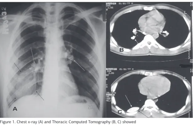
A 30 year-old man was admitted with the symptoms of chest pain. Opacity with smooth margins in right lower zone at chest x-ray. Thoracic computed tomography showed in right-sided a calcified pleural mass (Figure 1). The patient operated with the diagnosis of chest wall mass. In the operation we seen pleural mass that was to lie down on from 4. costovertebral junction to costophrenic sinus. The mass settled between
Figure 1. Chest x-ray (A) and Thoracic Computed Tomography (B, C) showed
Figure 2. Operative showed (A, B) the endothoracic fascia and the parietal
pleura. The lesion content was white-colored, dense mud was the consistency at the material (Figure 2). The studied frozen result of the lesion content was interpreted as benign tissue. The lesion was excised totally together with the contents. The patient’s general condition
improved after the removal of the mass.