J Bras Pneumol. 2011;37(3):416-418
Subglottic and mediastinal hemangioma in a child:
treatment with propranolol
Hemangioma subglótico e mediastinal em criança: tratamento com propranolol
Mauro Tamagno, Benoit Jacques Bibas, Helio Minamoto,
Fernanda Sobreiro Alfinito, Ricardo Mingarini Terra, Fabio Biscegli Jatene
To the Editor:
A 6-month-old girl with upper airway obstruction was evaluated in the emergency room. The patient had been born prematurely (at gestational week 34). Prior to the emergency room visit, she had been treated for gastroesophageal reflux and recurrent respiratory infection. Physical examination revealed laryngeal stridor. All laboratory test results were normal.
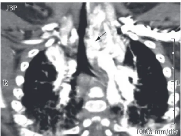
A CT scan of the chest, larynx, and trachea showed a heterogeneous, highly vascularized mass in the left hemithorax (Figure 1). However, there was no cardiac malformation. Rigid bronchoscopy performed under general anesthesia revealed a large pulsatile mass that obstructed approximately 80% of the larynx and trachea (Figure 2a; Video 1*). On the basis
of the radiological and endoscopic findings, a diagnosis of hemangioma was made. Because of the risk of bleeding, we decided not to perform a biopsy.
Corticosteroid therapy was initiated (betamethasone, 0.5 mg/kg per day). After two weeks of treatment with the corticosteroid, the patient showed no clinical improvement. The corticosteroid therapy was discontinued, and treatment was started with propranolol (1 mg/ kg per day). To avoid potential complications, cardiac function and blood glucose levels were monitored. After 5 days of treatment, a clinical response was observed, and the stridor lessened. The dose of propranolol was gradually increased to 2 mg/kg per day. After 3 months of treatment, the patient again underwent rigid bronchoscopy (Figure 2b; Video 2†). There was endoscopic
improvement, the lesion showing a reduction in size and tracheal lumen obstruction being less than 20%. We found no granulation tissue
* Video available in the online version of the Brazilian Journal of Pulmonology (www.jornaldepneumologia.com.br/)
† Video available in the online version of the Brazilian Journal of Pulmonology (www.jornaldepneumologia.com.br/)
or tracheal stenosis, the mucosa of the larynx and trachea being normal in appearance. At this writing, the patient was still being treated with propranolol, and the treatment was scheduled to continue for 1 year. We observed neither cardiac arrhythmias nor any other side effects of propranolol.
Hemangioma is the most common vascular neoplasm in children.(1) It generally develops
during the first weeks of life and grows rapidly for the next 3-6 months; this is followed by involution, which begins at approximately 12 months of age and can be complete after 3-7 years.(1,2) Because most hemangiomas resolve
spontaneously, intervention is rarely necessary. Nevertheless, treatment is required in 10-15% of the cases, especially when the hemangioma is life-threatening and can impair physiological function.(2) Prednisolone (at doses of 2-5 mg/kg
per day) has been used as the first-line treatment for hemangiomas.(1) However, prolonged
corticosteroid therapy has been associated with a number of side effects.(1,2)
Although subglottic hemangioma is rare, it can lead to respiratory failure and is therefore life-threatening.(1,3) Various treatment modalities
are available for patients with subglottic hemangioma.(1) Tracheostomy has been proposed
as a treatment option and can be used until the hemangioma resolves spontaneously, thereby avoiding the risks of complex laryngotracheal surgery.(3) However, the rate of
tracheostomy-related complications is high in children; one such complication is accidental decannulation, which leads to asphyxia.(3) Laser therapy and
cryotherapy both significantly increase the risk of laryngeal and tracheal stenosis secondary to tissue healing. In children, laryngotracheal resection is a major surgical procedure that is associated with high rates of morbidity and mortality.(3,4)
J Bras Pneumol. 2011;37(3):416-418
417
significantly in size after the initiation of treatment with propranolol for obstructive cardiomyopathy.(5) Subsequent case reports have
shown that propranolol is effective in treating hemangiomas of various topographies. In many of those cases, propranolol was used as an adjuvant after other clinical therapies and surgical treatments had failed.(1,3,4) The remarkable clinical
response and minimal side effects have increased interest in the use of propranolol as a first-line treatment for hemangiomas.(4) Initial studies
of the use of propranolol as monotherapy for airway hemangiomas have produced excellent results.(1,3,4)
Possible explanations for the therapeutic effect that propranolol has on hemangiomas include immediate vasoconstriction (evidenced by a change in the color and texture of the hemangioma); decreased production of VEGF and basic FGF; and stimulation of apoptosis of capillary endothelial cells.(4) However,
propranolol can have serious side effects, such as bradycardia and hypotension; it can also mask the clinical signs of heart failure and impair cardiac performance.(6,7) Propranolol can
also exacerbate the clinical manifestations of hypoglycemia, which is associated with long-term neurological sequelae.(6)
Because of the possible side effects of propranolol, a strict protocol has been proposed,(4,6) including echocardiography, as
well as the monitoring of cardiac function (including arterial pressure and HR) and blood The pharmacological treatment of
subglottic hemangioma is based on the use of corticosteroids, which can be administered orally or by intralesional injection. The use of interferon and vincristine is reserved for lesions that are life-threatening.(2) None of these treatments are free
of side effects. Prolonged use of corticosteroids can result in growth retardation, peptic ulcer, diabetes mellitus, avascular necrosis of the hip, spontaneous fractures, and immunosuppression, as well as other metabolic and developmental problems.(1,2)
Propranolol is a nonselective beta blocker that has previously been used in the treatment of hemangiomas.(1,5) In one report, a nasal
hemangioma in a child was reported to decrease Figure 1 - CT scan of the chest showing a highly vascularized mass in the left hemithorax (arrow). Note mediastinal and tracheal shift to the right.
418
J Bras Pneumol. 2011;37(3):416-418
Helio Minamoto Attending Physician, Department of Thoracic Surgery, University of São Paulo School of
Medicine Hospital das Clínicas, São Paulo, Brazil
Fernanda Sobreiro Alfinito Resident in Thoracic Surgery, University of São Paulo School of
Medicine Hospital das Clínicas, São Paulo, Brazil
Ricardo Mingarini Terra Attending Physician, Department of Thoracic Surgery, University of São Paulo School of
Medicine Hospital das Clínicas, São Paulo, Brazil
Fabio Biscegli Jatene Full Professor,
Department of Thoracic Surgery, University of São Paulo School of
Medicine Hospital das Clínicas, São Paulo, Brazil
References
1. Truong MT, Chang KW, Berk DR, Heerema-McKenney A, Bruckner AL. Propranolol for the treatment of a life-threatening subglottic and mediastinal infantile hemangioma. J Pediatr. 2010;156(2):335-8.
2. Léauté-Labrèze C, Sans-Martin V. Infantile hemangioma [Article in French]. Presse Med. 2010;39(4):499-510. 3. Denoyelle F, Leboulanger N, Enjolras O, Harris R, Roger
G, Garabedian EN. Role of Propranolol in the therapeutic strategy of infantile laryngotracheal hemangioma. Int J Pediatr Otorhinolaryngol. 2009;73(8):1168-72. 4. Maturo S, Hartnick C. Initial experience using propranolol
as the sole treatment for infantile airway hemangiomas. Int J Pediatr Otorhinolaryngol. 2010;74(3):323-5. 5. Léauté-Labrèze C, Dumas de la Roque E, Hubiche
T, Boralevi F, Thambo JB, Taïeb A. Propranolol for severe hemangiomas of infancy. N Engl J Med. 2008;358(24):2649-51.
6. Siegfried EC, Keenan WJ, Al-Jureidini S. More on propranolol for hemangiomas of infancy. N Engl J Med. 2008;359(26):2846; author reply 2846-7.
7. Hiraki PY, Goldenberg DC. Diagnóstico e tratamento do hemangioma infantil. Rev Bras Cir Plást. 2010;25(2):388-97.
glucose levels, during the first 48 h of treatment. Propranolol is administered every 8 h, the initial dose being 0.5-1.0 mg/kg. If vital signs and blood glucose levels remain normal, the dose is gradually increased (maximum, 2-3 mg/kg per day).(4) The appropriate length of propranolol
treatment in children has yet to be determined; however, it is recommended that the treatment be given only within the first year of life, i.e., while the hemangioma is in the proliferating phase.(4)
Propranolol has proven effective in treating hemangiomas of the head and neck.(5) Initial
findings suggest that propranolol is effective as a first-line treatment for subglottic hemangiomas that cause significant airway obstruction.
(1,3,4,7) Propranolol is also a useful alternative in
children, many of whom experience side effects when treated with corticosteroids or develop complications after tracheostomy, as well as after multiple local treatments. Subglottic hemangioma is relatively rare, and long-term prospective data are therefore scarce. However, it is expected that the ideal dose and the side effects of propranolol will be better understood as its use becomes more widespread.(4)
Acknowledgments
We would like to thank Professor Paulo Francisco Guerreiro Cardoso for reviewing the manuscript, as well as for his valuable comments.
Mauro Tamagno Resident in Thoracic Surgery, University of São Paulo School of
Medicine Hospital das Clínicas, São Paulo, Brazil
Benoit Jacques Bibas Preceptor for the Department of Thoracic Surgery, University of São Paulo School of