Submitted19 July 2016 Accepted 24 October 2016 Published6 December 2016
Corresponding author
Mohd Basyaruddin Abdul Rahman, [email protected]
Academic editor Christopher Cooper
Additional Information and Declarations can be found on page 9
DOI10.7717/peerj.2714
Copyright 2016 Mohtar et al.
Distributed under
Creative Commons CC-BY 4.0
OPEN ACCESS
Expression and characterization of
thermostable glycogen branching enzyme
from
Geobacillus mahadia
Geo-05
Nur Syazwani Mohtar1, Mohd Basyaruddin Abdul Rahman1,2,
Raja Noor Zaliha Raja Abd Rahman3, Thean Chor Leow3, Abu Bakar Salleh3and Mohd Noor Mat Isa2
1Faculty of Science, Universiti Putra Malaysia, Serdang, Selangor, Malaysia 2Malaysia Genome Institute, Kajang, Selangor, Malaysia
3Faculty of Biotechnology and Biomolecular Sciences, Universiti Putra Malaysia, Serdang, Selangor, Malaysia
ABSTRACT
The glycogen branching enzyme (EC 2.4.1.18), which catalyses the formation ofα -1,6-glycosidic branch points in glycogen structure, is often used to enhance the nutritional value and quality of food and beverages. In order to be applicable in industries, enzymes that are stable and active at high temperature are much desired. Using genome mining, the nucleotide sequence of the branching enzyme gene (glgB) was extracted from theGeobacillus mahadiaGeo-05 genome sequence provided by the Malaysia Genome Institute. The size of the gene is 2013 bp, and the theoretical molecular weight of the protein is 78.43 kDa. The gene sequence was then used to predict the thermostability, function and the three dimensional structure of the enzyme. The gene was cloned and overexpressed inE. colito verify the predicted result experimentally. The purified enzyme was used to study the effect of temperature and pH on enzyme activity and stability, and the inhibitory effect by metal ion on enzyme activity. This thermostable glycogen branching enzyme was found to be most active at 55 ◦C, and the half-life
at 60◦C and 70◦C was 24 h and 5 h, respectively. From this research, a thermostable
glycogen branching enzyme was successfully isolated fromGeobacillus mahadiaGeo-05 by genome mining together with molecular biology technique.
SubjectsBiotechnology, Molecular Biology
Keywords 1-4-alpha-glucan branching enzyme, His-patch thioredoxin,Geobacillussp, Glycogen branching enzyme, Genome mining
INTRODUCTION
enzyme also used in bread as an anti-staling agent, produce low viscosity and high molecular weight starch, use for paper coating and even warp sizing textile fibers to make the fibers stronger (Van der Maarel et al., 2002). Studies of GBE are also emerging with therapeutic applications; for example, against tuberculosis and glycogen branching enzyme deficiency disease (Pal et al., 2010;Garg et al., 2007; Bruno et al., 1993). The thermostable GBE is very practical in industries, but the production of this enzyme in its thermophilic host is very low. Therefore, recombinant DNA technologies, such asEscherichia colicloning and expression systems, were often utilized in order to maximize enzyme production. TheE. colisystem is often preferred, as this system is easy to manipulate, capable of producing enzyme rapidly and reasonably cheap.
‘Genome mining’ is a term given to a technique that uses basic bioinformatics tools and databases to search for genes with a specific function, such as enzymes, natural products and metabolites, from genome sequences of numerous kinds of organisms (Van der Maarel et al., 2002;Ferrer, Martínez-Abarca & Golyshin, 2005;Challis, 2008). This technique exploits the readily accessible public databases that store gene and genome sequences; for example, GenBank at the National Center for Biotechnology Information (http://www.ncbi.nlm.nih.gov), the UCSC Genome Browser (http://genome.ucsc.edu) and the Ensembl Genome Browser (http://www.ensembl.org) (Corre & Challis, 2007; Schattner, 2009).
For this research, a thermophilic bacterium,Geobacillus mahadiaGeo-05, was sampled from Sungai Klah Hot Springs, Sungkai, Perak, Malaysia at 90◦C and therefore it was
postulated that this bacterium species would produce thermostable glycogen branching enzyme that is active at high temperature. The objectives of this research are to isolate and characterize glycogen branching enzyme gene (glgB) fromGeobacillus mahadiaGeo-05.
MATERIALS AND METHODS
Genome mining
The genome sequence ofGeobacillus mahadiaGeo-05 used in this research was contributed by Malaysia Genome Institute. KnownglgBnucleotide sequences from otherGeobacillus sp. were obtained from GenBank and were used in sequence alignment softwares, local BLAST and ClustalW, to locate the position of the open reading frame (ORF) of glgB in the G. mahadiaGeo-05 genome (Hall, 2010; EMBL-EBI, 2010; NCBI, 2010). glgB sequences ofGeobacillussp. obtained from GenBank that were used areBacillussp. NBRC 15315 (AB294568),Geobacillus stearothermophilus(M35089),Geobacillussp. Y412MC10, Geobacillussp. Y412MC61 (CP001794) andGeobacillus thermodenitrificansNG80-2. The similarity of amino acid sequence of GBE fromGeobacillus mahadiaGeo-05 compared to GBE from the otherGeobacillussp. are 97%, 81%, 51%, 99% and 91%, respectively.
Microorganisms and media
TheGeobacillus mahadiaGeo-05 used in this research was contributed by the Malaysia Genome Institute (DSMZ accession number: DSM 29729).G. mahadiaGeo-05 was grown in nutrient broth and nutrient agar (Merck). The bacteria were cultivated at 60◦C for 18 h.
Cloning and expression
TheglgBfromG. mahadiaGeo-05 were amplified using polymerase chain reaction (PCR). The forward primer has additional four bases at the 5′end to prepare the insert for cloning
reaction into pET102/D-TOPOR vector (Invitrogen). Forward primer: 5′–CACCATG
CGA TCC AGC TTG ATT GC–3′; Reverse primer: 5′–TCA ATG ATC CGG TAC TTC
CC–3′. Amplification process was carried out in a reaction mixture containing 20–50 ng
DNA template, 0.2µM forward and reverse primers, 0.2 mM dNTP mix, 1.2 UPfuDNA polymerase and 1×PfuBuffer with MgSO4.The genes were amplified using a thermocycler (MyCyclerTM, BioRad) with the temperature program of predenaturation at 95◦C for
5 min; 35 cycles of 30 s denaturation at 95◦C, 30 s annealing at 57◦C and 4 min extension
at 72◦C; followed by final elongation step at 72◦C for 7 min and hold at 10◦C. Fresh PCR
products were cloned into pET102/D-TOPOR vector from ChampionTMpET Directional TOPOR Expression Kit expressed inE. coliBL21 StarTM(DE3).
Expression was done in 200 mL LB broth containing 100µg/mL ampicillin in 1 L shake flask, incubated at 37◦C with 250 rpm shaking in INFORS HP (Ecotron) incubator shaker.
The expression was induced with 0.75 mM IPTG when optical density A600nmreached 0.5 for 8 h. After induction, cell culture was centrifuged at 12,000×g for 20 min at 4◦C.
Protein purification
The cell pellet was resuspended in 10 mL of 50 mM sodium phosphate buffer (pH 7.0), sonicated (Branson Digital Sonifier; 2 min with 30 s lapse; amplitude: 30%) and protein aggregates was separated from soluble protein by centrifugation (12,000×g, 20 min, 4◦C).
Recombinant GBE (GBE-05) (soluble protein) was purified by affinity chromatography technique using Äkta Explorer (GE Healthcare). The cleared cell lysate was loaded into 1 mL HisTrap HP column (GE Healthcare) at flow rate of 1 mL/min. The column was then washed with 20 column volume of binding buffer (20 mM sodium phosphate, 0.5 M NaCl, 30 mM imidazole, pH 7.4) and the bound enzyme was eluted with elution buffer (20 mM sodium phosphate, 0.5 M NaCl, 0.5 M imidazole, pH 7.4) by a linear gradient. Eluted protein fractions were pooled and subjected to buffer exchange using 30,000 mwco spin column (Millipore) to the buffer that was used for the assay and analysed using SDS-PAGE. SDS-PAGE (12% running gel, 6% stacking gel) was done using Laemmli’s method (Laemmli, 1970). The sample (10µL) was loaded into the gel and run at 180 volts for 1 h. The gel was then stained with Coomassie Brilliant Blue R-250 solution. The protein content was determined by Quick StartTM Bradford protein assay (Biorad).
Iodine stain assay
Enzyme solution in 50 mM sodium phosphate buffer, pH 7.0 (50 µl) was incubated with 50 µl of substrate at 50 ◦C for 30 min. The substrate was 0.1% amylose from
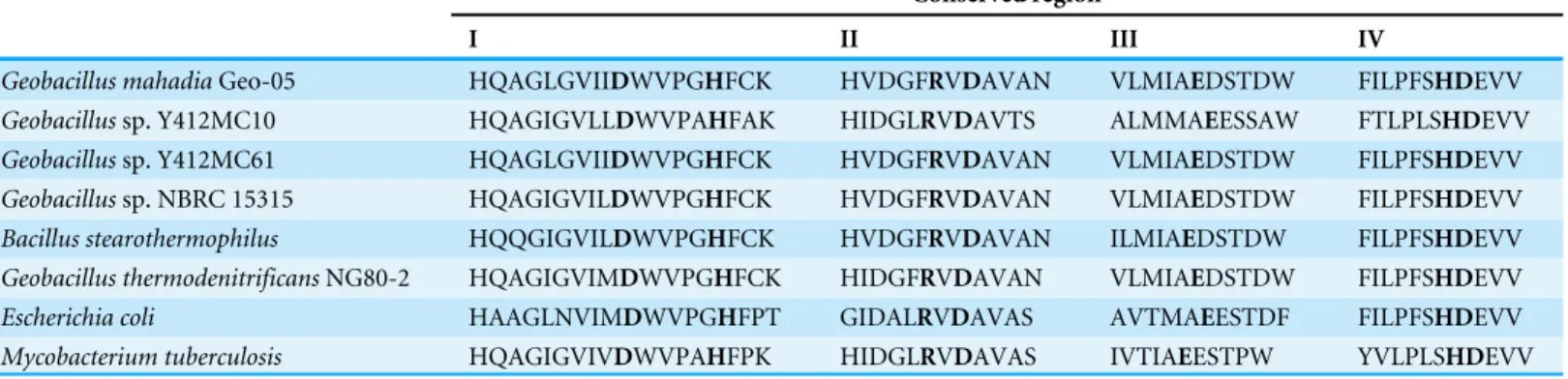
Table 1 Conserved regions in glycogen branching enzyme fromGeobacillusspp.,Escherichia coliandMycobacterium tuberculosis.
Conserved region
I II III IV
Geobacillus mahadiaGeo-05 HQAGLGVIIDWVPGHFCK HVDGFRVDAVAN VLMIAEDSTDW FILPFSHDEVV Geobacillussp. Y412MC10 HQAGIGVLLDWVPAHFAK HIDGLRVDAVTS ALMMAEESSAW FTLPLSHDEVV Geobacillussp. Y412MC61 HQAGLGVIIDWVPGHFCK HVDGFRVDAVAN VLMIAEDSTDW FILPFSHDEVV Geobacillussp. NBRC 15315 HQAGIGVILDWVPGHFCK HVDGFRVDAVAN VLMIAEDSTDW FILPFSHDEVV Bacillus stearothermophilus HQQGIGVILDWVPGHFCK HVDGFRVDAVAN ILMIAEDSTDW FILPFSHDEVV Geobacillus thermodenitrificansNG80-2 HQAGIGVIMDWVPGHFCK HIDGFRVDAVAN VLMIAEDSTDW FILPFSHDEVV Escherichia coli HAAGLNVIMDWVPGHFPT GIDALRVDAVAS AVTMAEESTDF FILPFSHDEVV Mycobacterium tuberculosis HQAGIGVIVDWVPAHFPK HIDGLRVDAVAS IVTIAEESTPW YVLPLSHDEVV
Notes.
The conserved amino acids are in bold.
in 10 mL of distilled water), 0.5 mL of 1 M HCl and diluted to 130 mL in distilled water. One unit (U) of enzyme activity was defined as the decreased of A660nmreading by 1% per minute. The decreased of A660nm reading represents the amylose-iodine complex (Shinohara et al., 2001).
Enzyme characterization
The effect of temperature on GBE-05 activity was studied at temperatures from 30◦C to
80◦C with 5◦C intervals. The enzyme thermostability test was done by incubating the
enzymes at 40◦C–80◦C for 24 h with 4 h intervals. After the incubation, the enzyme was
immediately cooled in an ice bath prior to assay. GBE activity was assayed at 50◦C, pH
7.0. The effect of pH on GBE-05 activity was studied at pH 4–pH 10. GBE-05 activity was assayed in 50 mM acetate buffer for pH 4–6, 50 mM potassium phosphate buffer for pH 6–8, 50 mM Tris-Cl buffer for pH 8–9 and50 mM glycine-NaOH for pH 9–10. The effect of pH on GBE-05 stability was studied by incubating the enzyme in the buffers mentioned at 25◦C for 1 h. GBE activity was assayed at 50◦C, pH 7.0. To study the effect
of metal ions on GBE-05 activity, GBE-05 was treated with 1 mM and 5 mM of metal ions (Mg2+, Ca2+, Fe2+, Mn2+, Zn2+ and Cu2+) for 30 min at 25◦C and immediately assayed
after the treatment at 50◦C, pH 7.0.
Nucleotide sequence accession number
The nucleotide sequence data reported in this paper are registered with the GenBank nucleotide sequence databases under accession numberKC951870.
RESULTS AND DISCUSSION
Genome mining
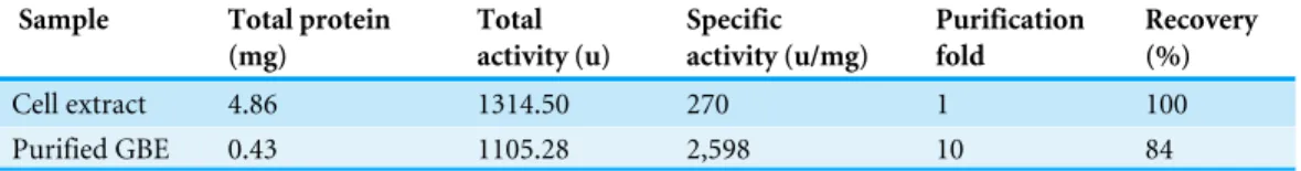
Table 2 Purification of GBE fromGeobacillus mahadiaGeo-05 using affinity chromatography.
Sample Total protein
(mg) Total activity (u) Specific activity (u/mg) Purification fold Recovery (%)
Cell extract 4.86 1314.50 270 1 100
Purified GBE 0.43 1105.28 2,598 10 84
roles in the catalysis and substrate binding. Three of the conserved residues are the catalytic residues; Asp313 in region II, Glu356 in region III and Asp424 in region IV. Four other conserved residues; Asp243and His248in region I, Arg311in region II and His423in region IV are responsible for substrate binding (Abad et al., 2002;Van der Maarel et al., 2003).
Protein purification
GBE-05 produced by pET102/D-TOPOR expression vector has His-Patch thioredoxin
fused to the protein. His-Patch thioredoxin is a mutated thioredoxin that has a metal binding domain, which has been shown to have high affinity for divalent cations and therefore, the fusion protein can be purified using metal chelating resins like nickel sepharose (Lu et al., 1996). The recovery of protein obtained after the purification process was high with the enzyme activity increased by ten fold (Table 2). The SDS-PAGE result shows a single band for the purified enzyme (pooled eluted fractions) in lane 3, which means that the enzyme was successfully purified (Fig. 1). The theoretical molecular weight of GBE was 78 kDa and with the addition of His-Patch thioredoxin (13 kDa), the expected size of the recombinant protein would be 91 kDa.
Enzyme characterization
GBE-05 was generally active at 45◦C–60◦C and enzyme activity was highest when assayed
at 55◦C (Fig. 2). This optimum temperature of GBE-05 was higher than GBEs isolated
fromG. stearothermophilusandA. gottschalkii, which has the optimum temperature of 50◦C (Takata et al., 1994;Thiemann et al., 2006). However, GBEs isolated from extreme
thermophilic bacteria,Rhodothermus obamensis,R. marinusandA. aeolicusshowed higher optimum temperature, that is between 65◦C–80◦C (Shinohara et al., 2001;Van der Maarel
et al., 2003;Yoon et al., 2008). These bacteria produce enzymes that are active at higher temperature comparatively to their optimal growth temperatures.
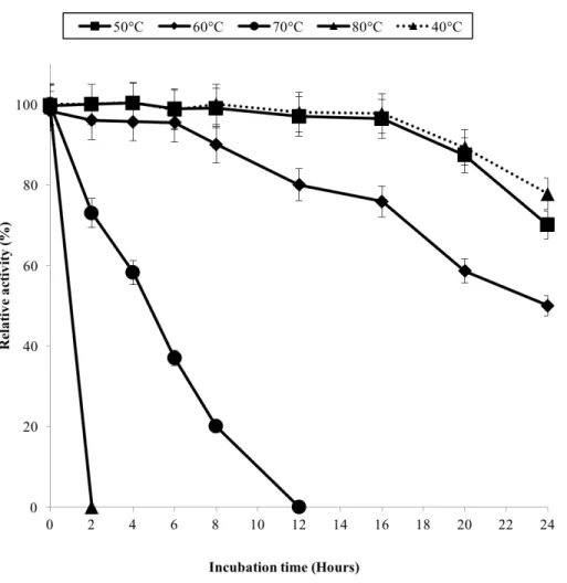
The half-life of the enzyme at 60◦C was 24 h while at 70◦C, 5 h (Fig. 3). GBE-05 is
more stable compared to GBE fromG. stearothermophilusthat has lost 20% of enzyme activity at 60 ◦C in just 30 min andA. gottschalkiithat has a half-life of only 55 min
at 55 ◦C (Takata et al., 1994;Thiemann et al., 2006). Since GBE-05 does not have any
Figure 1 SDS-PAGE of purified enzyme.M: Broad Range Prestained Protein Marker (Nacalai). Lane 1: Crude enzyme. Lane 2: Protein in flowthrough fractions. Lane 3: Purified enzyme
Figure 3 Effect of temperature on enzyme stability.GBE was incubated at 40◦C–80◦C prior to enzyme assay. Enzyme assay was done at 50◦C. 100% of activity is 793 U/mg using iodine stain assay. Note: Error bars represent means±5% for triplicate determinations.
temperature stability. The reason behind this is that the hydrophobic interactions between the aromatic groups are responsible for the stability of a thermophilic protein, while the deamination of thermolabile amino acids (asparagine and glutamine) resulted in the inactivation of enzymes at elevated temperature (Vieille et al., 2001).
GBE-05 displayed relatively high activity in broad pH range, where more than 60% of enzyme activity remained when assayed at pH 5–pH 9 (Fig. 4A), and was found to be most active at pH 6. The stability test shown that the enzyme was stable between pH 5–pH 9 where more than 50% of enzyme activity remained after the 30 min of pH treatment (Fig. 4A). It is important for GBE-05 to be active and stable in wide range of pH if this enzyme were to be applied industries.
Metal ions had different effects on GBE-05 activity but none of the metal ions experimented upon enhanced the enzyme activity (Fig. 5). Two alkaline earth metals of group 2 elements (Mg2+ and Ca2+) were tested to have no effect on enzyme activity.
However, GBE activity was slightly lowered to 73% when the concentration of Ca2+
Figure 4 (A) Effect of pH on enzyme activity. (B) Effect of pH on enzyme stability.Note: data repre-sents mean±SE (n=3).
seems to enhance the activity of GBE by 15% forR. marinus(Garg et al., 2007;Yoon et al., 2008). Four transition metals (Mn2+, Fe2+, Cu2+and Zn2+) were also tested out. 1 mm
Mn2+did not affect enzyme activity but the activity was decreased by 14% in 5 mM Mn2+.
Mn2+also showed slight inhibition on GBE activity isolated fromAnaerobranca gottschalkii
andR. marinus(Thiemann et al., 2006;Yoon et al., 2008). Zn2+ and Cu2+ repressed the
enzyme activity as only 40% and less remained. These metal ions also appear to restrain GBE activity from other bacteria,A. gottschalkii,R. marinusandM. tuberculosis(Thiemann et al., 2006;Garg et al., 2007;Yoon et al., 2008). 5 mM of Fe2+inhibits the enzyme by 60%,
Figure 5 Effect of metal ion on enzyme activity.Enzyme activity was assayed with two concentrations of metal ions, 1mM and 5 mM. 100% of activity is 641 U/mg using iodine stain assay. Note: error bars repre-sent means±5% for triplicate determinations
CONCLUSIONS
In conclusion, GBE-05 is stable and active at high temperature and therefore is very applicable in industries. The results of genome mining and computational prediction complement the results obtained from wet laboratory experiments. The vast information on genome sequence together with latest development in structural prediction software and algorithms enables scientists to compute data from genes to protein structure and function accurately.
ACKNOWLEDGEMENTS
We thank Malaysia Genome Institute forGeobacillus mahadiaGeo-05 bacterial strain and genome sequence.
ADDITIONAL INFORMATION AND DECLARATIONS
Funding
Grant Disclosures
The following grant information was disclosed by the authors: Genetics and Molecular Biology Initiatives.
Malaysia Genome Institute: 08-05-MGI-GMB002.
Competing Interests
The authors declare there are no competing interests.
Author Contributions
• Nur Syazwani Mohtar conceived and designed the experiments, performed the experiments, analyzed the data, contributed reagents/materials/analysis tools, wrote the paper, prepared figures and/or tables, reviewed drafts of the paper.
• Mohd Basyaruddin Abdul Rahman and Raja Noor Zaliha Raja Abd Rahman conceived and designed the experiments, analyzed the data, contributed reagents/materials/analysis tools, reviewed drafts of the paper.
• Thean Chor Leow and Abu Bakar Salleh conceived and designed the experiments, analyzed the data, contributed reagents/materials/analysis tools.
• Mohd Noor Mat Isa contributed reagents/materials/analysis tools.
Data Availability
The following information was supplied regarding data availability: GenBank. Accession number:KC951870.
REFERENCES
Abad MC, Binderup K, Rios-Steiner J, Arni RK, Preiss J, Geiger JH. 2002.The X-ray
crystallographic structure ofEscherichia colibranching enzyme.Journal of Biological Chemistry277:42164–42170DOI 10.1074/jbc.M205746200.
Bruno C, Servidei S, Shanske S, Karpati G, Carpenter S, McKee D, Barohn RJ, Hiranoi
M, Rifai Z, DiMauro S. 1993.Glycogen branching enzyme deficiency in adult
polyglucosan body disease.Annals of Neurology33:88–93.
Burley SK, Petsko GA. 1985.Aromatic-aromatic interaction: a mechanism of protein
structure stabilization.Science229:23–28DOI 10.1126/science.3892686.
Challis GL. 2008.Genome mining for novel natural product discovery.Journal of
Medicinal Chemistry51:2618–2628DOI 10.1021/jm700948z.
Corre C, Challis GL. 2007.Heavy tools for genome mining.Chemistry & Biology 14:7–9
DOI 10.1016/j.chembiol.2007.01.001.
EMBL-EBI. 2010.ClustalW.Available athttp:// www.ebi.ac.uk/ Tools/ msa/ clustalw2/
(accessed on 1 July 2010).
Ferrer M, Martínez-Abarca F, Golyshin PN. 2005.Mining genomes and ‘‘metagenomes’’
for novel catalysts.Current Opinion in Biotechnology16:588–593
DOI 10.1016/j.copbio.2005.09.001.
Garg SK, Alam MS, Kishan KVR, Agrawal P. 2007.Expression and characterization of
α-(1,4)-glucan branching enzyme Rv1326c ofMycobacterium tuberculosisH37Rv.
Hall T. 2010.BioEdit.Available athttp:// www.mbio.ncsu.edu/ bioedit/ page2.html
(accessed on 22 March 2010).
Kawabata Y, Toeda K, Takahashi T, Shibamoto M, Kobayashi M. 2002.Preparation
of highly branch starch by glycogen branching enzyme fromNeurospora crassa N2-44 and its characterization.Journal of Applied Glycoscience49:273–279
DOI 10.1128/JB.00390-06.
Kim WS, Kim J, Krishnan H, Nahm BH. 2005.Expression ofEscherichia colibranching
enzyme in caryopses of transgenic rice results in amylopectin with an increased degree of branching.Planta220:689–695DOI 10.1007/s00425-004-1386-3.
Kortstee A, Vermeesch A, De Vries B, Jacobsen E, Visser R. 1996.Expression of
Escherichia colibranching enzyme in tubers of amylose-free transgenic potato leads to an increased branching degree of the amylopectin.The Plant Journal10:83–90
DOI 10.1046/j.1365-313X.1996.10010083.x.
Laemmli UK. 1970.Cleavage of structural proteins during the assembly of the head of
bacteriophage T4.Nature227:680–685.
Lee C, Le Q, Kim Y, Shim J, Lee S, Park J, Lee K, Song S, Auh J, Lee S, Park K. 2008.
Enzymatic synthesis and properties of highly branched rice starch amylose and amylopectin cluster.Journal of Agriculture and Food Chemistry 56:126–131
DOI 10.1021/Jf072508s.
Lu Z, Diblasio-smith EA, Grant KL, Warne NW, Lavallie ER, Collins-racie LA, Follettie
MT, Williamson MJ, Mccoy JM. 1996.Histidine patch thioredoxins.The Journal of
Biological Chemistry 271:5059 –5065DOI 10.1074/jbc.271.9.5059.
NCBI. 2010.BLAST.Available athttp:// blast.ncbi.nlm.nih.gov/ Blast.cgi(accessed on 1
July 2010).
Pal K, Kumar S, Sharma S, Garg SK, Alam MS, Xu HE, Agrawal P, Swaminathan
K. 2010.Crystal structure of full-lengthMycobacterium tuberculosisH37Rv
glycogen branching enzyme: insights of N-terminal?-sandwich in substrate speci-ficity and enzymatic activity.Journal of Biological Chemistry285:20897–20903
DOI 10.1074/jbc.M110.121707.
Schattner P. 2009.Genomics made easier: an introductory tutorial to genome data
mining.Genomics93:187–195DOI 10.1016/j.ygeno.2008.10.009.
Serrano L, Bycroft M, Fersht AR. 1991.Aromatic-aromatic interactions and protein
stability investigation by double-mutant cycles.Journal of Molecular Biology
218:465–475DOI 10.1016/0022-2836(91)90725-L.
Shinohara ML, Ihara M, Abo M, Hashida M, Takagi S, Beck TC. 2001.A novel
ther-mostable branching enzyme from an extremely thermophilic bacterial species, Rhodothermus obamensis.Application of Microbiology and Biotechnology57:653–659
DOI 10.1007/s00253-001-0841-3.
Takata H, Akiyama T, Kajiura H, Kakutani R, Furuyashiki T, Tomioka E, Kojima I,
Kuriki T. 2010.Application of branching enzyme in starch processing.Biocatalysis
Takata H, Takaha T, Kuriki T, Okada S, Takagi M, Imanaka T. 1994.Properties and active center of the thermostable branching enzyme fromBacillus stearothermophilus. Applied and Environmental Microbiology 60:3096–3104.
Thiemann V, Saake B, Vollstedt A, Schäfer T, Puls J, Bertoldo C. 2006.Heterologous
expression and characterization of a novel branching enzyme from the thermoal-kaliphilic anaerobic bacteriumAnaerobranca gottschalkii.Applied Microbiology and Biotechnology13:60–71DOI 10.1007/s00253-005-0248-7.
Van der Maarel MJEC, Vos A, Sanders P, Dijkhuizen L. 2003.Properties of the glucan
branching enzyme of the hyperthermophilic acterium Aquifex aeolicus.Biocatalysis and Biotransformation21:199–207DOI 10.1080/10292920310001618528.
Van der Maarel MJE, Van der Veen B, Uitdehaag JC, Leemhuis H, Dijkhuizen L. 2002.
Properties and applications of starch-converting enzymes of theα-amylase family.
Journal of Biotechnology94:137–155DOI 10.1016/S0168-1656(01)00407-2.
Vieille C, Epting KL, Kelly RM, Zeikus JG. 2001.Bivalent cations and amino-acid
composition contribute to the thermostability ofBacillus licheniformisxylose isomerase.European Journal of Biochemistry268:6291–6301
DOI 10.1046/j.0014-2956.2001.02587.x.
Yoon S-A, Ryu S-I, Lee S-B, Moon T-W. 2008.Purification and characterization of
branching specificity of a novel extracellular amylolytic enzyme from marine hyperthermophilic rhodothermus marinus.Journal Microbiology and Biotechnology