ISSN 1557-4555
© 2009 Science Publications
Corresponding Author: E.S. Soliman, Department of Pathobiology, College of Veterinary Medicine, Nursing and Allied Health, Tuskegee University, Alabama, United States
Stressors Influence on Salmonella enterica Serovar Enteritidis
Colonization in Broilers
1
E.S. Soliman,
1E. Taha,
1K.D. Infante,
1K. Laboy,
2M.A. Sobieh and
1P.G. Reddy
1Department of Pathobiology, College of Veterinary Medicine,
Nursing and Allied Health, Tuskegee University,
Alabama, United States
2
Department of Animal Hygiene, Zoonoses and Animal Behavior,
College of Veterinary Medicine, Suez Canal University, Egypt
Abstract: Problem statement: Poultry industry usually exposing birds to a variety of actions and stressors includs fasting for gastrointestinal emptying before transportation and where birds are often exposed to high environmental temperature during the summer months. These environmental stressors may have influences on bird performance and susceptibility to pathogenssuch as Salmonella enteritidis
by altering the intestinal micrbiota and changes in the gut integrity. Approach: This research was conducted to show that acute stressors in the poultry production can induce changes in the normal intestinal microbiota and epithelium structure and execratory functions, which may cause an increase in the opportunities of attachment of Salmonella enteritidis. Results: Experiments were conducted to determine the influence of 24 h feed withdrawal with 24 h exposureto high temperature (30°C) on intestinal characteristicsof broilers. Attachment of Salmonella enteritidis to ileal tissuewas determined using an in vitro ileal loop assay. Changes in commensally intestinal microbial populations were determined using gel electrophoresis and alterations in ileal morphology were determined histologically. The results showed that attachmentof Salmonella enteritidis to ileal tissues increased by 1.5logs (9.05 log10 Vs 7.59 log10Salmonella enteritidis/g ofileal tissue; p = 0.0006) in broilers fasted for 24 h also, ileal tissues from birds subjected to 30°C for 24 h hadincreased the attachment of
Salmonella enteritidis (8.77log10 Vs 8.50 log10Salmonella enteritidis/g of ileum; p =0.01) compared with birds held at 23°C. Exposure to 30°Cfor 24 h also altered the microbial structure in the ileumand cecum. Where subjecting birds to 30°C for 24 h reduced the crypt depth (6.0 Vs 7.8 µ m, respectively; p = 0.002), but it hadno effect on villus height or villus: Crypt ratio. Conclusion: The findings of the experiment explained the mechanisms by which stressors alters the normal intestinal characterization and induces susceptibility to enteric infection. Future work should focus on the use of prophylactic measures to reduce the stress conditions causing alteration of the intestinal microbiota and changes in gut integrity like considering the probiotic organisms the offer a promising solution for reducing pathogen colonization when fed orally.
Key words:Salmonella enteritidis, heat stress, microbial, colonization, broilers
INTRODUCTION
Salmonella is the leading cause of bacterial food-borne diseases in the United States and causes approximately 1.2 million casesof human Salmonellosis each year. The most commonly implicated source of food-borne Salmonellosis through consumption of undercooked poultry products, Antunes et al.[1]. Environmental stresshas been shown to be a factor that may induce colonization of food animals by enteric pathogens, facilitate horizontal transmission of pathogens between animals, increase pathogen
shedding and contribute in carcass contamination during processing[8,16]. Stress is an important consideration in poultry production;because birds are routinely subjected to stressors specially feed withdrawal and temperature fluctuations during transportation[7,22]. Broilers are subjected to fasting to reduce the volume of intestinal contents before slaughter and thus minimize the risk of carcass contamination during processing; however, feed withdrawal has been associated with increased
stressor encounteredin seasonal environments, during the summer months and is also associated with increased intestinal colonizationand fecal shedding of pathogens in poultry[2]. The gastrointestinal tract is particularlyresponsive to stressors, which can cause a variety of changesas alteration of the normal, protective microbiota, Bailey and Coe[3,4] and decreased integrity of the intestinal epithelium[21]. The commensal intestinal populations can protect the host from pathogen colonization by competing for epithelial binding sites and nutrients, strengtheningthe intestinal immune response and by producing antimicrobial bacteriocins[11]. Therefore, stress-inducedthe integrity of the gut epithelium reduces innate protective mechanisms and may increase the potential for pathogens such as Salmonellato bind to and colonize the intestinal epithelium. Such colonizationin poultry will increase the risk of carcass contamination during processing and will increase the potential for
Salmonella to translocate to the reproductive tract, where it can contaminateeggs during formation.
MATERIALS AND METHODS
Birds: During feed withdrawal and heat stress experiments, 960 male and 308 broilers were housed to 24 floorpens by rate 40 birds/pen [23]. Birds were raised to 40-42 days of ageon a standard corn-soybean meal diet. Initial ambient temperature was held at 35°C for the newly hatched chicks and then gradually decreased to 22°Cby 21st day and held at 22°C for the duration of the experiment.
Stress conditions: (a) Feed Withdrawal: At 40 days old, 10 birds were randomly chosen, sacrificed in gas chamber and sampled. Feed was withdrawn (0 h) from theremaining flock and birds were kept on litter and given access to water for 4 h before being placed in transport crates for20 h. After 24 h of feed withdrawal, 10 additional birds were sacrificed and tissues were sampled.(b) Heat stress study: At 42 days, 20 birds were randomly chosen and 10 were immediately sacrificed and sampled, whereas the remaining 10 were subjectedto 30°C temperature in floor pens for 24 h, withfull access to feed and water, before euthanasia and sampling.
Intestinal sampling: (a) Feed Withdrawal: A 10 cm section of the ileum was taken from each bird for an ilealchallenge assay and was gently flushed with 0.05 M PBS.Tissue sections were immediately placed in ice-cold Dulbecco’sModified Eagle Medium + L-glutamine (DMEM) (Mediatech) and kept on ice until
used for an in vitro Salmonella challenge assay as described below. (b) Heat Stress Study:Intestinal tissue and contents were obtained immediately after euthanasia and were collected as following: (1) 10 cm section of the ileum was taken for an in-vitro Salmonella challenge assay; (2) 3 cm ileal section, 13 cm from the ileo-cecal junction, was collected for gel electrophoresis analysis of microbial structure. Ileal tissues wereopened and contents were gently removed, placed in microfuge tubes and immediately frozen at -20°C. Ileal tissueswere gently flushed with 0.05 M PBS and were frozenat -20°C; (3) 2 cm section of ileal tissue, 16 cmfrom the ileo-cecal junction, was collected for analysis ofintestinal morphology, flushed with PBS and fixed in 10% neutralbuffered formalin for 48 h and (4) 4 cm tissue section ofthe center of the cecum and cecal contents were obtained for Gel electrophoresis analysis.
Ileal loop assay for attachment of Salmonella enteritidis: (a) Challenge microorganism: Salmonella enterica
serovar Enteritidis obtained and transferred with a kanamycin-resistance plasmid to allowselection in the presence of kanamycin. The stock culture was grownin Luria Bertani (LB) broth containing 50 µg of kanamycin/mL(LB-kan) and stored with 20% (vol/vol) added glycerol. Fresh cultures were grown statically overnight in LB-kan broth, transferredto fresh LB-kan broth and grown overnight for the challenge study. Bacterial cells were harvested by centrifugation at 6,000×g at 4°C for 15 min and were washed 3 times in equalvolumes of sterile PBS. Cells were re-suspended in DMEM to an Optical Density (OD)600 of 0.4 (approximately 1×10⁶ cells mL−1). The inoculum was serially diluted and plated on LB broth to obtain the actual number ofcells in the inoculum.(b) Ileal Loop Assay: In the organ culture[14], the ileal sections were removed from DMEM, sealed atone end with 35 mm dialysis clamps and inoculated with approximately6 mL of Salmonella enteritidis culture suspended in DMEM. The open end of the ileal section was sealed with dialysis clamps,the exterior was rinsed with PBS and the ileal loops were incubatedin 100 mL of DMEM for 1 h at 37°C in a water bathin a 10% CO2 atmosphere. After incubation, ileal contents were removed, the interior and exterior of each section was rinsedwith PBS, tissues were homogenized, serially diluted in buffered peptone broth and plated on LB agar plates (Remel, Fisher Int.) containing 50 µg mL−1 kanamycin. Plates were incubated at 37°Cfor 24 h and were enumerated for
Intestinal morphology: After fixation in 10% neutral buffered formalin, a single 0.5 cmsample was cut from each ileal section, dehydrated with increasing concentrations (70, 80, 95 and 100%) of ethanol, cleared with xylene (Thermo Sci Fisher Products, Fisher Int.) and placed into polyfin embedding wax. Tissue sections (5 µ m) were cut, floated onto slides, stained with hematoxylin (Thermo Sci Acros Organics, Fisher Int.) and eosin (Thermo Sci Acros Organics, Fisher Int.) and measured for villus height and crypt depth using light microscopy and a micrometer. Measurements for villilength were taken from the tip of the villus to the valley between individual villi and measurements for crypt depth were taken from the valley between individual villi to the basolateral membrane. Eight villi and villus-associated crypts were measured for each sample. Morphology data were analyzed using the GLMprocedure in SAS.
DGGE: Genomic DNA was isolated from intestinal digesta and tissue samples using the Ultraclean Fecal DNA kit (Applied Biosystem), sampleswere diluted 1:1 with sterile distilled water and 0.25 g of the diluted sample was added to a bead beating tube containing beads, bead solution and lysis solution. Cells were lysed bya combination of detergent and mechanical action using a standard vortex. From the lysed cells, the released DNA was bound to a silica spin filter. The filter was washed and DNA was eluted using DNase-free Tris buffer.The DGGE was performed according to previously described methodswith modification[13], using bacteria-specific PCR primers to conserved regions flankingthe variable V3 region of 16S rDNA. Each PCR reaction mixture contained 0.02 nmol of reverse primer (534r):5'-ATT ACC GCG GCT GCT GG-3' and 0.02 nmol of forward primer with a GC clamp(341FGC): 5'CGC CCG CCG CGC GCG GCG GGC GGG GCG GGG GCA CGG GGG GCC TAC GGG AGG CAG CAG-3', 3.75 units of Taq DNA Polymerase, 5-10 ng of template DNA, 10×DNA Polymerase Buffer (containing 10 mM Tris-HCl, 50 mM KCl and 0.1% Triton X-100) and 25 mMMgCl2. Amplifications were performed using a cephid smart cycler using the followingprogram: (1) denaturation at 95°C for 5 min; (2) subsequentdenaturation at 95°C for 1 min; (3) annealing at 65°Cfor 1 min; (4) extension at 72°Cfor 1 min;(5) steps 2-4 repeated for 30 cycles; (6) denaturationat 95°C for 1 min; (7) annealing at 55°C for 1 min; (8)extension at 72°C for 1 min; (9) steps 6-8 repeated for 7cycles; (10) extension at 72°C for 7 min; (11) 4.0°C final holding temperature. Polyacrylamide gels (8% acrylamide-bisacrylamide ratio 37:5:1) were cast with a 40-60% urea: Deionized formamide
gradient. The100% denaturing acrylamide contained 7 M urea and 40% deionizedformamide. Amplified DNA was mixed with a 20% volume of 5x loading buffer (0.025% (wt/vol) bromophenol blue, 0.025% (wt/vol) xylenecyanol, 47% (vol/vol) 0.1 M EDTA and 47% (vol/vol) glycerol) and 20 µL was loaded into each sample well (20-well comb). Gels were placed in a DCode Universal Mutation Detection System and electrophoresed in 0.5x Tris-acetate-EDTAbuffer at 60 for 10 min at 200 V, followed by 16 h at 70 V.Gels were silver stained[19].Fragment pattern relatedness was determined using Bionumerics Software, which determined the number of bands per sample and similarity coefficients for banding patterns between pairs of samples. A distance matrix was calculated using the DICE function and dendrograms were constructed from this matrix using the Unweighted Pair GroupMeans Average (UPGMA) function. The degree of similarity of banding patterns between pairs of samples was represented asa similarity coefficient. All DGGE data were analyzed usingthe Mixed Model of SAS. Similarity coefficientsbetween pairs of samples were segregated by treatment and similaritycoefficients across treatments were used as an estimate of similarity assuming no treatment effect. Significance was determined using p-value<0.05.
RESULTS
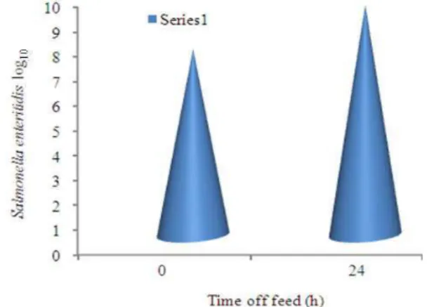
Effect of 24 h feed withdrawal on ileal susceptibility to Salmonella enteritidis attachment: Intestinal tissues from fasted birds were more susceptible to pathogen attachment than tissues from control birds, with a1.5 log increase (p = 0.01) in Salmonella enteritidis
associatedwith the ileal tissue of fasted birds compared with non fastedcontrols (Fig. 1).
Table 1: Denaturing gradient gel electrophoresis similarity coefficients within control (23°C) and acute heat-stress (30°C for 24 h) treatments and across treatments (cross-products)
Treatment
---
Sample 22°C 30°C Cross-products SEM
Ileal contents 68.0a 55.4b 41.7c 2.4
Ileal tissue 73.7a 72.5a 46.3b 2.3
Cecal contents 58.5a 60.4a 52.4b 1.5
Cecal tissue 53.4b 64.7a 44.5c 2.8
a-c: Means within rows with different superscript letters are
significantly different (p<0.05)
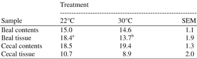
Table 2: Influence of heat stress on number of bands present in various intestinal systems of broilers as determined by gel electrophoresis
Treatment
---
Sample 22°C 30°C SEM
Ileal contents 15.0 14.6 1.1
Ileal tissue 18.4a 13.7b 1.9
Cecal contents 18.5 19.4 1.3
Cecal tissue 10.7 8.9 2.0
a,b: Means within rows with different superscript letters are
significantly different (p<0.05)
Influence of an acute high temperature on intestinal susceptibility to Salmonella enteritidis adhesion, microbial populations and morphology of the small intestine of broilers: Broilers placed in the 30°C room showed behavioral signsof heat stress such as panting and spreading of wings. In theileal loop assay, numbers of Salmonella enteritidis associatedwith the ileum were greater (p = 0.0006) in heat-stressed birds(8.77 log10 cfu g−1) compared with non stressed birds (8.50 log10 cfu g−1 of ileum). Amplicon profiles for bacteria inthe intestinal contents and tissues revealed differences in banding patterns between heat-stressed and non stressed birds.Birds held at 30°C exhibited lower (p = 0.0001) similarity coefficients for microbial communities in ileal contents than did birds at 22°C. In all intestinal samples, Table1, the similarity coefficients calculated across the 30 and22°C treatments (cross-product) were lower (p = 0.0001) than the similarity coefficients within individual treatments. The ileal tissue of heat-stressed animals contained fewer (p= 0.0251) amplicon fragments (bands) than the ileal tissue ofnon stressed birds, Table 2.
DISCUSSION
The ileal loop assays demonstrated that stress due to 24 h feedwithdrawal and exposure to high temperatures is associated withincreased susceptibility of intestinal tissues to Salmonella enteritidis colonization. Intestinal tissues from fasted birds had significantly greater
attachment of Salmonella than did tissues from control birds, Fig. 1. Ramirez et al.[18] inthe study reported here, numbers of Salmonella enteritidis associatedwith the ileum were greater in heat-stressed birds (8.77 log10 cfu g−1)compared with non stressed birds (8.50 log10 cfu g−1 ileum), indicating that stress may contribute to increased intestinal colonization by Salmonella. Moreover heat stress may have damaging effects on mucosal structure[20]. In addition, heat shock proteins, whose expression can be induced by high ambient temperature and other environmental stressors, Lindquist and Craig[9], may act as epithelial surface receptors for pathogen binding[24].
There is a linear reduction in the mucus lining the intestinaltract over a 24 h fasting, as well as changes in intestinal morphology[23]. Hinton et al.[6] showed that there were increases in intestinal Enterobacteriaceae and cecal aerobes with a concurrent decrease in lactic acid bacteriain broilers subjected to a 24 h feed withdrawal. Neurohormones associated with stress can increase growth and virulence factor expression in microbes including Escherichia coli, Yersinia enterocolitica and
Pseudomonas aeruginosa in vitro[17]. Release of norepinephrine in the intestinal tract increased the number of gram-negativebacteria within the lumen[10]. One possible limitation of analyzing similarity coefficients within treatments is that each treatment analysis is separate; thus, one may end up with numerically comparable similarity coefficients, but banding patterns within treatment may differ, as the data fromileal tissue, Table 1 shows the dendrogram shows that banding patterns were highly similar within eachtreatment, with similarity coefficients of 73.7 and 72.5 forbirds at 22 and 30°C, respectively. However, the banding patterns are obviously different between treatments.
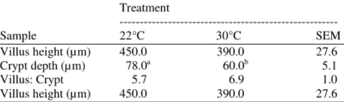
Table 3: Influence of heat stress on small intestinal morphological characteristics
Treatment
---
Sample 22°C 30°C SEM
Villus height (µm) 450.0 390.0 27.6
Crypt depth (µm) 78.0a 60.0b 5.1
Villus: Crypt 5.7 6.9 1.0
Villus height (µm) 450.0 390.0 27.6
a,b: Means within rows with different superscript letters are
significantly different (p<0.05)
The ileal tissue from birds held at 30°C contained fewer amplicon fragments than the ileal tissue from birds at 22°C, Table 2, indicatingthat exposing birds to high temperatures for 24 h caused a reduction in microbial species associated with the ileal wall. Influence of 24 h heat stress on ileal morphology was evaluated.In birds subjected to heat stress, crypt depth was reduced (p = 0.002) compared with non stressed birds (60 Vs 78 µm,respectively; Table 3. Villus height and the villus: Crypt ratiowere unchanged in response to 24 h heat stress.
Birds subjected to 30°C for 24 h had reduced crypt depth compared with birds at 22°C. Villus height and the villus: Crypt ratio were unchangedin birds exposed to 30°C this may be attributed to the short duration of the stressor andthe resistance of the ileum to structural change compared with other regions of the small intestine; Yamauchi et al.[26].It is likely that changes in cell proliferation would be observedfirst in the stem cells of the crypt rather than the villusbecause of the high proliferative activity of the crypt; Yamauchi et al.[26].Morphological changes in response to fasting occur more rapidly in the proximal two-thirds of the small intestine than in the ileum. In feed withdrawal studies with chickens, the structure of the duodenum and jejunum mucosa changes rapidly, often within 36 h of the onset of stress, whereas the ileum maintains its normal morphology longer and requires extended periods of stress to influence its structure; Yamauchi et al.[26]. The findings from the 24 h heat stressstudy suggest that stressors could act in several ways to increase intestinal susceptibility to in vitro Salmonella enteritidis attachment; by disrupting the normal protective microbiota and altering intestinal morphology, stressors create an opportunity for pathogens to colonize the intestine. Probiotic organisms such as lactobacilli, some strains of E. coliand yeast offer promise for reducing pathogen colonization when fed orally[15,25]. When administered consistently, probiotic organisms can colonize and form a nichein the intestine and may be a useful dietary treatment if administered before feed withdrawal or transportation
and processing. In addition, non digestible oligosaccharides, or probiotics, have been shown to enhance intestinal growth of probiotic or beneficial commensal organisms[5].The fermentation of prebiotic complex carbohydrates by intestinalmicrobes produces volatile fatty acids, which promote epithelial cell proliferation and renewal[12]. Suchactivity may enhance integrity of the intestinal epithelium.Therefore, dietary supplementation of the flock with prebiotics before periods of anticipated stress may also limit the damage to the intestinal epithelium elicited by stressors.
CONCLUSION
The study revealed that acute stressors can invoke significant changes in the normal intestinal microbiota, intestinal morphology and in-vitro susceptibility for
Salmonella Enteritidids attachment to the ileum in the broilers. Stressors can increase the intestinal susceptibility to in-vitro Salmonella Enteritidids
attachment by disrupting the normal protective microbiota or by altering intestinal morphology and as end result it create an opportunity for pathogens to colonize in the intestine. Also acute stressors can evoke alteration in the mucous production and composition that can affect the attachment capability of both commensal and pathogenic micro-organisms.
All the information that were concluded can be important to be considered in the poultry industry as a step during designing on-farm strategies aiming to reduce the pathogen contamination in poutry.
REFERENCES
1. Antunes, P., C. Réu, J.C. Sousa, L. Peixe and N. Pestana, 2003. Incidence of Salmonella from poultry products and their susceptibility to antimicrobial agents. Int. J. Food Microbiol., 82: 97-103. DOI: 10.1016/S0168-1605(02)00251-9
2. Bailey, J.S., 1988. Integrated colonization control of Salmonella in poultry. Poult. Sci., 67: 928-932. http://www.ncbi.nlm.nih.gov/pubmed/3413018 3. Bailey, M.T. and C.L. Coe, 1999. Maternal
separation disrupts the integrity of the intestinal microflora in infant rhesus monkeys. Dev.
Psychobiol., 35: 146-155.
http://www.ncbi.nlm.nih.gov/pubmed/10461128 4. Bailey, M.T., G.R. Lubach and C.L. Coe, 2004.
Prenatal stress alters bacterial colonization of the gut in infant monkeys. J. Pediatr. Gastroenterol. Nutr., 38: 414-421.
5. Bouhnik, Y., L. Raskine, E. Vicaut, C. Neut and B. Flourie et al., 2004. The capacity of nondigestible carbohydrates to stimulate fecal bifido-bacteria in healthy humans: A double-blind, randomized, placebo-controlled, parallel-group, dose-response relation study. Am. J. Clin. Nutr., 80: 1658-1664.
http://cat.inist.fr/?aModele=afficheN&cpsidt=16336653 6. Hinton Jr., A., R.J. Buhr and K.D. Ingram, 2000.
Physical, chemical and microbiological changes in the ceca of broiler chickens subjected to incremental feed withdrawal. Poult. Sci., 79: 483-488.
http://www.ncbi.nlm.nih.gov/pubmed/10780642 7. Humphrey, T., 2006. Are happy chickens safer
chickens? Poultry welfare and disease susceptibility. Br. Poult. Sci., 47: 379-391. DOI: 10.1080/00071660600829084
8. Isaacson, R.E., L.D. Firkins, R.M. Weigel, F.A. Zuckermann and J.A. Di Petrio, 1999. Effect of transportation and feed withdrawal on shedding of Salmonella typhimurium among experimentally infected pigs. Am. J. Vet. Res., 60: 1155-1158. http://cat.inist.fr/?aModele=afficheN&cpsidt=10208999 9. Lindquist, S. and E.A. Craig, 1988. The heat-shock
proteins. Ann. Rev. Genet., 22: 631-677. DOI: 10.1146/annurev.ge.22.120188.003215
10. Lyte, M. and M.T. Bailey, 1997. Neuroendocrine-bacterial interactions in a neurotoxin-induced model of trauma. J. Surg. Res., 70: 195-201. http://cat.inist.fr/?aModele=afficheN&cpsidt=2754 463
11. MacDonald, T.T. and G. Monteleone, 2005. Immunity, inflammation and allergy in the gut.
Science, 307: 1920-1925. DOI:
10.1126/science.1106442
12. Macfarlane, G.T., H. Steed and S. Macfarlane, 2008. Bacterial metabolism and health-related effects of galacto-oligosaccharides and other probiotics. J. Applied Microbiol., 104: 305-344. DOI: 10.1111/j.1365-2672.2007.03520
13. Muyzer, G. and K. Smalla, 1998. Application of Denaturing Gradient Gel Electrophoresis (DGGE) and Temperature Gradient Gel Electrophoresis (TGGE) in microbial ecology. Antonie Van Leeuwenhoek, 73: 127-141. DOI: 10.1023/A:1000669317571
14. Naughton, P.J., L.L. Mikkelsen and B.B. Jensen, 2001. Effects of nondigestible oligosaccharides on
Salmonella enterica serovar Typhimurium and nonpathogenic Escherichia coli in the pig small intestine in vitro. Applied Environ. Microbiol., 67: 3391-3395. DOI: 10.1128/AEM.67.8.3391-3395.2001
15. Parvez, S., K.A. Malik, S.K. Ah and H.Y. Kim, 2006. Probiotics and their fermented food products are beneficial for health. J. Applied Microbiol., 100: 1171-1185.
http://www.ncbi.nlm.nih.gov/pubmed/16696665 16. Poppe, C., 1999. Epidemiology of Salmonella enterica
serovar Enteritidis. In: Salmonellaenterica Serovar
Enteritidis in Humans and Animals: Epidemiology, Pathogenesis and Control, Saeed, A.M., R.K. Gast, M.E. Potter and P.G. Wall (Eds.). Wiley-Blackwell, Ames, ISBN: 0813827078, pp: 3-18. 17. Rahman, H., R. Reissbrodt and H. Tschape, 2000.
Effect of norepinephrine on growth of Salmonella
and its enterotoxin production. Indian J. Exp. Biol., 38: 285-286.
http://www.ncbi.nlm.nih.gov/pubmed/10927876
18. Ramirez, G.A., L.L. Sarlin, D.J. Caldwell, C.R. Yezak Jr.
and M.E. Hume et al., 1997. Effect of feed withdrawal on the incidence of Salmonella in the crops and ceca of market age broiler chickens.
Poult. Sci., 76: 654-656.
http://poultsci.highwire.org/cgi/content/abstract/76/ 4/654
19. Sambrook, J., E.F. Fritsch and T. Maniatis, 1989. Molecular Cloning: A Laboratory Manual. 2nd Edn., Cold Spring Harbor Laboratory Press, Woodbury, New York. http://orton.catie.ac.cr/cgi-bin/wxis.exe/?IsisScript=LIBRO.xis&method=post &formato=2&cantidad=1&expresion=mfn=006164 20. Sengupta, A. and R.K. Sharma, 1993. Acute heat stress in growing rats: Effect on small intestinal morphometry and in vivo absorption. J. Therm.
Biol., 18: 145-151.
http://cat.inist.fr/?aModele=afficheN&cpsidt=4842 037
21. Soderholm, J.D., D.A. Yates, M.G. Gareau, P.C. Yang, G. MacQueen and M.H. Perdue, 2002. Neonatal maternal separation predisposes adult rats to colonic barrier dysfunction in response to mild stress. Am. J. Physiol. Gastrointest. Liver Physiol.,
283: G1257-G1263. DOI:
10.1152/ajpgi.00314.2002
22. St-Pierre, N.R., B. Cobanov and G. Schnitkey, 2003. Economic losses from heat stress by US livestock industries. J. Dairy Sci., 86: E52-77. http://jds.fass.org/cgi/content/abstract/86/13_suppl/ E52
24. Wampler, J.L., K.P. Kim, Z. Jaradat and A.K. Bhunia, 2004. Heat shock protein 60 acts as a receptor for the Listeria adhesion protein in Caco-2 cells. Infect. Immun., 72: 931-936. DOI: 10.1128/IAI.72.2.931-936.2004
25. Wehkamp, J., J. Harder, K. Wehkamp, B.W.V. Meissner and M. Schlee et al., 2004. NF-κB-and AP-1-mediated induction of human beta defensin-2 in intestinal epithelial cells by Escherichia coli Nissle 1917: A novel effect of a probiotic bacterium. Infect. Immun., 72: 5750-5758. http://www.pubmedcentral.nih.gov/articlerender.fc gi?artid=517557