P . b . b . G Z 0 2 Z 0 3 1 1 0 8 M , V e r l a g s p o s t a m t : 3 0 0 2 P u r k e r s d o r f , E r s c h e i n u n g s o r t : 3 0 0 3 G a b l i t z
Homepage:
www.kup.at/
mineralstoffwechsel
Online-Datenbank mit
Autoren- und Stichwortsuche
P . b . b . G Z 0 2 Z 0 3 1 1 0 8 M , V e r l a g s p o s t a m t : 3 0 0 2 P u r k e r s d o r f , E r s c h e i n u n g s o r t : 3 0 0 3 G a b l i t z
Indexed in SCOPUS/EMBASE/Excerpta Medica
www.kup.at/mineralstoffwechsel
Österreichische Gesellschaft für Orthopädie und Orthopädische Chirurgie
Österreichische Gesellschaft für Rheumatologie Offizielles Organ der
Österreichischen Gesellschaft zur Erforschung des Knochens und Mineralstoffwechsels
Member of the
Wissenschaftliche Herbsttagung der
Österreichischen Gesellschaft zur
Erforschung des Knochens und
Mineralstoffwechsels (ÖGEKM) - 21.
November 2009, Wien. Abstracts von
Vorträgen und Postern
Journal für M ineralstoffwechsel &
M uskuloskelettale Erkrankungen
NEUES AUS DEM VERLAG
Abo-Aktion 2016
Wenn Sie Arzt sind, in Ausbildung zu einem ärztlichen Beruf, oder im Gesundheitsbereich
tätig, haben Sie die Möglichkeit, die elektronische Ausgabe dieser Zeitschrift kostenlos zu
beziehen.
Die Lieferung umfasst 4–6 Ausgaben pro Jahr zzgl. allfälliger Sonderhefte.
Das e-Journal steht als PDF-Datei (ca. 5–10 MB) zur Verfügung und ist auf den meisten der
marktüblichen e-Book-Readern, Tablets sowie auf iPad funktionsfähig.
聺
Bestellung kostenloses e-Journal-Abo
P
Besuchen Sie unsere
zeitschriftenübergreifende Datenbank
聺
Bilddatenbank
聺
Artikeldatenbank
聺
Fallberichte
P
P
P
Die meistgelesenen Artikel:
Journal für Gastroenterologische und
Hepatologische Erkrankungen
Journal für Klinische Endokrinologie und Stoffwechsel
P
P
J MINER STOFFWECHS 2009; 16 (Sonderheft 1)
3
Einfluss von Kollagenmembranen,
Knochenersatz-material, Carboxymethylcellulose und
Hydroxy-propylmethylcellulose auf die Differenzierung von
Osteoklasten und Osteoblasten
H. Agis, M. Magdalenko, B. Beirer, K. Stögerer, G. Watzek, R. Gruber Abteilung für Orale Chirurgie, Medizinische Universität Wien und Austrian Cluster for Tissue Regeneration
Zur Förderung der Knochenregeneration werden Kollagenmembra-nen und granuläre Knochenersatzmaterialien eingesetzt. Für mikro-invasive Operationstechniken sind derzeit spritzbare Knochenersatz-materialien in Entwicklung. Mögliche TrägerKnochenersatz-materialien für das granuläre Knochenersatzmaterial sind Carboxymethylcellulose (CMC) und Hydroxypropylmethylcellulose (HPMC). In welchem Ausmaß die Biomaterialien die der Knochenregeneration zugrunde liegenden zellbiologischen Prozesse beeinflussen, ist unklar. Ziel dieser Studie war es deshalb, den Effekt von Kollagenmembranen, Knochenersatzmaterial, CMC sowie HPMC auf die Differenzierung der knochenaufbauenden Osteoblasten und knochenabbauenden Osteoklasten zu klären. Hierzu wurden in vitro murine Knochen-markszellen in der Gegenwart von Differenzierungsfaktoren für Osteoklasten bzw. Osteoblasten mit Kollagenmembran Bio-Gide®,
dem Knochenersatzmaterial Bio-OSS®, CMC und HPMC inkubiert.
Es wurde der Einfluss auf die Differenzierung und Vitalität der Osteo-klasten und Osteoblasten bestimmt. Der Effekt der Biomaterialien auf die Osteoklastogenese und Resorptionsaktivität wurde über die Bildung von mehrkernigen tartratresistente saure Phosphatase-posi-tiven Zellen und über die resorbierte Fläche quantifiziert. Weiters wurde die tartratresistente saure Phosphatase-Aktivität im Zellkultur-überstand bestimmt. Der Einfluss auf die Bildung von osteoblastären Zellen wurde über die Bildung von für alkalische Phosphatase posi-tiven Kolonien und die alkalische Phosphatase-Aktivität im Kultur-überstand bestimmt. Mittels MTT-Tests wurde auf Änderungen der Vitalität geprüft. Unsere Ergebnisse zeigen, dass die Bildung von osteoklastären Zellen sowohl auf der Oberfläche der Bio-Gide® als
auch in Gegenwart von CMC reduziert ist. Dieser Effekt geht mit einer Reduktion der tartratresistente saure Phosphatase-Aktivität im Überstand einher. Inkubation mit Überständen von Bio-OSS® und
HPMC führte in unserem Modell zu keiner signifikanten Reduktion der Osteoklastogenese. Die Resorptionsaktivität der osteoklastären Zellen wurde durch CMC reduziert. Vorhandene osteoklastäre Zellen zeigten keine Änderung der Resorptionsaktivität in Gegenwart von Bio-Gide®. Die Osteoblastogenese wurde weder durch Bio-Gide®,
CMC noch HPMC reduziert.
Zusammengefasst zeigen unsere Daten, dass invitro sowohl Kollagen-membranen als auch CMC die Bildung von osteoklastären Zellen, nicht aber von Osteoblasten reduzieren können. Diese Ergebnisse legen die Vermutung nahe, dass Kollagenmembranen und CMC auch
in vivo die Resorption hemmen können. Ob sich diese Vermutung bewahrheitet, müssen weiterführende Studien zeigen.
Steigerung der pro-angiogenen Kapazität
parodontaler Fibroblasten durch pharmakologische
Stabilisierung von HIF-1: Eine In-vitro-Studie
H. Agis, K. Mukaddam, G. Watzek, R. Gruber
Abteilung für Orale Chirurgie, Medizinische Universität Wien und Austrian Cluster for Tissue Regeneration
Abbau des Knochens und des parodontalen Ligaments im Zuge von Periimplantitis und Parodontitis kann letztlich zum Verlust von Im-plantaten und Zähnen führen. Die Stimulation der Geweberegenera-tion im Parodont durch Steigerung der pro-angiogenen Kapazität parodontaler Fibroblasten ist hier eine vielversprechende therapeu-tische Option. Ziel dieser Studie ist es, zu klären, ob durch pharma-kologische Stabilisation von Hypoxia Inducible Factor-1 (HIF-1) in parodontalen Zellen die Produktion von Vascular Endothelial Growth Factor gesteigert werden kann. Hierzu wurden humane Fibroblasten der Gingiva und des parodontalen Ligaments isoliert und mit den HIF-1-Stabilisatoren Dimethyloxaloylglycine, Desferrioxamine, L-Mimosine und CoCl2 stimuliert. Um den Einfluss der
HIF-1-Stabili-satoren auf die Bildung von pro-angiogenen Faktoren zu testen, wurde die Produktion von Vascular Endothelial Growth Factor auf der mRNA- und auf der Proteinebene mittels qPCR und ELISA gemes-sen. Um mögliche toxische Wirkungen zu bestimmen, wurde der Einfluss auf Vitalität, Proliferation und Proteinsynthese mittels MTT-Test und über den Einbau von 3[H]Thymidin und 3[H]Leucin
be-stimmt. Die Ergebnisse zeigen, dass effektive Konzentrationen von Dimethyloxaloylglycine, Desferrioxamine, L-Mimosine und CoCl2
erreicht werden können, welche die Bildung von Vascular Endothelial Growth Factor fördern. Ob die erhöhte Produktion von Vascular Endothelial Growth Factor durch parodontale Zellen auch eine Stei-gerung der Angiogenese und der parodontalen Regeneration bewirkt, ist derzeit Gegenstand weiterführender Studien.
Formation of 1,25-Dihydroxyvitamin D from
25-Hydroxyvitamin D by Human Osteoblast-Like
Osteosarcoma Cells
M. Benedikt, N. Schweighofer, W. Karl, A. Fahrleitner-Pammer, H. P. Dimai, H. Dobnig
Klinische Abteilung für Endokrinologie und Nuklearmedizin, Universitäts-klinik für Innere Medizin, Medizinische Universität Graz
Introduction As opposed to the kidney’s 1alphaOHase which can be up regulated in case of low vitamin D status by an increase in parathyroid hormone levels low 25-hydroxyvitamin D [25(OH)D] substrate can probably not be compensated in various other cell sys-tems because here PTH may not increase 1alphaOHase activity. The question has become a highly relevant one whether the osteoblast by itself similar to other cell systems in the body also has the ability to convert 25(OH)D to 1,25-dihydroxyvitamin D [1,25(OH)2D]. It is generally thought that 1,25(OH)2D derives from renal conversion of 25(OH)D by the 25-hydroxyvitamin D-1α-hydroxylase
(1alpha-OHase). At a later stage 1,25(OH)2D is inactivated by the enzyme 1,25-dihydroxyvitamin D-24-hydroxylase (24OHase).
Wissenschaftliche Herbsttagung der
Österreichischen Gesellschaft zur Erforschung des
Knochens und Mineralstoffwechsels (ÖGEKM)
21. November 2009, Wien
Abstracts von Vorträgen und Postern*
* Reihung alphabetisch nach Erstautor
Aim of the study To show whether human osteoblast-like osteo-sarcoma cells (HOS-cells) also have the ability to convert the 25(OH)D into 1,25(OH)2D as has been suggested previously by Atkins et al.
Methods We studied gene expression and regulation of
1alphaO-Hase (Cyp27B1) and 24O1alphaO-Hase (Cyp24A1) by real time PCR-analy-sis. To do that we incubated HOS-cells in EMEM supplemented with 1% BSA with 100 and 1000 nM 25(OH)D for 48 hours. We extracted mRNA with the RNeasy Mini Kit (Qiagen) and used 1 µg RNA for reverse transcription (cDNA reverse transcription kit from Applied Biosystems). In addition we measured 1,25(OH)2D levels in the culture supernatant by an Elisa Kit from IDS. 1,25(OH)2D levels were normalized with protein amount.
Results Real Time (RT) PCR-analysis demonstrated that CYP27B1
is expressed by HOS-cells under basal conditions and up regulated by 1.44 fold (p = 0.004) when cells are treated with 100 nM of 25(OH)D. RT PCR-analysis further showed that the expression level of the cata-bolic 24OHase was also increased by 1.66 fold (p = 0.07) following treatment with 100 nM 25(OH)D and by 3.42 (p = 0.001) with 1000 nM 25(OH)D. After HOS-cells were treated with 1000 nM 25(OH)D for 48 hours 1,25(OH)2D in the cell supernatant increased by 35.3 fold compared to ethanol treated control cells.
Conclusion Our findings show that treatment of HOS-cells with 25(OH)D leads to 1) generation of active 1,25(OH)2D metabolite and 2) to the induction of gene expression of Cyp24A, coding for an enzyme that inactivates 1,25(OH)2D. Further investigations need to be done to elucidate regulators of the 1alphaOHase and to further characterize activating and inactivating effects of this process on downstream biologic responses.
An Improved Technique for Inducing Compression
Fractures of Vertebral Bodies In Vitro
E. Dall’Ara1, R. Schmidt2, D. Pahr1, P. Varga1, J. Patsch3, Y. Chevalier1,
F. Kainberger3, P. Zysset1
1Institute of Lightweight Design and Structural Biomechanics, Vienna
University of Technology, 2Department of Traumatology, 3Department of
Diagnostic Radiology, Medical University of Vienna, Austria
Vertebral compression fracture is a common medical problem in os-teoporotic individuals. The computer tomography- (CT-) based finite element- (FE-) method may be used to predict vertebral stiffness and failure load in vivo, but needs to be improved through refined validation with experimental tests in vitro.
The aim of this study was to develop a novel technique for inducing realistic compression fractures in human vertebral bodies in vitro and to make a preliminary comparison between the fracture load prediction of a voxel FE-method based on a previously published one [1] and the volumetric bone mineral density (vBMD). First, the cortical endplates were removed from 37 vertebral bodies (T12-L5) extracted from 10 donors (7 males and 3 females with age 44–82). The obtained slices were polished to obtain plane and paral-lel loading surfaces. Afterwards, each slice was scanned with a clin-ical CT and the vBMD was evaluated using a calibration phantom. The vertebral slices were carefully positioned in the testing system and loaded in compression beyond fracture up to a large deforma-tion. Rotation of the upper loading plate was allowed by means of a ball joint. To circumvent testing device compliance, the displacement and the angles of rotation were measured directly on the loading plates with 3 sensors. A FE-method simulating the experimental boundary conditions was applied for computing both stiffness and failure load of the vertebral slices. Samples with osteophytes or big calcifications were intentionally not excluded. In agreement with clinical observation, most of the vertebrae underwent an anterior wedge fracture. The failure loads measured in this study (2.3–9.2 kN) were consistent with values found in the literature [2]. The high val-ues of stiffness obtained in this study (16.8–54.8 kN/mm) are in line with the reduced sample thickness and the improved testing proto-col. As expected, the FE-method predicted both stiffness and failure load substantially better than vBMD as the correlation coefficients (R²) improve from 0.27 to 0.51 and from 0.34 to 0.82, respectively.
In conclusion, an improved technique for generating compression fractures was developed and successfully applied to a large set of human vertebrae. The obtained results will be exploited to identify the best FE-modeling strategies to predict vertebral failure load in vivo.
Literatur:
1. Chevalier et al. Spine 2008. 2. Crawford et al. Bone 2003
Imaging Research Network in Osteology Vienna
(IRNO): Ein neuer Ansatz zur interdisziplinären
Forschung mit hochauflösenden bildgebenden
Verfahren
J. Deutschmann1, A. Valentinitsch1, B. Patzak2, J. Patsch1, C.
Schüller-Weidekamm1, F. Kainberger1, H. Resch3
1Universitätsklinik für Radiodiagnostik, Medizinische Universität Wien; 2Pathologisch-anatomisches Bundesmuseum Wien; 3II. Medizinische
Abteilung mit Gastroenterologie und Rheumatologie, Krankenhaus der Barmherzigen Schwestern Wien
Einleitung Zur optimalen Beantwortung osteologischer
Fragestel-lungen erscheint es notwendig, österreichische Forschungseinrich-tungen mit entsprechendem Schwerpunkt in einem Netzwerk zu ver-binden.
Material und Methoden Durch gemeinsame Projekte wird vorerst
die Zusammenarbeit der Universitätsklinik für Radiodiagnostik der Medizinischen Universität Wien, dem Vienna micro-CT Lab des Departments für Anthropologie der Universität Wien und der TU Wien sowie dem Krankenhaus der Barmherzigen Schwestern Wien vertieft. Die hochauflösenden Untersuchungsmodalitäten Scanco Xtreme CT, Viscom µCT und Scanco µCT40 der Partnerinstitutionen werden entsprechend den Gerätespezifikationen zur Beantwortung osteologischer Fragestellungen eingesetzt. Als Startprojekt wurde ein Schädel mit dem Erkrankungsbild einer Leontiasis im AKH Wien und auf der Anthropologie Wien mit CT und dem Viscom µCT unter-sucht. Der Schädel wurde für die Untersuchungen aus dem Bestand des Pathologisch-Anatomischen Bundesmuseums Wien entliehen.
Resultate Eine Kooperation mit dem Vienna micro-CT Lab des Departments für Anthropologie der Universität Wien ermöglichte eine hochauflösende µCT des Schädels mit dem Erkrankungsbild der Leontiasis. Da sich aus der Literatur der Verdacht ergibt, dass es sich bei der Leontiasis und der Sklerosteose bzw. der van Buchem-Erkrankung um dieselben Pathologien handeln könnte, hat sich das Startprojekt des IRNO mit der knöchernen Mikroarchitektur dieses Schädels beschäftigt. Die µCT-Bilddaten zeigen eindrucksvoll eine starke Zunahme der Knochenmasse des gesamten Schädels, wobei der trabekuläre Knochen teilweise verdickt und verdichtet erscheint.
Schlussfolgerung Die ersten Ergebnisse zeigen, dass ein derartiges Netzwerk technisch und organisatorisch machbar ist und Synergie-effekte einer interdisziplinären Kooperation genutzt werden können.
Virtuelle Präparation von zwei fossilen Affen aus
Afrika
J. Deutschmann1, B. Viola2, A. Stadlmayr2, G. Weber2, H. Seidler2 1Universitätsklinik für Radiodiagnostik, Medizinische Universität Wien; 2Department für Anthropologie, Universität Wien
Einleitung Der Sinus maxillaris ist ein wichtiges Merkmal, um
fos-sile Affen taxonomisch einzuordnen. Da durch Fossilisationsprozesse häufig die Schädelhohlräume mit Gesteinsmatrix verfüllt sind, muss für eine genaue Untersuchung der paranasalen Morphologie diese Matrix virtuell vom umliegenden Knochen getrennt werden.
dien-J MINER STOFFWECHS 2009; 16 (Sonderheft 1)
5
ÖGEKM-Herbsttagung – Abstracts
ten Schwellenwerte, die mittels eines Half-maximum-height-Proto-kolls anhand von Grauwertprofilen der Knochen-Matrix-Grenze er-mittelt wurden. Zusätzlich zu dieser virtuellen Präparation wurden im Vienna micro-CT Lab des Departments für Anthropologie, Univer-sität Wien, Mikro-Computertomographien (µCT) der Schädel heuti-ger Primaten angefertigt, um deren nasale und paranasale Morpho-logie mit jener der Fossilien zu vergleichen.
Resultate Die Analyse der Grauwertprofile zeigte einen deutlichen
Grauwertgradienten im Bereich der Knochen-Matrix-Grenze. Die mithilfe der ermittelten Schwellenwerte segmentierten und virtuell präparierten Bilddaten zeigen, dass keines der beiden Fossilien einen Sinus maxillaris besitzt. Die µCT lieferte qualitativ hochwertige Ergebnisse mit einer Auflösung von 200–300 µm isotropischer Voxel-größe und hoher Detailerkennbarkeit.
Schlussfolgerung Die virtuelle Präparation auf Grundlage einer
schwellenwertbasierten Segmentierung von CT-Daten ist zwar auf-wändig, lässt sich jedoch gut durchführen und ist eine hilfreiche Methode zur Untersuchung der paranasalen Morphologie der Fossi-lien. Aufgrund der Tatsache, dass keines der Fossilien einen Sinus maxillaris besitzt, besteht kein Widerspruch zur derzeitigen Bestim-mung als aff. Theropithecus und ?Parapapio (cf.) ado.
Vitamin D-Status internistischer Akutpatienten:
Eine prospektive Kohortenstudie
C. Friedl, H. Dobnig, T. R. Pieber, S. Pilz, J. C. Piswanger-Sölkner, C. Stiegler, H. Warnkross, A. Fahrleitner-Pammer
Klinische Abteilung für Endokrinologie und Nuklearmedizin, Universi-tätsklinik für Innere Medizin, Medizinische Universität Graz
Einleitung Der Vitamin D-Mangel ist immer noch eine unerkannte Pandemie. Ein Vitamin D-Defizit hat bekanntlich negative Auswir-kungen auf die muskuloskelettale Gesundheit und wird im Zusam-menhang mit zahlreichen Erkrankungen wie kardio- und zerebro-vaskulären, malignen sowie immunologischen, neuropsychiatrischen und pulmonalen Krankheiten diskutiert. Einige Studien zeigen darüber hinaus eine erhöhte Mortalität in Vitamin D-defizitären Po-pulationen.
Ziel dieser prospektiven Kohortenstudie war die Evaluierung des Vitamin D-Status sowie die Erfassung einer bestehenden Substituti-onstherapie bei allen internistischen Akutpatienten, die an der Bet-tenstation der Klinischen Abteilung für Endokrinologie und Nuklear-medizin der Medizinischen Universität Graz stationär aufgenommen werden mussten.
Methoden Von November 2008 bis Februar 2009 wurde im Rah-men des routinemäßigen Aufnahmelabors (Blutbild, Elektrolyte, Leber- und Nierenfunktionsparameter, Serumproteine, Glucose, CRP, Schilddrüsenparameter) bei 238 Akutpatienten auch die Serumkon-zentration von 25-Hydroxyvitamin D [25(OH)D ng/ml] sowie von biochemischen Knochenumbaumarkern (Parathormon, knochenspe-zifische alkalische Phosphatase, Osteocalcin, Osteoprotegerin, tar-tratresistente saure Phosphatase, β-Crosslaps) bestimmt. Bei einem
Normbereich von 30–60 ng/ml wurde ein Vitamin D-Serumspiegel als „normal“, von 20–29 als „insuffizient“ und bei Werten < 20 ng/ ml als „defizient“ bezeichnet.
Ergebnisse 53 % (n = 126) der Patienten waren männlich mit einem mittleren Alter von 67 ± 15. Mit 74 ± 18 Jahren waren die Frauen (n = 112) signifikant älter (p = 0,003). Der 25(OH)D-Serumspiegel lag mit einem Mittelwert von 19,2 ± 11,1 ng/ml im defizienten Bereich, wobei die Spiegel unabhängig von Geschlecht, Alter, Nieren-und Leberfunktion waren.
Lediglich 22 (9 %) Patienten erhielten zum Zeitpunkt der Aufnahme eine Vitamin D-Substitution. Mit einem Mittelwert von 34,8 ± 12,6 25(OH)D hatten diese eine niedrig-normale und signifikant höhere 25(OH)D-Serumkonzentration (∆: +17,2 ng/ml; p < 0,001) vergli-chen mit der übrigen Population.
Nur 23 (10 %) der restlichen 216 Patienten hatten normale 25(OH)D-Serumspiegel (38,6 ± 8,1), 42 (18 %) waren Vitamin D-insuffizient (MW 24,2 ± 3,1) und 151 (63 %) hatten ein Vitamin D-Defizit (12,5 ± 3,9).
Schlussfolgerung Unsere Daten zeigen, dass ein Vitamin D-Man-gel bei internistischen Patienten ein gewichtiges Problem darstellt. 81% aller internistischen Akutpatienten, die zur stationären Behand-lung aufgenommen werden mussten, hatten insuffiziente Vitamin D-Spiegel, ein klares Defizit bestand bei fast zwei Drittel. Aufgrund der bekannten negativen Effekte eines Vitamin D-Mangels, nicht zuletzt im Kontext zahlreicher internistischer Krankheitsbilder, sollte ein generelles Screening des Vitamin D-Status aller internistischen Patienten mit konsekutiver Substitution zur Routine werden.
Osteocyte Morphology in Bone from MT1-MMP
Knock-Out Mice – Is the Osteocyte Cell Body
Involved in Mechanosensing?
E. V. Gruber1,2, R. Kulkarni2, T. D. Chae2,3, V. Everts2, A. D. Bakker2,
J. Klein-Nulend2
1Medical University of Vienna, Bernhard Gottlieb School of Dentistry,
Bone Research Group, Vienna, Austria; 2Department for Oral Cell Biology,
ACTA-University of Amsterdam and VU University Amsterdam, Research Institute MOVE, Amsterdam, The Netherlands; 3Columbia University
College of Dental Medicine, New York, USA
Bone, as a living tissue, undergoes constant remodeling in order to adapt to varying mechanical loads. This process is thought to be orchestrated by osteocytes, which sense the mechanical load and direct the executive cells of bone remodeling, osteoblasts and osteo-clasts. The ability to react to mechanical loads is likely depending on the osteocyte cell processes, but it has also been suggested that the cell body can sense mechanical loads and that osteocytes adapt their morphology accordingly. In the diaphysis of long bone for in-stance, the osteocyte cell body bears alignment to the principal load-ing direction. However, it is still largely unknown to what extend the osteocyte cell body is involved in sensing mechanical loads in vivo. Deletion of the gene for matrixmetalloproteinase MT1-MMP results in severely affected osteocyte processes, hence a severly affected mechanosensitivity of osteocytes can be assumed in MT1-MMP knock-out mice. If the osteocyte cell body is not involved in mech-anosensing, then the osteocytes in bone of MT1-MMP knock-out mice should no longer bear alignment in the direction of the principal mechanical load. The aim of the present study was to investigate the involvement of the cell body in mechanosensing of osteocytes in vivo, using long bone of MT1-MMP knock-out mice.
Histomorphometric analysis was performed to examine osteocyte morphology, by measuring the length-to-width ratio of osteocyte lacunae in MT1-MMP knock-out mice compared to heterozygous and wild-type mice (control) aged between 14 and 50 days. Furthermore, we distinguished between diaphysis and metaphysis to account for possible differences in loading patterns on osteocyte morphology. The diaphysis is mainly unidirectionally loaded, and elongated osteo-cyte lacunae aligned in one (principal loading) direction are expected, while the metaphysis mainly multidirectionally loaded, and more round osteocyte lacunae are expected with no alignment in any par-ticular direction.
Increased Matrix Mineralization in the Immature
Femoral Head Following Ischemic Osteonecrosis
J. G. Hofstaetter1,2 , P. Roschger1, K. Klaushofer1, H. K. W. Kim3 1Ludwig-Boltzmann-Institute of Osteology, Hanusch Hospital of WGKK
and AUVA Trauma Centre Meidling, 4th Medical Dept. Hanusch Hospital,
Vienna, Austria; 2Department of Orthopaedic Surgery, Vienna General
Hospital, Medical University of Vienna, Austria; 3Texas Scottish Rite
Hos-pital for Children, UT Southwestern Medical Center, Dallas, Texas, USA.
Traditionally, it is believed that structural failure of the ischemic epi-physis as well as changes in radiodensity seen in the early stage of Legg-Calve-Perthes disease, is due to the repair process. However, little is known if matrix properties are altered following ischemic in-jury of the juvenile femoral head. Purpose of this study was to deter-mine the matrix deter-mineralization density, an important determinant of material quality and strength, of the proximal femoral epiphysis in an experimental animal model of Perthes disease.
Ten piglets were surgically induced with femoral head ischemia and euthanized at 4 and 8 weeks following surgery. Contralateral, unop-erated femoral heads were used as controls. Bone and calcified car-tilage mineralization density distribution parameters were determined using quantitative backscattered electron imaging (qBEI) in the epi-physeal calcified articular cartilage, subchondral bone and central trabecular bone region. Histological as well as radiographic assess-ment was also performed.
In the necrotic calcified epiphyseal cartilage matrix, a significant in-crease in the mean degree of mineralization (CaMean: +24 %, p < 0.0001)
as well as the homogeneity of mineralization (CaWidth: –21%, p < 0.05)
and a significantly reduced amount of low mineralized matrix (CaLow: –49 %, p < 0.0001) were already present at 4 weeks post
ischemia induction. Similar changes, but more moderate, were also seen in the subchondral bone region. In contrast, in the necrotic central trabecular region significant changes in matrix mineralization were found at 8 weeks (CaMean: +4 %, p < 0.05; CaWidth: –22 %, p < 0.05;
CaLow: –8 %, p < 0.05) but not at 4 weeks post-ischemia induction.
Our findings indicate that the process of matrix mineralization con-tinues in necrotic calcified articular cartilage and bone following femoral head ischemia, which leads to a higher and more homogenous mineralized tissue matrix altering its intrinsic material properties. This may also explain the increased radiodensity seen in the early stage of Perthes disease prior to the initiation of the repair process.
From Fracture and Bone Markers to Bone
Histo-morphometry and Gene Expression Level –
A Back-Breaking Work
W. Karl1, H. Dobnig1, T. R. Pieber1, H. P. Dimai1, A. Ferstl-Rohrbacher1,
J. Gerdova1, C. Muschitz2, H. Resch2, A. Fahrleitner-Pammer1 1Division of Endocrinology and Nuclear Medicine, Medical University of
Graz, Austria; 2II. Medical Department, BHS Vienna, Austria
Introduction, Background and Aims Osteoporotic fractures
commonly occur independently from BMD (Bone Mineral Density) and bone turnover. However, experts still rely on these surrogate parameters in diagnosis and monitoring of the disease. Bone remod-eling is a lifelong process. The RANKL/RANK/OPG triangle is the central regulatory mechanism that coordinates the osteoclast-/osteo-blast-communication and its overall bone turnover dynamics is among other markers reflected by osteocalcin and bALP (bone-specific alkaline ahosphatase) on the bone formation- and TRAP5b (tartrat-resistant acid phosphatase type 5b) (Table 1) on the bone resorption
side.
Material and Methods We analyzed 19 patients with spontaneous
fractures. Besides blood sampling for analysis of bone markers, bone biopsies of the iliac crest were obtained for histomorphometric and molecular workup. We isolated RNA and synthesized cDNA to meas-ure mRNA levels from genes related to bone metabolism (OPG, RANKL, osteocalcin, bALP and TRAP5b).
Results The results of quantitative-PCR (qPCR) analysis are given
in Table 1, showing significant correlations between
mRNA-expres-sion levels (“m”) of RANKL and OPG, mRANKL/mOPG-ratio with mTRAP5b, mOsteocalcin and mbALP (Table 1).
Serum formation markers (“s”) were correlated to each other: sbALP vs sOsteocalcin (r = 0.58; p < 0.05) bALP vs TRAP5b (r = 0.56; p < 0.05). However, there were no significant correlations between serum concentrations of sOPG, sRANKL, sOsteocalcin, sTRAP5b and sRANKL/sOPG-ratio.
On the other hand qPCR of local bone marker expressions were as-sociated with histomorphometric indices as shown in Table 2.
BV/TV, bone volume fraction (bone volume/total volume, %); BS/ BV, specific bone surface (bone surface/bone volume, mm2/mm3); ES/BS, eroded surface (eroded surface/bone surface, %); Tb. N., trabecular number (number of trabecular/mm); Tb. Sp. trabecular separation (mm).
In contrast to the PCR results serum markers and the serum sRANKL/ sOPG-ratio were not associated with histomorphometric findings. Likewise, histomorphometric indices were not correlated to any serum parameter assessed.
Summary/Conclusion At least in a population of patients with
fragility fractures serum markers did not seem to reflect histomor-phometric findings. However, quantitative-PCR results were related to bone microarchitecture assessed by histomorphometric analysis. Future analysis will primarily focus on molecular/histomorphometric analysis of bone in a healthy population sample in order to better understand the relationship between local bone turnover dynamics or microarchitecture with expression levels of relevant bone genes.
Unterschiedliche Mineralisationsstörungen als
Ursache für Kniegelenkspathologien (Genua vara)
bei Kindern: Zwei Kasuistiken
K. Nawrot-Wawrzyniak1, P. Roschger1, N. Fratzl-Zelman1, A. Nader1,2,
G. Mueller3, A. Krebs4, A. Al Kaissi1, K. Klaushofer1
1Ludwig-Boltzmann-Institut für Osteologie, Hanusch-KH der WGKK und
AUVA UKH Meidling, Wien; 2Institut für Pathologie und Mikrobiologie,
Hanusch-KH der WGKK, Wien; 3Abteilung für Kinder- und
Jugendheil-kunde, Landeskrankenhaus Feldkirch; 4Ambulanz der Abteilung für
Kin-der- und Jugendorthopädie, Orthopädisches Spital Speising, Wien
Fehlstellungen der Beinachsen sind im Kindesalter häufig und können auf unterschiedlichen Ursachen beruhen. Wir präsentieren zwei Kin-der mit Genu varum, einen 15-jährigen Jungen mit X-chromosomal
Table 1: Karl W et al. Correlations of mRNA levels of various bone relevant markers (n = 19; Spearman’s rho).
Marker Correlation Significance mRANKL vs mOPG r = 0.89 p < 0.0001 mRANKL/mOPG vs mOsteocalcin r = 0.48 p < 0.05 mRANKL/mOPG vs mbALP r = 0.47 p < 0.05 mRANKL/mOPG vs mTRAP5b r = 0.58 p < 0.01
Table 2: Karl W et al. Correlation of qPCR-results vs histomorphometric parameters (n = 14; Spearman’s rho)
mRNA Marker BV/TV BS/BV ES/BS Tb. N. Tb. Sp. mRANKL r = 0.65 r = – 0.68 r = – 0.63
p < 0.05 p < 0.01 p < 0.05 mbALP r = – 0.62 r = – 0.58
p < 0.05 p < 0.05
mTRAP5b r = 0.54 r = – 0.57 r = – 0.57 r = 0.56 r = – 0.62 p < 0.05 p < 0.05 p < 0.05 p < 0.05 p < 0.05 mRANKL/mOPG r = 0.64 r = – 0.63 r = 0.62 r = – 0.69
J MINER STOFFWECHS 2009; 16 (Sonderheft 1)
7
ÖGEKM-Herbsttagung – Abstracts
dominanter hypophosphatämischen Rachitis (XLH) und ein 10-jäh-riges Mädchen mit unklarer Genese. Beide Kinder hatten normale Ca-Spiegel im Serum. Während der Osteotomie wurden Becken-kammbiopsien mit schriftlichem Einverständnis der Eltern entnom-men.
Struktur- und statische Parameter des Knochenanbaus und -abbaus, sowie die Knochenmineraldichteverteilung (BMDD) (mittels rück-gestreuter Elektronenbildung – qBEI) wurden in Knochenbiopsien bestimmt. Beim Patienten mit XLH fanden wir eine dramatische Zunahme in Osteoidparametern (OS/BS +251 %; OV/BV +1535 % und O.Th +521 %) im Vergleich zur gesunden Kontrollgruppe. Im Fall der Patientin mit unklarer Genese waren diese Parameter deut-lich reduziert (OS/BS –70 %; OV/BV –77 % und O.Th. knapp an der unteren Grenze der Norm). Das qBEI-Bild von mineralisierten Tra-bekeln war bei beiden Kindern verändert. Die BMDD-Kurven zeigten eine Linksverschiebung und damit verbundene Untermineralisierung (CaMean –10 % und –6 %; CaPeak –2 % und –5 %). Zusätzlich war auch
eine inhomogenere Mineralisation erkennbar (CaWidth +92 % und +27 %).
Im Fall von XLH fanden wir eine Zunahme vom sowohl nieder mi-neralisierten (CaLow +358 %) wie auch hoch mineralisierten (CaHigh
+210 %) Knochenanteil. Im Fall des Genu varum unklarer Genese war das CaLow zwar auch erhöht (+118 %), aber das CaHigh deutlich
reduziert (–48 %).
Zusammenfassend zeigten beide Kinder eine schwere Reduktion des Mineralgehaltes in der Knochenmatrix, die wahrscheinlich die Ur-sache der verminderten Knochenfestigkeit ist und in Folge die beob-achteten Beindeformitäten verursachte. Allerdings, sowohl die Histo-morphometrie als auch die Form und Position der BMDD-Kurve lassen auf unterschiedliche Mechanismen der gestörten Matrixmi-neralisierung schließen. In einem Fall liegt eine Form einer Osteo-malazie mit Reduktion des Volumens der mineralisierten Matrix und sehr heterogener Mineralisation vor, im anderen ein normales Kno-chenvolumen mit geringerem Mineralisierungsgrad.
CYP24A1 Splice Variants – Implications for the
Antitumorigenic Actions of 1,25-(OH)
2D
3in
Colorectal Cancer
T. Nittke1, H. Horváth2, Z. Khabir1, E. Kallay1
1Department of Pathophysiology, Medical University Vienna, Austria; 21st Department of Medicine, Semmelweis University Budapest, Hungary
Epidemiological studies indicate that vitamin D insufficiency is in-volved in the etiology of colorectal cancer (CRC). The vitamin D hormone 1,25-dihydroxyvitamin D3 (1,25-D3) exerts strong
anti-pro-liferative effects, induces differentiation and apoptosis mediated by the vitamin D receptor. In CRC and other types of cancer the antitu-morigenic actions of 1,25-D3 are often restricted severely. It seems
that unbalanced high levels of the 1,25-D3 catabolizing enzyme,
CYP24A1, might be a determinant of vitamin D resistance in tu-mors. Several reports have shown the existence of CYP24A1 isofor-ms. In our study we have examined if CYP24A1 splice variants (SV) are expressed in colon cancer cells.
For this purpose, we screened the entire coding sequence of CYP24A1 in colon cancer cell lines and CRC tumor specimens with several primer combinations.
Indeed, we could identify two 5’- and one 3’-SV in the colon cancer cell lines examined. The 5’-SVs have lost exons 1 and 2 coding for the mitochondrial translocation domain. The truncated enzyme can not translocate into the mitochondrion and therefore it is not func-tional. Whereas the 3’-SV misses exon 10 coding for the hem bind-ing domain, resultbind-ing most likely in a non-active isoform. However, all SV still contain the sterol binding domain and therefore they could bind 25-D3 and/or 1,25-D3. Thus, they might reduce availability of
1,25-D3 either by competing with CYP27B1 for substrate or by
se-questering the active hormone.
Our data shows the presence of alternative SV of CYP24A1 also in colorectal tumors. Translation of these variants would lead to a dys-functional enzyme without catalytic activity that still binds its sub-strates. Therefore they might compete for substrate with the
synthesiz-ing and catabolizsynthesiz-ing enzymes of vitamin D. The pathophysiological significance of these isoforms have to be investigated .
Demographic and Paleopathologic Analyses of Two
Late Roman Burial Grounds from Lower Austria
P. Pail1,2
1Natural History Museum Vienna, Austria; 2Department of
Pathophysio-logy, Medical University Vienna, Austria
Objective Purpose of the study was to reconstruct living conditions during the Late Roman period in Lower Austria. First, we investi-gated possible social differences by focusing on demographic and biological parameters. Subsequently, differential diagnoses of path-ological, degenerative and traumatic conditions were performed. Nutritional conditions, immunological and degenerative diseases were assessed as well. Therefore, the individuals of the two burial grounds Saladorf/Tullnerfeld (n = 28) and Mannersdorf/Leithage-birge (n = 32) were analyzed. Both represent civil populations. Whereas Saladorf is located at the country side, Mannersdorf is situated on the main trade route to the Eastern part of the Roman Empire which could suggest a high stress level for this population.
Methods Analyses were performed according to the guidelines of the “Global History of Health Project Europe“. For age and sex esti-mations, established anthropological methods were used.
Results Both populations showed obvious distinctions with refer-ence to demographic parameters, physical stress, body height and nutrition. They did not vary regarding to modifications that are in-duced by infectious diseases.
In Mannersdorf, symptoms of malnutrition such as numerous for-mations of dental enamel hypoplasies – combined with a shorter body height – and the high frequency of traumatic changes may refer to great physical stress. In this regard, the population of Saladorf dif-fered clearly from the population of Mannersdorf. Moreover, the degenerative diseases and some stress indicators – as porotic hyper-ostosis – seemed to indicate an intraserial social differentiation in Saladorf. Thence, a group of individuals was nourished on more pro-tein- and sugar-based food and showed no markers of physical stress.
Conclusions With respect to Mannersdorf, a settlement of “veterans of Carnuntum” could be supposed. In Saladorf, there may have lived a small group of civilians who probably occupied a Villa rustica.
Osteolytic and Osteoblastic Alterations in Calvaria
from Vienna of the 18
thCentury
P. Pail, D. Schamall, J. Patsch, P. Pietschmann
Institute of Pathophysiology, Medical University of Vienna, Austria
Objective In the course of systematic excavations, skeletal remains
that exhibit alterations of bone turnover may be identified. Among recently discovered skeletal fragments out of 380 individuals – from a probable plague pit – of Vienna two outstanding cases with con-spicuous pathologies are presented.
Methods Analyses were performed according to the guidelines of the “Global History of Health Project Europe”. For age and sex esti-mations, established anthropological methods were used.
Results The first individual was determined as an early adult female.
Multiple irregular lytic lesions with a diameter up to 4 cm were noted at several regions of the calvarium. The second individual was aged approximately 30–35 years; the sex could not be unequivocally de-termined. In addition to osteolytic also osteoblastic alterations mainly at the frontal bone were seen. Furthermore, cribra orbitalia and en-docranial appositions were identified. In both cases postmortal ori-gins of the findings definitely could be excluded.
Discussion/Conclusion The findings of the calvarium of the first
In historic skeletal remains severe manifestations of diseases that nowadays rarely are encountered can be identified successfully.
Influence of Age and Gender on Associations of
Body Mass Index with Bone Mineral Density,
Circulating Calcium-Regulating and Sex Hormones,
and Bone Turnover Markers
T. Puntus1, J. Meran1, B. Schneider2, M. Peterlik3, S. Kudlacek1 1Medizinische Abteilung, Lehrkrankenhaus Barmherzige Brüder, Wien; 2Institut für Medizinische Statistik, Medizinische Universität Wien; 3Institut für Pathophysiologie, Medizinische Universität Wien
Background Osteoporosis as a result of an imbalance between
bone resorption and bone formation is characterized by a diminished bone mass and deterioration of bone microarchitecture which both cause an increased risk of atraumatic fractures. There is evidence that total body fat mass is positively related to bone density and in-versely related to fracture risk. The endocrine function of adipose tissue is well accepted since it was shown that the so-called “adipo-kines”, such as leptin and adiponectin, are engaged in a complex cross-talk with bone tissue. However, the mechanisms underlying the bone-fat mass relationship are not fully understood.
Methods We therefore analyzed data from a large cohort of healthy Austrian men and women, aged 19–79 years, to study possible asso-ciations of body mass index (BMI) with bone mineral density (BMD) measured by DEXA at different sites of the skeleton (total, spine, femoral neck, Ward’s triangle, trochanter), bone resorption and for-mation markers, calcium nutrition and vitamin D status.
Results Significant positive correlations between age and BMI
were found in men (n = 387; p < 0.0002) as well as in premenopausal (n = 432; p < 0.0001) and postmenopausal women (n = 160; p < 0.02). Importantly, total BMD increased significantly with BMI (p < 0.0001) in men and in women, independent of their menopausal status. We found a negative correlation of 25-hydroxyvitamin D and BMI in women (premenopausal: p < 0.0001; postmenopausal: p = 0.01) but not in men. Serum PTH levels in women increased in parallel with BMI (p < 0.0005), whereas no association was found in men. Sur-prisingly, we found that a high BMI had significant negative effects on serum estrogen and testosterone in postmenopausal women and in men, respectively. Significant inverse relations between BMI and all bone turnover markers measured, viz. osteocalcin, P1NP, cross-laps and cathepsin K, were found in premenopausal women. In men, only osteocalcin and crosslaps showed a significant decrease with increasing BMI. In postmenopausal women, respective associations, with the exception of crosslaps, were far from reaching statistical significance.
Sensitivitätsanalyse der Steifigkeit des
unidirektio-nalen Fibrillenverbunds in lamellarem Knochen
A. G. Reisinger, D. H. Pahr, P. K. Zysset
Institut für Leichtbau und Strukturbiomechanik, Technische Universität Wien
Die makroskopischen mechanischen Eigenschaften von Knochen-gewebe werden von dessen Zusammensetzung und Morphologie auf niedrigen Längenskalen bestimmt. Der grundlegende hierarchische Aufbau bis zur Nanoebene ist bekannt, während die Eigenschaften der einzelnen Bestandteile und die Prinzipien deren Interaktion noch großteils unerforscht sind. Dies gilt im Speziellen für die zwei un-tersten Längenskalen des Knochens, die man als Verbundwerkstof-fe, bestehend aus mehreren Phasen, betrachten kann. Hydroxyapa-titplättchen und Kollagenmoleküle formen mineralisierte Fibrillen – lange Fasern mit einem Durchmesser unter 1 µm. Diese Fibrillen aggregieren zu unidirektional ausgerichteten Bündeln oder Lamellen, in denen sie in einer extrafibrillären Matrix eingebettet sind. Diese Matrix besteht aus Proteinen wie Osteopontin und aus Hydroxyapatit. Die Morphologie und die Materialeigenschaften dieser Grundbestand-teile sind mit experimentellen Mitteln nur schwer und mit großem
Aufwand zu erfassen. Eine nach wie vor offene Frage ist, wie die Steifigkeit der Fibrillen und jene des unidirektionalen Fibrillenver-bunds von den Eigenschaften der Grundbestandteile abhängen. Zur Behandlung dieses Problems wurde ein Multiskalenmodell, ba-sierend auf der Mori-Tanaka-Mean-Field-Methode erstellt, das es ermöglicht, die Sensitivität der Steifigkeit des Fibrillenverbundes aufgrund von Veränderungen der morphologischen und materiellen Eigenschaften dessen Bestandteile abzuschätzen.
Die Mori-Tanaka-Mean-Field-Methode ist ein kontinuum-mikrome-chanischer Zugang zur Berechnung des vollständigen anisotropen Steifigkeitstensors eines 2-Phasen-Composites. Die Phase der Ein-schlüsse besteht aus unidirektionalen, im Raum zufällig positionier-ten Spheroiden, die perfekt mit der umgebenden Matrixphase ver-bunden sind (Abb. 1a).Dieser Zugang wird auf zwei miteinander
verbundenen Längenskalen verwendet: Zuerst werden die Steifig-keiten von Fibrillen und extrafibrillärer Matrix berechnet. Im zwei-ten Schritt werden diese Resultate als Eingangsgrößen für das Mo-dell des Fibrillenverbunds herangezogen (Abb. 1b). Durch die
Veränderung eines Eingangsparameters und der Beobachtung des Effekts auf das Resultat kann die Sensitivität der Steifigkeit auf die-sen Eingangsparameter ermittelt werden.
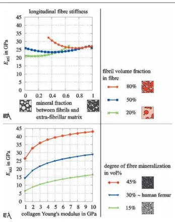
Die Ergebnisse zeigen nun: Nimmt man an, dass sich ca. 50–75 % des gesamten Minerals in den Fibrillen befindet, so ist der longitudinale Elastizitätsmodul nur gering vom Volumenanteil der Fibrillen im Verbund abhängig. Auch die Verteilung des Minerals zwischen Fibril-len und extrafibrillärer Matrix zeigt in diesem Bereich wenig Ein-fluss (Abb. 2a). Im Gegensatz dazu ist die Sensitivität der
Steifig-keit hoch für Veränderungen der Gewebemineralisierung und der Kollagensteifigkeit (Abb. 2b).
Betrachtet man die absoluten Steifigkeitswerte für den Fibrillenver-bund, zeigt sich, dass das Modell hier durchaus realistische Ergeb-nisse, beispielsweise für eine menschliche Knochenlamelle, liefert. Das Modell bestätigt den Mineralanteil als Hauptbestimmungsgröße für die Gewebesteifigkeit. Eine andere sensitive Bestimmungsgröße ist der Elastizitätsmodul von Kollagen. In der Literatur werden hierfür Werte zwischen 1–8 GPa angegeben, was – laut des vorliegenden Modells – Schwankungen der Gewebesteifigkeit von 100 % ergäbe. Andererseits scheint es egal zu sein, ob der Fibrillenverbund viele oder wenige Fibrillen enthält. Dieser Effekt stammt von den vergleichsweise ähnlichen Eigenschaften von Fibrillen und Matrix im Modell.
Es ist wichtig zu betonen, dass das verwendete Modell einige Ein-schränkungen besitzt: Es verwendet einen kontinuum-mechanischen Zugang für sehr kleine Volumen, dessen Eigenschaften nur mehr von wenigen Molekülbindungen bestimmt werden. Außerdem können einige Merkmale der realen Strukturen im Modell nicht abgebildet werden. Diese Einschränkungen verringern vermutlich die Genauig-keit des Modells bei der Voraussage der SteifigGenauig-keitseigenschaften
Abbildung 1: Reisinger AG et al. Methoden: (a) Schema eines Mori-Tanaka-Modells. Unidirektional ausgerichtete Spheriode sind in einer homogenen Matrix perfekt gebun-den. Die anisotropen elastischen Eigenschaften dieses Composites werden berechnet.
(b) Volumenelemente von Fibrillen, extrafibrillärer Matrix und Fibrillenverbund. Die Fi-brillen sind unidirektional in die extrafibrilläre Matrix eingebettet und ergeben mit dieser den Fibrillenverbund. Diese Strukturen werden mit der Mori-Tanaka-Methode modelliert.
J MINER STOFFWECHS 2009; 16 (Sonderheft 1)
9
ÖGEKM-Herbsttagung – Abstracts
von mineralisiertem Knochengewebe. Wir glauben, dass es zur Studie von kritischen Parametern dennoch geeignet ist.
Qualitative and Quantitative Differential Diagnosis of
Individuals With and Without Impaired Mineralisation
Exemplified by Historic and Contemporary Skeletal
Remains. Using the Radiological, Histological and
Histomorphometric Spectrum of Applications
D. Schamall1,*, C. Reiter2, J. Muhsil3, M. Teschler-Nicola1,4
1Department of Anthropology, Natural History Museum Vienna, Austria; 2Department of Forensic Medicine, Medical University of Vienna, Austria; 3Department of Education and Public Relation, Natural History Museum
Vienna, Austria; 4Department of Anthropology, University of Vienna,
Austria
Investigators on diseases with disturbed mineralisation of bony tis-sue are generally satisfied with radiological and light-microscopical techniques on decalcified thin-sections. The present study conducted innovative examination methods on human lumbar vertebral bodies (affected and non-affected by a known bone disease) to assess their methodological usability. Furthermore, it was intended to upgrade the hitherto existing access in gaining diagnoses of mineralisation insufficiencies in the human skeleton; moreover, a comparative mor-phological and morphometrical evaluation on cancellous bone tissue was carried out.
The analyses were accomplished on individuals with pathologic-ana-tomically verified alterations due to rickets respectively osteomalacia. From the historical collection of skeletons of the Federal Patholog-ic-Anatomical Museum (Vienna) 61 specimens with known age-at-death, sex, and pathological data were provided. Additionally, one individual from a recently deceased contemporary from the Depart-ment of Forensic Medicine of the Medical University (Vienna) was examined.
Bone structure and mineralisation were evaluated by so-called “non-invasive” and ““non-invasive” methods. As “non-“non-invasive” methods, con-ventional radiography and computed tomography (CT) were applied. Additionally, on a trial basis, quantitative CT and Dual-Energy X-Ray Absorptiometry (DEXA) were accomplished. Subsequently, a fraction of the convolute was scrutinised by “invasive methods” us-ing undecalcified stained thin-ground sections and microradiographs. Afterwards, some preparations were analysed in the “backscattered electron-mode” as well as in the “secondary electron-mode” in a scanning electron microscope (SEM). Finally, the established histo-morphometric parameters were measured from microradiographs. The applied techniques provided differential diagnoses, which were in most cases in accordance with the documented protocols among the museum specimens. The etiology of the contemporary individual could be allocated to renal insufficiency due to exceeding ingestion of phosphoric acid additions to soft drinks. Concluding, the poten-tial benefit of these techniques for clinical tasks and taphonomically altered (pre)historical skeletal remains is discussed.
Colles’ Fracture Load Can Be Accurately Predicted
by Finite Element Models of Ultra-Distal Sections
of the Radius
P. Varga1, D. H. Pahr1, S. Baumbach2, P. K. Zysset1
1Institute of Lightweight Design and Structural Biomechanics, Vienna
University of Technology, Austria; 2Medical University of Vienna, Austria
Occurring earlier that other osteoporotic fracture, distal radius fractures may be precursors to identify patients at risk early in time. High reso-lution peripheral quantitative computed tomography (HR-pQCT) provides in vivo access to the trabecular architecture of the peripheral skeleton like the wrist. Following successful in vitro validation studies, HR-pQCT based anatomy specific micro finite element (µFE) models may succeed to densitometry and morphological analysis in predicting fracture risk.
An experimental model of Colles’ fracture was developed and ap-plied on 21 embalmed radii in a previous study. The distal 1/3 of the
bones were cut, scanned using HR-pQCT and tested mechanically in compression until failure with well defined boundary conditions [1]. The goals of the present study were to investigate 1) if a distal section of the human radius provides an adequate model to estimate full bone strength, 2) if the standard HR-pQCT section of the distal radius is the best predictor and 3) if patient specific µFE models can better predict Colles’ fracture load than the density-based and mor-phological indices.
Two distinct 9 mm thick image sections of each radius were selected for analysis: one corresponding to the region of the clinical protocol and a second one, ultradistal, located adjacent to the subchondral endplate. Standard morphological indices were evaluated using the software of the HR-pQCT system. µFE models of the slices were built from the segmented HR-pQCT images and linear analyses of axial compression were performed. Ultimate load was estimated with a tissue strain-based criterion [2].
Exceptional prediction of the experimental fracture load was achieved using the µFE method (R2 = 0.96). Correlation between the
experi-mental strength and all the investigated measures was indeed higher for the most distal region compared to the standard section. As ex-pected, the µFE method provided a superior prediction of fracture load compared to morphological parameters such as trabecular BV/TV (R2 = 0.82) or cortical thickness (R2 = 0.26). Density-based measures
were closely, but not as good predictors as µFE (aBMD: R2 = 0.92,
BMC: R2 = 0.94).
These results underline the outstanding capabilities of the HR-pQCT-based µFE method and support the use of a 9 mm wrist section in the clinical evaluation of fracture risk. They also suggest the need for shifting the region of analysis distally, adjacent to the endplate.
References:
1. Varga et al. J Biomech 2009; 42: 1726–31. 2. Pistoia et al. Bone 2002; 30: 842–8.
Abbildung 2: Reisinger AG et al. Ergebnisse: Longitudinaler E-Modul des Fibrillen-verbundes (fibre), abhängig von (a) Fibrillenvolumenanteil im Fibrillenverbund und Mineralverteilung zwischen Fibrillen und extrafibrillärer Matrix, (b) Mineralvolumen-anteil im Fibrillenverbund und Kollagensteifigkeit.
a)
Vitamin D Deficiency, Hyperparathyroidism and
Bone Turnover in Patients Listed for Liver
Transplan-tation (LTX): Results of a Cross-Sectional Study
D. Wagner1, H. Dobnig2, D. Kniepeiss1, F. Iberer1, K. H. Tscheliessnigg1,
T. R. Pieber2, M. Trauner3, A. Fahrleitner-Pammer2
1Department of Transplantation Surgery, 2Endocrinology and
Nuclear-medicine, 3Department of Gastroenterology and Hepatology, Medical
University of Graz, Austria
Background Although transplantation bone disease is a common
complication following liver transplantation (LTX), screening and preventive measures during the pre-transplant period are not part of routine patient care. Aim of the current analysis was to evaluate bone metabolism of liver transplant candidates.
Methods Currently 60 patients (mean age 59 ± 7) with end stage
liver disease (mean MELD 13 ± 4) who are in evaluation for LTX were included into the present study. Blood sampling was performed in the morning following an overnight fast. Aside of routine param-eters 25-hydroxyvitamin D [vitD], parathyroid hormone [iPTH], bone
specific alkaline phosphatase [bALP], osteocalcin [OC], tartrate re-sistant alkaline phosphatase 5b [TRAP5b] and serum crosslaps [sCTX] levels were analyzed.
Results Mean VitD serum level of the patient cohort was 17.8 ±
11ng/ml. Only 14 % of the patients had levels within the normal range (30–65). Nearly two thirds (64.3 %) of the patients had levels below 20 ng/ml, indicative of a high prevalence of vitamin D defi-ciency. Serum VitD levels were negatively correlated to iPTH val-ues [r = –0.58; p = 0.001], and 54 % of the patients had secondary hyperparathyroidism (sHPT). Patients with sHPT had comparable kidney function and bone formation markers (OC, bALP) when com-pared to those without sHPT, however, had significantly higher sCTX (p = 0.01) and TRAP5b (p = 0.005) levels, indicating a negative bal-ance of bone turnover.
Conclusion Vitamin D deficiency and sHPT are common in patients
J MINER STOFFWECHS 2009; 16 (Sonderheft 1)
11
ÖGEKM-Herbsttagung – Abstracts