Prevalence of Microorganisms and
Immunoglobulins in Children with Tonsillar
Hypertrophy and Adenoiditis
Henrique Prestes Miramontes
1Djalma José Fagundes
2Julia Coelho Lima e Jurgielewicz
3Haroldo Prestes Miramontes Neto
4Renan Gianotto de Oliveira
3Gustavo Gianotto de Oliveira
3Maria Rosa Machado de Souza
51MD; Extension Course Student, Universidade Anhembi Morumbi
-UAM, São Paulo, SP, Brazil
2PhD; Universidade Federal de São Paulo - UNIFESP, São Paulo, SP,
Brazil
3MS, Universidade Anhembi Morumbi - UAM, São Paulo, SP, Brazil 4MS, Universdidade Cidade de São Paulo - UNICID, São Paulo, SP, Brazil 5PhD; Hospital Central de Guaianases, São Paulo, SP, Brazil;
Universidade Anhembi Morumbi - UAM, São Paulo, SP, Brazil
Int Arch Otorhinolaryngol 2014;18:311–315.
Address for correspondence Mr. Henrique Prestes Miramontes, Medical Sciences at the Universidade Anhembi Morumbi-UAM, Av. Prof. Olavo Avalone, 910, Colina das Estrelas, São Paulo, SP, Brazil (e-mail: [email protected]).
Introduction
Beginning in the second trimester, tonsils form in the fetus at various locations. Their main function is to produce lympho-cytes.1Tonsils are named based on their location: the palatine tonsils (located between the glossopalatine and pharyngo-palatine arches), the lingual tonsil (located at the posterior third of the tongue), the tubal tonsils (eustachian tubes), the
pharyngeal tonsil, and the adenoid (adjacent to the choanae and the pharyngeal ostium of the eustachian tubes). All tonsillar lymph nodes form Waldeyer’s ring, which represents 3 to 5% of the entire lymphatic system.2,3Excessive infection caused by microorganisms or the allergic process can lead to hyperactivity of the tonsils and pharynx, which can increase its volume, thereby hindering the passage of air to the
Keywords
►
child
►
tonsillectomy
►
immunoglobulins
►
immune system
Abstract
Introduction:
Benign idiopathic tonsillar hypertrophy (HBI) may affect a child
’
s quality
of life and sleep. Several studies have sought to relate the clinical features of HBI with the
infectious and/or immunologic changes that occur.
Objective:
To increase the knowledge of the etiology of HBI.
Data Synthesis:
From 2012 to 2013 we conducted a retrospective observational study
of 101 children with HBI who underwent tonsillectomies at Ambulatory ENT General
Hospital of the East Zone of São Paulo City, a region with a poor socioeconomic
population. Preoperative serologic results were available to con
fi
rm mononucleosis,
cytomegalovirus, anti-streptolysin O (ASLO) and immunoglobulins. The mean patient
age was 5.8 years (55% male, 45% female). Using the Mann-Whitney
U
test, we identi
fi
ed
signi
fi
cant gender differences in the parameters of immunoglobulins (Ig) M (IgM), IgA,
and IgE. Forty-seven percent of the patients had increased ASLO levels, and 37% had
increased IgE levels.
Conclusion:
An evaluation of a patient
’
s serologic parameters and laboratory results
may be relevant to the etiology and prevention of HBI. Based on the results obtained
from the study sample, the identi
fi
cation of etiologic agents and causative factors
remain a public health challenge that affects the quality of life of children.
received
May 13, 2013
accepted
October 14, 2013
published online
May 15, 2014
DOI http://dx.doi.org/ 10.1055/s-0033-1364174.
ISSN 1809-9777.
Copyright © 2014 by Thieme Publicações Ltda, Rio de Janeiro, Brazil
choanae.4The literature is controversial regarding the pro-cess of benign idiopathic tonsillar hypertrophy. Some authors relate this process to changes in the production of other immunoglobulins and to infections caused by nonspecific pathogens in the lymphoid tissue or mononucleosis, cyto-megalovirus (CMV), and toxoplasmosis. Identifying the mi-crobiota that colonize the tonsils has become increasingly important both for the connection with recurrent infections and the possible association with the hypertrophy of the tonsils and pharynx.5,6
Adenotonsillectomy may be necessary for patients with respiratory disorders caused by hypertrophy of the tonsils and adenoids; these patients show significant improve-ments in their oxygen saturation and quality of life after surgery.7,8In view of this evidence, we aimed to evaluate the relationship between tonsillar hypertrophy, adenoids, microorganisms, and immunoglobulins in patients with surgical indications.
Objectives
The primary objective of the study was to identify the relationship between tonsillar hypertrophy and adenoids using serologic indicators (i.e., mononucleosis, toxoplasmo-sis, CMV, anti-streptolysin O [ASLO] and serum immunoglo-bulins [Ig] A [IgA], IgG, IgM, and IgE) in children between ages 1 and 10 years who underwent or were indicated for tonsillectomy.
Gender differences in the levels of immunoglobulins were also investigated.
Materials and Methods
This retrospective, observational study was performed from July 2012 to April 2013 by reviewing the medical records of children between the ages of 1 and 10 years who were indicated for adenotonsillectomy surgery at the ear, nose, and throat clinic at General Hospital of the East Zone of São Paulo City (a poor socioeconomic region).
Inclusion and Exclusion Criteria
The following inclusion criteria were applied. Children were enrolled if they complained of upper airway tract obstruction, difficulty swallowing, mouth breathing, snoring, sleep apnea, and recurrent tonsillar infections in the area where the surgical treatment was indicated. All children with the symptoms described above underwent an endoscopic naso-pharynx examination and were included in the study if they showed tract obstruction of air50% because of tonsillar hypertrophy. At that time, samples were collected for labora-tory and serological tests. Patients with incomplete medical records were excluded from the study.
Laboratory and Serology Tests
According to the Ambulatory ENT General Hospital of the East Zone protocol, preoperative serology tests were requested for mononucleosis, toxoplasmosis, CMV, and serum immunoglo-bulins (i.e., ASLO, IgA, IgG, IgM, and IgE).
All tests were performed in a laboratory that was out-sourced by the hospital. Nephelometry was used to examine the serum immunoglobulin levels (i.e., IgA, IgG, IgM, and IgE); serology was used for toxoplasmosis; chemiluminescence was used for CMV; and passive hemagglutination was used for mononucleosis serology. The ASLO test was used as the parameter for the streptolysin O dosage.
Statistical Analysis
Data were tabulated and analyzed using STATA version 12.1 (Stata Corp.,Texas, USA). The variables related to age and immunoglobulin levels were analyzed using the mean and standard deviation, and categorical variables were deter-mined by absolute and relative frequencies (the Friedman test). The results were stratified by gender and compared using the Mann-Whitney U test to determine significant differences, andpvalues<0.05 were considered significant.
Results
Description Sample
This study evaluated 101 children between the ages of 1 and 10 years (55.4% male, 44.5% female) with tonsillar hypertro-phy who were undergoing adenotonsillectomy. The mean age was 5.8 ( 2.19) years. Of the participants, <5% were
between 1 and 3 years old and45% were between 5 and 7 years old (►Table 1).
Serum Test Results
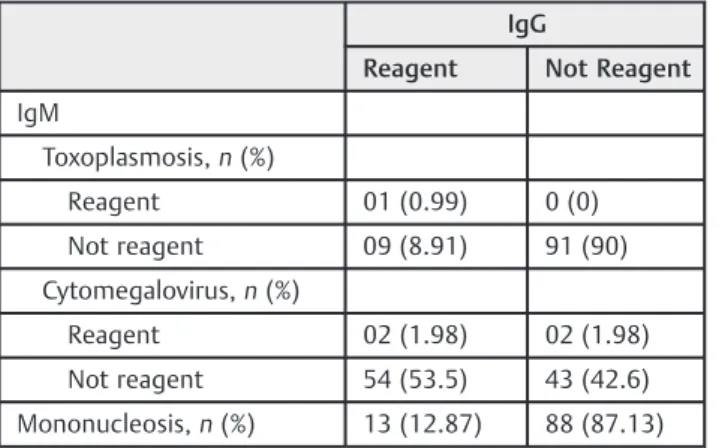
According to the preoperative serologic tests, 90 and 43% of the children had never had contact with toxoplasmosis and CMV, respectively. However, 54% of the children had immu-nity to CMV, indicating that at some point of their lives, they had contact with the agents. However, only 9% of the toxo-plasmosis cases showed immunity, although none had acute illness. As shown in ►Table 2, we did not observe acute
toxoplasmosis infections, but two children (1.98%) were seropositive for CMV.
Regarding mononucleosis, 13% of the children had re-agents, meaning that they had contact with the Epstein-Barr virus (EBV) or had active infections by the agent. The immunoglobulin levels were compared with the reference values and are displayed by the relative frequencies (►Table 3). Immunoglobulins play an important role in
detecting humoral immune response to infectious and aller-gic processes. Thus, we observed that33% of the patients had increased IgG levels, 30% had increased IgA levels, and
Table 1 Profiles of children undergoing adenotonsillectomy
n %
Gender
Male 56 55.4
Female 45 44.6
Age (meanSD) 5.822.19 Max: 10 and min: 1
37% had increased IgE levels. The mean and standard devia-tion for these immunoglobulins were 1162251, 14086, and 253186, respectively. Regarding IgM, only 3.96% of the patients had increased values, which had little correlation with tonsillar hypertrophy.
All patients showed symptoms, including complaints of recurrent respiratory infections or signs of sleep apnea, and there was at least one obstruction of the nasal pharynx (50%). Despite the increase in immunoglobulins that correlated with tonsillar hypertrophy, some patients did not show the same symptoms. When comparing the gender results, we found that the IgM values were significantly lower for boys. IgA and IgE values differed between genders also, which suggests a greater exposure to allergens in boys than girls (►Table 4).
ASLO is released by gram-positive bacteria that colonize the upper airway and is usually responsible for respiratory infections in children. Approximately 47% of the patients had increased ASLO levels.
Discussion
Tonsillectomy (with or without adenoidectomy) is the most common surgery among children. It is associated with im-proved quality of life and sleep, although its causes and its
effects on the immune system (e.g., reducing infections) are still controversial.9
As shown in this study, CMV and EBV agents have been cited in the literature as possible causes of tonsillar hypertro-phy. Using polymerase chain reaction, a study in Turkey noted the presence of some types of herpesviruses, including CMV, mononucleosis, and EBV, in children 2 to 9 years of age and a reported positivity of 25.2 to 11.3% for EBV and CMV among children with adenoid hypertrophy and adenoiditis.10
Another study conducted in China by Zhang and colleagues demonstrated a correlation between EBV and adenoid hyper-trophy; 51.9% of the children had EBV in tissues with adenoid and tonsillar hypertrophy, although it was absent in the
Table 2 Results of serologic tests for mononucleosis, IgG and IgM toxoplasmosis, and Cytomegalovirus
IgG
Reagent Not Reagent
IgM
Toxoplasmosis,n(%)
Reagent 01 (0.99) 0 (0)
Not reagent 09 (8.91) 91 (90)
Cytomegalovirus,n(%)
Reagent 02 (1.98) 02 (1.98)
Not reagent 54 (53.5) 43 (42.6)
Mononucleosis,n(%) 13 (12.87) 88 (87.13)
Abbreviation: Ig, immunoglobulin.
Table 3 Reference values for the assayed immunoglobulins
Reference values (mg/dL) n(%)
Serum dosage IgG
<420 01 (0.99)
420–1,240 67 (66.34)
>1,240 33 (32.67)
Serum dosage IgM
<50 01 (0.99)
50–300 96 (95.05)
>300 04 (3.96)
Serum dosage IgA
<18 04 (3.96)
18–160 67 (66.37)
>160 30 (29.7)
Serum dosage IgE
0.4 0 (0)
0.4–352 64 (63.37)
>352 37 (36.63)
Serum dosage ASLO
<250 54 (53.47)
>250 47 (46.53)
Abbreviation: ASLO, anti-streptolysin O; Ig, immunoglobulin.
Table 4 Gender comparison of the immunoglobulin and ASLO levels
Male (n¼56) Female (n¼45) 95% CI pa
Mean SD Mean SD
IgM 138.82 145.51 149.39 61.91 120–166 0.0235
IgG 1,161.34 244.47 1,162.29 263.33 1,112–1,211 0.6445
IgA 157.89 91.28 117.46 74.49 122–156 0.0072
IgE 609.23 729.3 204.14 282.15 308–548 0.0001
ASLO 268.12 163.12 234.41 212.05 216–289 0.1641
blood. This result aligns with our results, as our study only performed serology and did not investigate this agent in the tissue.11
Regarding the gender differences, Zhang et al found no statistical significance. These results differ from those in this study, which found that the values of IgM, IgA, and IgG differed between boys and girls.11 Other viral agents of epidemiologic importance were also cited in a cross-sectional study by Proença-Modena and colleagues, who reported high rates of common respiratory viruses (97.5% of children with chronic adenotonsillar disease and no history of acute respi-ratory symptoms).12 Human adenovirus was found more frequently (47%), followed by enterovirus (40%), rhinovirus (38%), bocavirus (30%), metapneumovirus (17%), and respira-tory syncytial virus (16%).12Our study should have consid-ered these viruses profiles; however, it has proven difficult to do so in daily practice.
Generally, in clinical practice, adenotonsillectomy reduces the incidences of respiratory infections and allergic obstruc-tion. The adverse impact of immune adenotonsillectomy surgery in children has been studied for years, but past reports have shown different postoperative immunoglobulin levels in the long and short term.9
IgG is the main immunoglobulin found in blood, corre-sponding to70 to 75% of the total immunoglobulin level. An IgG deficiency in children may be related to repeated respira-tory infections.13 In contrast, our findings suggest a 30% increase in IgG, which can be related to the organism response.
In their last publication of adenotonsillectomy, Santos and colleagues followed patients for 14 months over two visits. Thefirst follow-up visit occurred 1 to 2 months postsurgery, and the second occurred 12 to 14 months postsurgery. The immunoglobulin results were compared using the Friedman test (three groups), and they showed the preoperative value of IgG (p¼0.002) and the significant reduction of IgA (p¼0.026).9
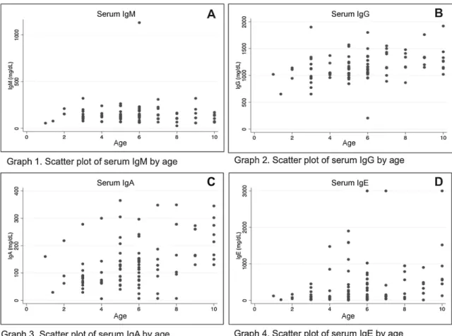
►Fig. 1A shows that the IgM values are low with little
variability; only one individual (age 6 years) had increased IgM values. We observed greater variability for IgG (►Fig. 1B),
with predominance among the benchmarks. Our findings, however, are similar to those found by Santos et al,9who reported normal levels in the preoperative and postoperative periods (1 to 2 months). However, after 12 months of follow-up, the IgA and IgG levels showed significant increases. The level of IgA immunoglobulin improved in 30% of the samples; its primary biological function is to protect against invading organisms, such as viruses and bacteria on mucosal surfaces, and inhibiting their adhesion to epithelial cells. IgA does not
fix complement; therefore, it can work against microorgan-isms without triggering the cascade of inflammation that damages the epithelial surfaces. However, it does not always protect against reinfection. It also promotes the decreased absorption of a variety of antigens or allergens (inhaled or ingested) that can trigger immune responses.14Note that we used the serum IgA and not the dosage salivate - salivary IgA levels. In a second step, it should be studied.
Note that the IgA (►Fig. 1.C) levels remained within the
parameters for all ages, and 30% of the patients showed increases. IgE (►Fig. 1D) appears to increase with age and
was increased by 37%. Griffin et al have reported a 20% increase in IgE in children undergoing adenotonsillectomy, but they found no evidence of symptom improvement after surgery, and their follow-up period was only 1 year.15
The etiology of tonsillar hypertrophy, as many studies have suggested, is related to the presence of infectious agents, such as viruses and bacteria that are commonly found in the microflora of the upper airway and in the humoral immune response to the body’s sensitivity to one or more allergens.
Identifying the main etiologic agents remains a public health challenge.
Conclusion
Based on our results, we found that there was no relationship between the positive serology for toxoplasmosis and tonsillar hypertrophy. However, we observed a positive relationship between the serology for CMV and tonsillar hypertrophy. Regarding mononucleosis, a low percentage (13%) of patients had positive serology.
Regarding ASLO, there was a 47% increase in the patients studied. Concerning the dosages of serum immunoglobulins in these patients, there was an increase in the serum levels of immunoglobulins IgA, IgE, and IgG but not an increase or decrease in IgM.
References
1 Huang SW, Giannoni C. The risk of adenoid hypertrophy in children with allergic rhinitis. Ann Allergy Asthma Immunol 2001;87(4):350–355
2 Marchesan IQ. Fundamentos em Fonoaudiologia: aspectos clínicos da motricidade oral. Rio de Janeiro: Guanabara Koogan; 1998: 23–36
3 Gross CW, Harrison SE. Tonsils and adenoids Pediatrics. Review 2000;21(3):75–78
4 Dalley AF, Moore KL. Anatomia Orientada para Clínica. 4a edição. Rio de Janeiro: Guanabara Koogan; 2001 pp. 851–867
5 Brodsky L, Moore L, Stanievich J. The role ofHaemophilus infl uen-zae in the pathogenesis of tonsillar hypertrophy in children. Laryngoscope 1988;98(10):1055–1060
6 Kielmovitch IH, Keleti G, Bluestone CD, Wald ER, Gonzalez C. Microbiology of obstructive tonsillar hypertrophy and recurrent tonsillitis. Arch Otolaryngol Head Neck Surg 1989;115(6): 721–724
7 Arrarte JL, Lubianca Neto JF, Fischer GB. The effect of adenotonsil-lectomy on oxygen saturation in children with sleep disordered breathing. J Bras Pneumol 2007;33(1):62–68
8 Soccol BB, Rocha RT, Henrique VP, Marchi RD. Avaliação do impacto da adenotonsilectomia sobre a qualidade de vida em crianças com hipertrofia das tonsilas palatinas e faríngeas. Rev Bras Otorrino-laringol 2009;75(1):64–69
9 Santos FP, Weber R, Fortes BC, Pignatari SS. Short and long term impact of adenotonsillectomy on the immune system. Braz J Otorhinolaryngol 2013;79(1):28–34
10 KarlıdağT, Bulut Y, KeleşE, et al. Presence of herpesviruses in adenoid tissues of children with adenoid hypertrophy and chronic adenoiditis. Kulak Burun Bogaz Ihtis Derg 2012;22(1):32–37 11 Zhang X, Li H, Liu X, et al. Study and analysis on the quantitative
detection of EBV-DNA in adenoidal hypertrophic and tonsillitis tissues of children. J Clin Otorhinolaryngol 2009;23(24):1108–11 12 Proença-Modena JL, Pereira Valera FC, Jacob MG, et al. High rates of detection of respiratory viruses in tonsillar tissues from children with chronic adenotonsillar disease. PLoS ONE 2012;7(8):e42136 13 Burtis CA, Ashwood ER. Textbook of Clinical Chemistry. 3 ed.
Philadelphia: Saunders; 1999
14 Rúpolo BS, Mira JGS, Kantor Junior O. Deficiência de IgA. J Pediatr (Rio J) 1998;74(6):433–440