ISABELLA SANTIAGO DE ABREU CARVALHO
EARLY SEX DISCRIMINATION IN Carica papaya L. BY
MOLECULAR CYTOGENETICS
Thesis presented to the
Universidade Federal de Viçosa, as part of the requirements of the Genetics and Breeding Graduate Program, for the attainment of the title Doctor Scientiae.
VIÇOSA
ii
To my dear parents, Messias and Cleonice, to my brother and friend
Guilherme, and to my beloved husband Daniel, for their unconditional love
and for supporting me all the time.
iii
ACKNOWLEDGEMENTS
Most of all, I thank God, the Lord of my life, for giving me great
strength and perseverance to keep going in hard times, and dear friends to
help me on this journey. To Him all honor and glory.
I thank Universidade Federal de Viçosa and the Genetics and
Breeding Graduate Program for providing me the opportunity to carry out and
conclude my doctorate, contributing to my academic and professional growth.
I also thank Coordenação de Aperfeiçoamento de Pessoal de Nível
Superior (CAPES) and Conselho Nacional de Desenvolvimento Científico e
Tecnológico (CNPq) for the financial support during my graduate study.
I am grateful to my advisor Prof. Dr. Carlos Roberto de Carvalho, for
all his scientific teachings, guidance and support over the past nine years of
my academic life.
I am very thankful to my family, who are all for me, my parents, my
brother and my husband, for their unconditional love and steady dedication to
me, and for supporting me whenever I need. In particular, to my husband
Daniel, my partner and best friend, for all his love, patience and
encouragement, especially in difficult times. I love them all.
I thank all my friends of the Laboratório de Citogenética e Citometria
Vegetal, Thaís, Guilherme, Fernanda, Paulo, Sirlei, Ana Paula, Denise,
Christiane, Andréa, Gabriella and Kassiana, for their friendship, pleasant
living together on work and for they have always cheered me up and valued
me. My special thanks to Fernanda for her great support in the conduction of
my experiments; without her help the execution of my research would have
been more difficult.
I thank to thesis committee members, Prof. Sérgio Motoike, Prof.
Wellington, Prof. Denise Bazzolli and Dr. Maria Andréia, for their attention to
this work, for spending their time on reading of it and for their suggestions.
I also thank to Caliman Agrícola S.A. and Instituto Capixaba de
Pesquisa, Assistência Técnica e Extensão Rural (Incaper), especially
Fabíola Lacerda, for providing me the plant material, essential to my
iv
BIOGRAPHY
ISABELLA SANTIAGO DE ABREU CARVALHO, daughter of Messias Moreira de Abreu Neto and Cleonice Carlos Santiago de Abreu, was
born in Timóteo, Minas Gerais, on May 12th, 1985.
In 2004, she started the college of Bachelor in Biological Sciences at
Universidade Federal de Viçosa (UFV), in Viçosa, MG, getting degree in
December, 2007. During graduation, she was subsidized with a scholarship
by PIBIC/CNPq of the Department of General Biology, when learned and
developed researches in plant cytogenetics and cytometry.
In March 2008, she joined the Graduate Program, in Master degree, in
Genetics and Breeding at UFV, subsidized by CAPES, submitting to the
defense of dissertation in February 2010. Over that time, she developed
researches in plant tissue culture, cytogenetics and cytometry. Besides, she
was awarded the Graduate Prize for best work in the area of Genetics,
Evolution and Improvement of Plants, presented at 54° Brazilian Congress of
Genetics, SBG.
In March 2010, she joined the Graduate Program, in Doctor degree, in
Genetics and Breeding at UFV, subsidized by CNPq, submitting to the
defense of thesis in June 2014. In that time, she learned and developed
v
SUMMARY
ABBREVIATION LIST ... vi
ABSTRACT ... vii
RESUMO ... ix
1. INTRODUCTION ... 1
2. LITERATURE REVIEW ... 4
2.1. General aspects of Carica papaya ... 4
2.2. Sex determination in papaya ... 5
2.3. Problems concerning papaya cultivation ... 12
2.4. Molecular markers for sex identification in papaya ... 13
3. MATERIALS AND METHODS ... 17
3.1. Plant material ... 17
3.2. Genomic DNA extraction ... 17
3.3. PCR amplification with SCAR primers ... 17
3.4. Selection of SCAR marker and probe labeling ... 19
3.5. FISH ... 19
4. RESULTS ... 21
4.1. PCR assays with SCAR markers ... 21
4.2. FISH ... 24
5. DISCUSSION ... 27
5.1. PCR assays with SCAR markers ... 27
5.2. FISH ... 29
6. CONCLUSIONS AND FUTURE PROSPECTS ... 31
vi
ABBREVIATION LIST
AFLP – amplified fragment length polymorphism
BAC – bacterial artificial chromosome
bp – base pairs
DAPI – 4’,6-diamidino-2-phenylindole
FISH – fluorescence in situ hybridization
FISHIS – fluorescence in situ hybridization in suspension
HSY – hermaphrodite-specific region of the Y chromosome
kb – kilobases
LG1 – linkage group 1
Mb – megabases
miRNA – micro RNA
MSY – male-specific region of the Y chromosome
MYA – million years ago
PCR – polymerase chain reaction
PRSV – papaya ringspot virus
RAPD – randomly amplified polymorphic DNA
SCAR – sequence characterized amplified region
sRNA – small RNA
SSC – saline-sodium citrate
vii
ABSTRACT
CARVALHO, Isabella Santiago de Abreu, D. Sc., Universidade Federal de Viçosa, June, 2014. Early sex discrimination in Carica papaya L. by molecular cytogenetics. Advisor: Carlos Roberto de Carvalho.
The papaya, Carica papaya L., is the most economically important species of
the family Caricaceae. Native of Central and South America, this herbaceous
and fruitful crop is cultivated mainly in tropical and subtropical regions
worldwide, and it is widely consumed for its edible fruit. C. papaya is
characterized as a polygamous species with three sex types: male, female
and hermaphrodite. Considering its preferred seminiferous propagation,
inherent problems of papaya crop refer to the segregation of sex types and
late sex detection. The sex identification is only possible after flowering, by
inspection of the flowers, since there is no recognized chromosomal
dimorphism or morphological difference between the three papaya sex types
in seedling stage. In order to save time, labor and financial resources, it is
desirable for growers that the sex type of this crop is known before
transplanting. Researchers in molecular biology have developed a number of
genetic markers in an attempt to distinguish the sex of papaya before
reaching reproductive maturity. In this study, it was investigated seven
sequence characterized amplified region (SCAR) markers previously
described in the literature, by the polymerase chain reaction (PCR)
technique, in two commercially important Brazilian varieties of C. papaya
(‘Golden’ and ‘Rubi’), and then, it was developed a fluorescence in situ
hybridization (FISH) protocol by using a chosen marker as probe. Thus, we
aimed to provide a molecular diagnosis of early sexing for the female and
hermaphrodite plants. Firstly, genomic DNA was isolated from female and
hermaphrodite young leaves of the both papaya varieties. After optimizing
PCR conditions, amplifications of the seven SCAR primers were carried out
in seven samples of each sex type from each variety. The fragments
obtained were analyzed by agarose-gel electrophoresis. Among the genetic
markers tested, three of them (NAPF-2, SDSP and SCARpm) generated
viii
papaya cultivars. Another SCAR marker (T12) produced a single sex-specific
fragment for only hermaphrodite plants from ‘Rubi’ cultivar. NAPF-2 marker
was selected to be used as FISH probe based on its consistently
polymorphic banding pattern. FISH analyses showed a single strong
fluorescent signal in ‘Golden’ hermaphrodite nuclei and many strong
fluorescent signals in ‘Rubi’ hermaphrodite nuclei, while no detectable or very
low intensity fluorescence signal was observed in female nuclei from both
cultivars. In conclusion, the present study investigated successfully, for the
first time, the discriminating potential of SCAR markers in two Brazilian
commercial papaya cultivars, and proposed a new, reliable, efficient and
relatively fast diagnostic method, based on FISH technique, for early
identification of hermaphrodite plants from cultivars ‘Golden’ and ‘Rubi’. In
future studies, this method could be improved for a large-scale screening in
commercial cultivation by flow cytometric approach after FISH in nuclei
ix
RESUMO
CARVALHO, Isabella Santiago de Abreu, D. Sc., Universidade Federal de Viçosa, junho, 2014. Early sex discrimination in Carica papaya L. by molecular cytogenetics. Orientador: Carlos Roberto de Carvalho.
O mamoeiro, Carica papaya L., é a espécie mais importante
economicamente da família Caricaceae. Nativa da América Central e do Sul,
esta espécie frutífera e herbácea é cultivada principalmente nas regiões
tropicais e subtropicais, e é amplamente consumida pelo seu fruto
comestível. C. papaya é caracterizada como uma espécie polígama com três
tipos sexuais: macho, fêmea e hermafrodita. Considerando que o mamão é
propagado preferencialmente via sementes, problemas inerentes desta
cultura se referem à segregação dos tipos sexuais e à detecção sexual
tardia. A identificação sexual só é possível após o florescimento, pela
inspeção das flores, uma vez que não há dimorfismo cromossômico
reconhecido nem diferenças morfológicas entre os três tipos sexuais do
mamoeiro no estágio de plântula. Com o objetivo de economizar tempo,
manejo e recursos financeiros, é desejável para os produtores que o tipo
sexual desta cultura seja conhecido antes do transplantio. Pesquisadores da
área de biologia molecular têm desenvolvido um grande número de
marcadores genéticos na tentativa de distinguir o sexo do mamão antes de
atingir a maturidade reprodutiva. Neste estudo, foram investigados sete
marcadores do tipo sequence characterized amplified region (SCAR)
previamente descritos na literatura, pela técnica de polymerase chain
reaction (PCR), em duas variedades brasileiras comercialmente importantes
de C. papaya (‘Golden’ e ‘Rubi’), e então, foi desenvolvido um protocolo de
fluorescence in situ hybridization (FISH), usando um marcador selecionado
como sonda. Dessa forma, objetivou-se prover um diagnóstico molecular de
sexagem precoce para plantas fêmeas e hermafroditas. Primeiramente, DNA
genômico foi isolado de folhas jovens coletadas desses dois tipos sexuais de
ambas as variedades de mamão. Após a otimização das condições de PCR,
amplificações dos sete primers SCAR foram realizadas em sete amostras de
x
por eletroforese em gel de agarose. Dentre os marcadores genéticos
testados, três deles (NAPF-2, SDSP e SCARpm) geraram fragmentos
sexo-específicos significativos em todas as amostras hermafroditas de ambos os
cultivares de mamão. Outro marcador SCAR (T12) produziu um único
fragmento sexo-específico para somente plantas hermafroditas do cultivar
‘Rubi’. O marcador NAPF-2 foi escolhido para ser usado como sonda FISH,
baseado em seu padrão de bandas consistentemente polimórfico. Análises
FISH mostraram um único e forte sinal fluorescente em núcleos de
hermafrodita do cv. ‘Golden’, e muitos e fortes sinais fluorescentes em
núcleos de hermafrodita do cv. ‘Rubi’, enquanto nenhum sinal fluorescente
detectável ou de baixíssima intensidade foi observado em núcleos de fêmea
de ambos os cultivares. Em conclusão, o presente trabalho investigou com
sucesso, pela primeira vez, o potencial discriminativo de marcadores SCAR
em dois cultivares comerciais brasileiros de mamão, e propôs um novo
método diagnóstico confiável, eficiente e relativamente rápido, baseado na
técnica de FISH, para a identificação precoce de plantas hermafroditas dos
cultivares ‘Golden’ e ‘Rubi’. Em estudos futuros, este método poderá ser
melhorado para um escaneamento em larga-escala na produção comercial
por meio de uma abordagem citométrica de fluxo após FISH em suspensão
1
1. INTRODUCTION
Carica papaya L. species is the most economically important fruit crop
of the family Caricaceae (BAJPAI & SINGH 2006, ZHANG et al. 2010,
SUDHA et al. 2013). It is cultivated worldwide mainly in tropical regions, for
its edible fruit (MING et al. 2007), and subtropical ones, for valuable papain
proteolytic enzyme production (PARASNIS et al. 2000, YU et al. 2009).
Papaya is referred to as the third major tropical fruit produced globally. Brazil
is the second largest papaya producer of the world, with a production of
about 1.5 million tonnes of fruits in 2012, according to FAOSTAT data. The
largest plantations of the main Brazilian cultivars, ‘Solo’ and ‘Formosa’, are
located in the states of Bahia, Espírito Santo and in other northeastern
regions of the country (IBGE 2012).
Papaya is a polygamous species with three sex forms, female, male
and hermaphrodite (YU et al. 2008, ZHANG et al. 2010), which are
differentiated by their inflorescence and fruit shape (MING et al. 2007). Due
to high productivity and pear-shaped fruits with lower ovarian cavity,
hermaphrodite plants are the major type for commercial cultivation in tropical
regions, including Brazil (MAGDALITA & MERCADO 2003, OLIVEIRA et al.
2007, YU et al. 2008, PINTO et al. 2013). Female plants are cultivated mainly
for papain production (PARASNIS et al. 1999, 2000, REDDY et al. 2012),
while males are useless for economic purposes (CHAVES-BEDOYA &
NUÑEZ 2007, URASAKI et al. 2002a, b).
Over the years, the intriguing sex determination system of C. papaya
has been subject of studies in evolutionary biology and molecular genetics
(LIU et al. 2004, MA et al. 2004, MING et al. 2007). Understanding the
molecular factors behind sex expression has great importance in both basic
and applied researches (GANGOPADHYAY et al. 2007). Since the 1930’s,
many hypotheses about the sex determination system in papaya have been
proposed: a single gene with three alleles (HOFMEYR 1938, STOREY
1938), a group of genes confined to a small region on the sex chromosome
(STOREY 1953), genic balance between sex chromosomes and autosomes
2
JIMÉNEZ 1967), a trans-regulatory element controlling flower organ
development (SONDUR et al. 1996). All these hypotheses are in agreement
on one aspect: there is a genetic factor that controls sex expression for male
and hermaphrodite plants, existing as heterozygous, and for females, as
homozygous.
More recent application of molecular techniques and biotechnology
has revolutionized the field of sex determination research in C. papaya.
Sex-linked DNA markers were developed by several research groups and linkage
maps of the papaya genome were constructed. Apart from these studies play
an important role in evolutionary biology researches, molecular markers and
methods have been widely used for identifying the papaya sex type at an
early developing stage to improve fruit production (MING et al. 2007). The
sex type identification is a limiting factor on cultivation of papaya, since there
are no distinguishing morphological features at the seedling stage or
heteromorphic sex chromosomes between the sex forms (PARASNIS et al.
1999, 2000, CHAVES-BEDOYA & NUÑEZ 2007, GANGOPADHYAY et al.
2007), and such identification is only possible after flowering (DEPUTY et al.
2002). The selection of the appropriate papaya sex type prior to planting in
the field is of great interest by growers, for reducing the production costs
(PARASNIS et al. 1999, SANTOS et al. 2003, COSTA et al. 2011, REDDY et
al. 2012).
A variety of molecular markers for sex types in papaya have been
developed: randomly amplified polymorphic DNA (RAPD) markers
(PARASNIS et al. 2000, LEMOS et al. 2002, URASAKI et al. 2002a,
CHAVES-BEDOYA & NUÑEZ 2007, OLIVEIRA et al. 2007, REDDY et al.
2012); sequence characterized amplified region (SCAR) markers, converted
from RAPD (PARASNIS et al. 2000, DEPUTY et al. 2002, URASAKI et al.
2002a, b, CHAVES-BEDOYA & NUÑEZ 2007); simple sequence repeat
(SSR) markers (PARASNIS et al. 1999, COSTA et al. 2011). These markers
were tested for the discrimination of sex type in different papaya cultivars
worldwide. However, the use of molecular markers in large-scale cultivation
is considered unfeasible, since this technology is expensive and intensive
3
Considering the economic importance in early sex identification of
papaya crop, the present study aimed to develop an alternative diagnostic
assay based on FISH technique, proposed to be reliable, efficient, less costly
and relatively faster than PCR approach, in order to be applied in large scale.
For this, we (i) investigated the discriminative potential of seven SCAR
markers previously described in the literature by PCR methodology, in two
commercially important Brazilian varieties of C. papaya, (ii) applied the PCR
methodology for construction of labeled probe from a selected marker, (iii)
developed a cytological protocol for obtaining nuclei suspension, used for
slide preparation, and (iv) applied FISH technique in these slides, aiming to
4
2. LITERATURE REVIEW
2.1. General aspects of Carica papaya
The common papaya, Carica papaya L., belongs to the small
dicotyledonous family Caricaceae, which comprises 35 species (including 32
dioecious, two trioecious and one monoecious species) spread over six
recently reclassified genera (VAN DROOGENBROECK et al. 2002, DREW
2003, MING et al. 2007, YU et al. 2008). Vasconcellea is the largest genus of
the family, comprising 21 species, followed by Jacaratia, Jarilla,
Cylicomorpha, Horovitzia, with seven, three, two and one species,
respectively. C. papaya is the only species of the genus Carica. These
herbaceous, shrubby or arborescent plants are native to Tropical America,
except the genus Cylicomorpha, which is originated from equatorial Africa
(BADILLO 1993).
C. papaya is by far the most economically important fruit crop of the
family Caricaceae. It is cultivated mainly in tropical and subtropical regions
worldwide (BAJPAI & SINGH 2006, ZHANG et al. 2010, SUDHA et al. 2013),
for its edible and nutritious fruit rich in vitamin A and C (CHANDRIKA et al.
2003, YU et al. 2008), as well as for its milky latex. This latter is the source of
papain, a commercially valuable proteolytic enzyme widely used in food
processing, medical applications, and textile, dairy, pharmaceutical and
cosmetic industries (PARASNIS et al. 1999, VAN DROOGENBROECK et al.
2002, MING et al. 2007, 2008, YU et al. 2009). According to FAOSTAT
(2014), papaya is referred to as the third major tropical fruit produced
globally. Global papaya production has grown significantly over the years,
mainly as a result of increased production in India, corresponding to 41.6% of
the world production during 2012. Brazil has been the second largest papaya
producer (12.2%), followed by Indonesia, Dominican Republic, Nigeria and
Mexico, and the second exporter in the world. In 2012, Brazil produced about
1.5 million tonnes of papaya fruits. The largest plantations of the main
5
(45.03%), Espírito Santo (31.93%) and in other northeastern regions of the
country (IBGE 2012).
Papaya is among the limited number of plant species that are
trioecious with three sex forms – female, male and hermaphrodite (YU et al.
2008, ZHANG et al. 2010). Male trees are characterized by long, pendulous,
many-flowered inflorescences bearing slender male flowers lacking a pistil,
except for occasional pistil-bearing flowers at the distal terminus. Female
trees have short inflorescences with few flowers bearing large functional
pistils without stamens. Hermaphroditic trees have short inflorescences
bearing bisexual flowers that can be sexually variable (MING et al. 2007).
Mainly in tropical regions as in Brazil, hermaphrodites are the major type for
fruit commercial cultivation because of their higher productivity (MAGDALITA
& MERCADO 2003, MING et al. 2007) and pyriform-shaped fruits (MA et al.
2004, YU et al. 2008, BLAS et al. 2012) with lower ovarian cavity (LEMOS et
al. 2002, PINTO et al. 2013). Conversely, female plants are preferred in
subtropical areas for their greater production under cool winter temperatures
(YU et al. 2009). Female flowers are stable at low temperature while
hermaphrodite flowers tend to fuse anthers to the carpels and produce
deformed carpellodic fruit (MING et al. 2007). Besides, females are chosen
for papain extraction, whose proteolytic activity and quantity obtained from
these dioecious fruits are superior to the hermaphrodite ones (PARASNIS et
al. 1999, 2000, REDDY et al. 2012). Male plants are useless for economic
purposes (CHAVES-BEDOYA & NUÑEZ 2007, URASAKI et al. 2002a, b).
2.2. Sex determination in papaya
Since sex represents a huge problem in evolutionary biology,
understanding the molecular factor(s) behind sex expression has immense
importance in both basic and applied researches (GANGOPADHYAY et al.
2007). The polygamous angiosperm species C. papaya (2n = 18), which has
male, female and hermaphroditic forms, offers several advantages for
genetic and evolutionary studies (LIU et al. 2004, MING et al. 2008, WEI &
6
relatively small genome of 372 megabases (Mb) (ARUMUGANATHAN &
EARLE 1991) and 2C = 0.65 pg (ARAÚJO et al. 2010), diploid inheritance
with nine pairs of chromosomes, a short generation time of 9 – 15 months,
continuous flowering of various flower types throughout the year, a unique
evolutionary process in female flowers, an intriguing primitive system of sex
determination and a well-established transformation system (LIU et al. 2004,
MING et al. 2008). Based on the knowledge and information available at the
time, sex determination in papaya has been a frequent subject of genetic
analyses (HOFMEYR 1938, 1967, STOREY 1938, HOROVITZ & JIMÉNEZ
1967, SONDUR et al. 1996, PARASNIS et al. 1999, LIU et al. 2004, MA et al.
2004, MING et al. 2007), because it is directly related to efficient commercial
fruit production (SILVA et al. 2007). Thus, various hypotheses about the sex
determination system in this species have been postulated over the years.
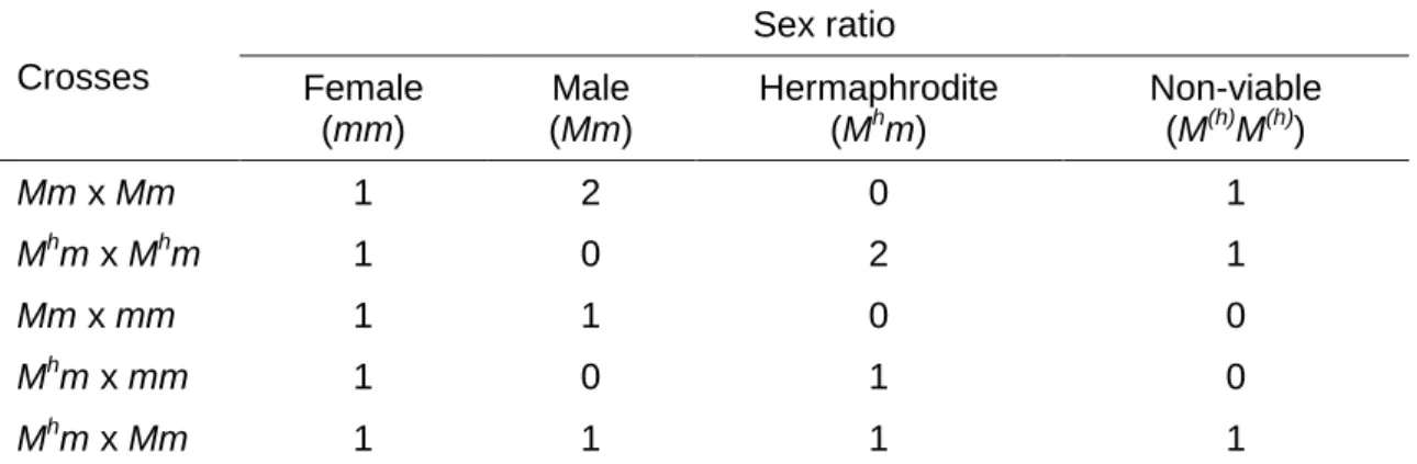
On the basis of segregation ratios from crosses among the three sex
types, STOREY (1938) and HOFMEYR (1938) each proposed originally that
sex determination in papaya is controlled by a single gene with three alleles:
M (or M1), Mh (or M2) and m. Male (Mm) and hermaphrodite (Mhm) individuals
are heterozygous, whereas females (mm) are homozygous recessive. The
dominant combinations of MM, MhMh and MMh are embryonic lethal, resulting
in a segregation ratio of 2:1 hermaphrodite to female from self-pollinated
hermaphrodite trees, and a 1:1 segregation of male to female, or
hermaphrodite to female, from cross-pollinated female trees by male’s pollen
or hermaphrodite’s pollen, respectively. When occasionally male trees are
selfed, it occurs a segregation ratio of 2:1 male to female, or if male’s pollen
fertilizes hermaphrodite’s pistil, a progeny of 1 male:1 hermaphrodite:1
female is generated (Table 1).
Later, STOREY (1953) revised the hypothesis to suggest that papaya
sex is regulated not by a gene, but rather by a complex of genes confined to
a small region on the sex chromosome within which recombination is
precluded. The different segments in this region are so closely linked
together that they behave as inherited unit factors. In addition to genes
differing in functionality of the androecium and gynoecium, this sex locus also
contains genes controlling several co-segregating secondary sexual
7
Table 1. Segregation ratio from crosses between the three sex forms in C. papaya
Crosses Sex ratio Female (mm) Male (Mm) Hermaphrodite (Mhm)
Non-viable (M(h)M(h))
Mm x Mm 1 2 0 1
Mhm x Mhm 1 0 2 1
Mm x mm 1 1 0 0
Mhm x mm 1 0 1 0
Mhm x Mm 1 1 1 1
Font: Storey (1938) and Hofmeyr (1938). Obs.: The Storey’s designation for genotypes was adopted considering its convenience to separate the hermaphrodite allele Mh from male allele M.
peduncle length), cross-over suppression (C) and lethality (L). Based on
Storey’s model, the genotypes of male, hermaphrodite and female were
given as in Table 2.
Table 2. Genotypes of the three sexes of papaya according to the Storey’s model
Sex type Genotype
Male Mp l C + sg / + + + sa +
Hermaphrodite + l C + sg / + + + sa +
Female + + + sa + / + + + sa +
Mp – long peduncles of male flowers, l – zygotic lethal factor, C – hypothetical factor for suppression of recombination at the sex determination region, sa – supressor of the androecium, sg – supressor of the gynoecium. Font: Storey (1953).
The gene sa controls the suppression of stamen development when it
is recessive homozygous as in female trees. The gene sg controls the
suppression of carpel development when it is recessive homozygous. The
author suggested that most male and hermaphrodite trees were
heterozygous for this second gene, because of sex reversal between
hermaphrodite and male flowers, influenced by environmental changes, often
observed in the field.
Alternatively, HOFMEYR (1967) published the genic balance theory
for papaya sex determination, aiming to explain the zygotic lethality in the
dominant homozygous condition and the differential sexual stability among
the three sexes. It was assumed that M1 (for males) and M2 (for
8
of slightly different lengths from which vital genes are missing. Therefore, any
combinations of M1M1, M1M2 or M2M2 would be lethal, while M1m and M2m
would be viable because an m “sex chromosome” is present in each
genotype. Because the regions of masculinity (M1 and M2) have slightly
different lengths, the sex types of papaya are the results of genic balance
between the sex chromosomes and autosomes.
Based on intergeneric hybridizations between Carica and
Vasconcellea species, HOROVITZ & JIMÉNEZ (1967) proposed a sex
determination system in C. papaya of XX – XY chromosomal type, even
though heteromorphic chromosomes have not been found in this species
(PARASNIS et al. 1999, URASAKI et al. 2002a, LIU et al. 2004,
CHAVES-BEDOYA & NUÑEZ 2007, YU et al. 2008, JUNIOR et al. 2010). Their
hypothesis is similar to that suggested by STOREY (1938) and HOFMEYR
(1938), by employing another terminology, namely: XX, XY and XY2 for female, male and hermaphrodite genotypes, respectively, where Y2 is the modified Y chromosome. As the combinations of Y(2) chromosomes are lethal, these researchers suggested that both chromosomes share the same
lethal factor.
An additional hypothesis to explain the genetic basis of sex
inheritance in C. papaya includes regulatory elements of the flower
development pathway (SONDUR et al. 1996). In this model, the dominant
male allele, designated SEX1-M, encodes a trans-acting regulatory factor
that induces male floral parts while suppressing carpel development. The
dominant hermaphrodite allele, SEX1-H, is considered intermediate with the
ability to give rise to stamens, but only reduces carpel size rather than
inhibiting it. The recessive female allele, sex1-f, was hypothesized as
incapable of promoting male structures. Functional carpels would develop in
heterozygous SEX1-H/sex1-f plants. The lethality of SEX1-M or SEX1-H
homozygotes could result from an additional required function present only in
the sex1-f allele. The variability observed in the secondary sexual
characteristics could result from environmental or allelic interaction effects on
the expression / function of SEX1. For instance, it is possible that the
interaction between SEX1-H and its target, either a promoter sequence or
9
SEX1-M and the target. This could account for the difference in carpel size
between males and hermaphrodite flowers, and for the sex reversal
sometimes seen in hermaphrodites but which is rare in males.
All the hypotheses mentioned above share the same foundation: a
genetic factor controlling sex expression for male and hermaphrodite that
exists as heterozygous, while the female’s is homozygous. Over the following
years, advances in genomic technology made it possible a lot of studies
could be performed for characterization of the genomic region involved in
papaya sex determination at the molecular level. Sex-linked DNA markers
were developed by several research groups and linkage maps of the papaya
genome were constructed (MING et al. 2007).
The first genetic linkage map of papaya using DNA molecular markers
was constructed by SONDUR et al. (1996). This map consisted of 61 RAPD
markers, of which the SEX1 flower sex determinant locus was mapped on
linkage group 1 (LG1), where it was flanked by two markers at 7 cM on each
side. Nevertheless, this low density map provided no clue whether there was
suppression of recombination at that locus (MING et al. 2007). Since then,
different DNA markers flanking the SEX1 locus have been developed,
increasing the understanding of the genetic structure of this region:
microsatellite (PARASNIS et al. 1999) markers, RAPD (LEMOS et al. 2002)
markers, and SCAR markers, converted from RAPD (DEPUTY et al. 2002,
PARASNIS et al. 2000, URASAKI et al. 2002a). However, these authors did
not report detailed data on linkage.
High-density genetic linkage mapping of the papaya genome has
revealed severe suppression of recombination around the sex determination
locus (MA et al. 2004), thus validating the STOREY’s (1953) hypothesis that
the region containing the gene complex of sex determination behaves as a
unique factor so that there is no crossing over inside. MA et al. (2004)
analyzed 1501 amplified fragment length polymorphism (AFLP) and
morphological markers, of which 225 co-segregated with sex types and were
included on linkage group 1 (corresponding to the LG1 of the RAPD map by
Sondur and co-workers). These data showed an extremely high level of DNA
10
Concurrently, a physical mapping and sample sequencing of bacterial
artificial chromosomes (BACs) in the sex determination region also showed
severe suppression of recombination and extensive divergence between
homologues, in addition to DNA sequence degeneration, nucleotide
insertions, deletions and substitutions. These findings provided evidence that
sex determination in papaya would be controlled by a pair of incipient sex
chromosomes, with a small male-specific region (MSY) that accounts for only
about 10% of the Y chromosome. It was also found that hermaphrodite and
male plants share nearly identical DNA sequences in the MSY region,
suggesting that the Y and Yh chromosomes might have originated from the same ancestral chromosome. The incipient sex chromosomes of papaya
may yield insights about earlier stages of sex chromosome evolution in plants
(LIU et al. 2004).
More recently, other research groups also constructed physical maps
for the hermaphrodite-specific Yh chromosome region (HSY) and its X counterpart (NA et al. 2012), and for MSY region as well (GSCHWEND et al.
2011), using BAC libraries. These mappings provide the foundation for
sequencing the sex specific regions of papaya. The quality of genome
information is enhanced, however, when complementary resources of each
map are integrated. In this sense, YU et al. (2009) integrated a BAC-based
physical map of papaya with high-density genetic map and genome
sequence. The integrated map allowed identifying recombination hotspots,
regions suppressed for recombination and estimating physical distances
between genetic markers.
Cytological features of papaya chromosomes have been also linked
with the existing genome sequence or with the genetic and physical maps,
improving the knowledge of papaya genome organization and evolution at
the chromosomal level (WAI et al. 2010). By physically allocating
MSY-specific BACs on hermaphrodite chromosome Y (Yh) and, thereafter, sequencing some of them, YU et al. (2007) placed the MSY near the
centromere of the chromosome Yh and observed extreme gene paucity, high density of retroelements and local sequence duplications in that region. WAI
et al. (2010, 2012) and ZHANG et al. (2010) integrated chromosomal traits
11
molecular markers-tagged BACs directly on papaya chromosomes by
fluorescence in situ hybridization (FISH). Representative BAC clones of all 12
linkage groups derived from genetic maps were reassigned to the nine
chromosomes of papaya, corresponding to its haploid number (WAI et al.
2010, ZHANG et al. 2010). WAI et al. (2012), in turn, constructed a molecular
cytogenetic map of the papaya sex chromosome (chromosome 1).
WANG et al. (2012) sequenced the HSY region, and its X counterpart,
yielding an 8.1 Mb HSY pseudo molecule, and a 3.5 Mb sequence for the
corresponding X region. In contrast to the Y chromosome in other organisms,
Y chromosomes in Caricaceae have higher genetic diversity than the X
chromosomes (WEINGARTNER & MOORE 2012). Sequence divergence
between HSY and X regions and the expansion of the HSY are mostly due to
retrotransposon insertions, inversions, recombination suppression and
numerous additional chromosomal rearrangements (WANG et al. 2012).
These authors inferred the oldest rearrangement event in papaya sex
chromosomes occurred about 7 million years ago (MYA) and, the most
recent, only 1.9 MYA. These data support theoretical models of early sex
chromosome evolution in papaya.
An alternative approach involving analyses of small RNA (sRNA)
libraries was explored by ARYAL et al. (2014), once it is known that sRNA
plays an important role in DNA methylation and gene silencing, suggesting its
participation in sex differentiation in plants. A total of 14 micro RNAs (miRNA)
were differentially expressed among male, female, and hermaphrodite
flowers, such as miR169 that regulates the genes in auxin signaling pathway
and, hence, in carpel development. The results indicate potential function of
these sRNAs in papaya sex determination.
All the studies mentioned above clarify the understanding of possible
evolutionary mechanisms that drive the divergence of the papaya sex
chromosomes, which will be better accessed by further researches
(WEINGARTNER & MOORE 2012, ARYAL & MING 2014). Concerning
commercial applications, knowing more about the sex determination gene
may eventually lead to engineering a true breeding hermaphrodite cultivar to
improve papaya fruit production (GSCHWEND et al. 2011, ARYAL & MING
12
2.3. Problems concerning papaya cultivation
The commercial cultivation of C. papaya is conducted conventionally
via seeds derived from open pollination (DREW 1987, PARASNIS et al.
1999, BHATTACHARYA & KHUSPE 2001, CHAVES-BEDOYA & NUÑEZ
2007). Although vegetative propagation methods, such as cuttings, grafting
and micropropagation in tissue culture, are available (MING et al. 2007), they
are laborious and expensive (MAGDALITA & MERCADO 2003, SAKER et al.
1999, REDDY et al. 2012). Being an alogamous species, the inherent
heterozygosity of papaya crop results in a mixture of genotypes with
considerable variation regarding yield, fruit quality and susceptibility to
various diseases (SAKER et al. 1999). Its most important pathogen is papaya
ringspot virus (PRSV), a devastating disease that has a detrimental impact
on both commercial papaya production and Caricaceae germplasm
conservation (MATSUMOTO et al. 2010). The introduction of the commercial
virus-resistant transgenic variety ‘SunUp’, in 1998, saved the Hawaii papaya
industry from collapse in the mid-1990s (MING et al. 2007, WEI & WING
2008, REDDY et al. 2012). Because PRSV is widespread in nearly all
papaya-growing regions, ‘SunUp’ could serve as a transgenic germplasm
source to be used in breeding of suitable cultivars resistant to the virus in
various parts of the world (MING et al. 2008).
Considering the trioecious nature of C. papaya, with male,
hermaphrodite and female forms, the sex type identification is another
limiting factor on cultivation of this crop. The lack of clearly distinct
morphological features at the seedling stage and heteromorphic sex
chromosomes have hampered the early sexing based on morphology or
cytology (MAGDALITA & MERCADO 2003, PARASNIS et al. 1999, 2000,
CHAVES-BEDOYA & NUÑEZ 2007, GANGOPADHYAY et al. 2007).
Therefore, the sex of papaya plants is uncovered only after flowering
(DEPUTY et al. 2002) which may take 5 – 8 months (PARASNIS et al. 1999,
2000, MA et al. 2004). Once hermaphrodite fruits are commercially preferred
over that of females, and hermaphrodite plants segregate usually into
13
are commonly planted in one hill, followed by uprooting of undesired female
trees, in order to guarantee a number of hermaphrodites evenly distributed in
the field. Such practice raises the production costs, by wastage of cultivation
area, labor, time and other resources (PARASNIS et al. 1999, LEMOS et al.
2002, URASAKI et al. 2002a, SANTOS et al. 2003, MA et al. 2004, COSTA
et al. 2011).
The selection of the appropriate papaya sex type at the juvenile forms,
prior to transplanting, would be beneficial especially for producers
(MAGDALITA & MERCADO 2003, PARASNIS et al. 1999,
CHAVES-BEDOYA & NUÑEZ 2007, REDDY et al. 2012). In order to overcome these
constraints in cultivation practice and to make it more profitable, there has
been a long-standing interest in developing strategies for identifying the
papaya sexes at the vegetative stage (PARASNIS et al. 2000). Molecular
markers for sex types in papaya that could be generated through DNA
analysis using polymerase chain reaction (PCR) technology have been seen
as a reliable strategy (MAGDALITA & MERCADO 2003).
2.4. Molecular markers for sex identification in papaya
Researches in molecular biology have led to the development of a variety of
DNA markers linked to the sex determination locus in papaya (MAGDALITA
& MERCADO 2003, PARASNIS et al. 1999, 2000, DEPUTY et al. 2002,
LEMOS et al. 2002, URASAKI et al. 2002a, b, CHAVES-BEDOYA & NUÑEZ
2007, OLIVEIRA et al. 2007, COSTA et al. 2011, REDDY et al. 2012) (Table
3), as a means to sex the plants prior to flowering. The advantages of DNA
markers include their abundance and stability, a relatively high rate of
polymorphism in many populations, usually routine technologies for scoring,
frequent comparability across species, clear dominance or codominance in
most situations, presence at all stages and tissue types of plant growth, lack
of epistasis and no detectable phenotypic effect (MA 2003,
1
4
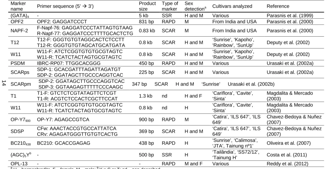
Table 3. DNA molecular markers used for predicting the sex type in C. papaya Marker
name Primer sequence (5’ 3’)
Product size
Type of marker
Sex
detectiona Cultivars analyzed Reference
(GATA)4 - 5 kb SSR H and M Various Parasnis et al. (1999)
OPF2 OPF2: GAGGATCCCT 831 bp RAPD M From India and USA Parasnis et al. (2000)
NAPF-2 F-Napf-76: GAGGATCCCTATTAGTGTAAG
R-Napf-77: GAGGATCCCTTTTGCACTCTG 0.83 kb SCAR M From India and USA Parasnis et al. (2000)
T12 T12-F: GGGTGTGTAGGCACTCTCCTT
T12-R: GGGTGTGTAGCATGCATGATA 0.8 kb SCAR H and M
‘Sunrise’, ‘Kapoho’,
‘Rainbow’, ‘SunUp’ Deputy et al. (2002)
W11 W11-F: ATCTCGGTGTGTGCGTAGTC
W11-R: TCATCTACTAGTGCGTAGTC 0.8 kb SCAR H and M
‘Sunrise’, ‘Kapoho’,
‘Rainbow’, ‘SunUp’ Deputy et al. (2002)
PSDM IBRC-RP07: TTGGCACGGG 450 bp RAPD H and M Various Urasaki et al. (2002a)
SCARps SDP-1: GCACGATTTAGATTAGATGT
SDP-2: GGATAGCTTGCCCAGGTCAC 225 bp SCAR H and M Various Urasaki et al. (2002a) SCARpm SDP-2: GGATAGCTTGCCCAGGTCAC
SDP-3: GGTAAGAGTTTTTCCCAAGC 347 bp SCAR H and M ‘Sunrise’ Urasaki et al. (2002b)
T1 T1-F: GTCTCTCGTATAGTTCTCGT
T1-R: ACGTCTCCACTCGCTTCCAT 1.3 kb nd H and F
‘Cariflora’, ‘Cavite’, ‘Sinta’
Magdalita & Mercado (2003)
W11 W11-F: ATCTCGGTGTGTGCGTAGTC
W11-R: TCATCTACTAGTGCGTAGTC 0.8 kb nd H
‘Cariflora’, ‘Cavite’, ‘Sinta’
Magdalita & Mercado (2003)
OP-Y7900 OP-Y7: AGAGCCGTCA 900 bp RAPD M
‘Catira’, ‘ILS 647’, ‘ILS 649’
Chavez-Bedoya & Nuñez (2007)
SDSP CFw: AAACTACCGTGCCATTATCA
CRv: AGAGATGGGTTGTGTCACTG 369 bp SCAR H and M
‘Catira’, ‘ILS 647’, ‘ILS 649’
Chavez-Bedoya & Nuñez (2007)
BC210438 BC210: GCACCGAGAG 438 bp RAPD H
‘Sunrise’, ‘Calimosa’,
‘JTA’, Tainung nº1’ Oliveira et al. (2007)
(AGC)5Yb - 500 bp SSR H
‘Tailândia’, ‘SS72/12’,
‘Tainung H’ Costa et al. (2011)
OPL-13 - - RAPD M and F Various Reddy et al. (2012)
a
15
In predicting the sex type of papaya, MAGDALITA & MERCADO
(2003) used two 20mer primer pairs, whose sequences were provided by
University of Hawaii, in the PCR amplification. Hermaphrodites were
distinguished by having two distinct bands (1.3 and 0.8 kilobases – Kb),
females had a single band (0.8 Kb), while males had no band. The noted
frequency of females, hermaphrodites and males, identified as such both by
PCR and field observation, in three varieties (‘Cavite’, ‘Cariflora’ and ‘Sinta’
hybrid) showed 100% accuracy in the prediction.
LEMOS et al. (2002) carried out RAPD assays to differentiate between
the sexual forms of three commercial C. papaya cultivars belonging to the
‘Solo’ group, grown in Brazil. From 152 RAPD primers tested, one (BC210)
showed polymorphic banding pattern between hermaphrodite and female
samples of all three cultivars. The efficiency of the BC210 molecular marker
in detecting sex differentiation early in the development of papaya seedlings
(one-month old) was evaluated. The amplification of sex-specific fragment
was accurately detected in only hermaphrodite samples. The same BC210
marker was successfully tested in other Brazilian commercial genotypes, two
varieties of the ‘Solo’ group and two hybrids of the ‘Formosa’ group
(OLIVEIRA et al. 2007). In a different approach, REDDY et al. (2012)
employed a RAPD marker to confirm the early identification of male and
female sex types based on their leaf morphology and rate of growth.
PARASNIS et al. (2000), DEPUTY et al. (2002), URASAKI et al.
(2002a, b) and CHAVES-BEDOYA & NUÑEZ (2007) selected sex-specific
RAPD markers and converted them into more reliable SCAR markers, by
cloning and sequencing the fragments and, then, designing longer primers. A
PCR- based sex diagnostic assay using the SCAR marker (NAPF-2)
developed by PARASNIS et al. (2000) revealed reliability of >90% on sex
identification of juvenile papaya plants. These researchers inferred that the
errors detected were mainly due to variations in concentration of the
extracted DNA. A greater accuracy (99.2%) was achieved when using two
other SCAR markers to discriminate hermaphrodite and male from female
plants at seedling stage (DEPUTY et al. 2002). URASAKI et al. (2002a)
developed a SCAR marker (SCARps) from the RAPD marker PSDM, and
16
plants. PSDM marker was further tested on early sexual detection and the
results were confirmed with 100% accuracy. Another SCAR marker,
designed SCARpm, was used in a multiplex-PCR assay by the same
researchers’ group (URASAKI et al. 2002b).
CHAVES-BEDOYA & NUÑEZ (2007) reported an interesting finding.
After screening 32 RAPD primers, one was amplified in male but not in
female and hermaphrodite samples. This marker was converted into SCAR,
which, in turn, amplified a 369-base pairs (bp) fragment from both male and
hermaphrodite but not from female plants. This fact could be explained as an
expected result of the higher specificity of SCAR marker since its primers are
twice longer than the RAPD primer.
In contrast to most, PARASNIS et al. (1999) employed highly
informative microsatellite and minisatellite probes to identify sex-specific
differences in papaya. Among these, only the microsatellite probe (GATA)4 demonstrated to be sex-specific in all the cultivars analyzed. Then, the
diagnostic potential of this specific marker was exploited to sex papaya
plants at the seedling stage (2-months old), generating unequivocal results.
COSTA et al. (2011) have also demonstrated the feasibility of SSR markers
on early molecular sexing in a variety of accessions belong to a Brazilian
germplasm collection. Only one marker in three papaya genotypes proved to
be discriminative.
Although molecular markers could be valuable tool for researchers to
employ in sex determining of experimental material, some authors contest its
practicability in routine sex testing by farmers, since that is an expensive and
17
3. MATERIALS AND METHODS
3.1. Plant material
Two commercially important Brazilian C. papaya cultivars, grown in
the state of Espírito Santo, were analyzed: cv. ‘Golden’ of ‘Solo’ group and
cv. ‘Rubi’ of ‘Formosa’ group, kindly provided by Caliman Agrícola S.A. and
Instituto Capixaba de Pesquisa, Assistência Técnica e Extensão Rural
(Incaper), respectively.
Leaf samples from seven female and seven hermaphrodite mature
plants of each cultivar were harvested. Male samples were not used in our
experiments since farmers do not cultivate them for commercial purposes.
Following collection, the leaf tissues were frozen in liquid nitrogen and stored
at –80°C till further use in both PCR and FISH techniques.
All the procedures were carried out at the Laboratório de Citogenética
e Citometria Vegetal, of the Departamento de Biologia Geral, Universidade
Federal de Viçosa (UFV).
3.2. Genomic DNA extraction
Genomic DNA of the samples was extracted by using GenEluteTM Plant Genomic DNA Miniprep kit (Sigma®). DNA concentration and purity were determined spectrophotometrically, and its integrity was further checked
by agarose-gel electrophoresis.
3.3. PCR amplification with SCAR primers
Seven SCAR markers from previous studies were investigated
regarding the sex discrimination potential in those above papaya cultivars, by
PCR assays. The papain gene marker was used as positive control of PCR
18
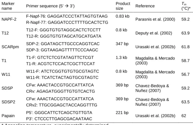
Firstly, annealing temperatures for each primer pair and PCR
conditions were optimized. Some information about the molecular markers
used is summarized in Table 4. Each PCR mixture consisted of 100 ng
genomic DNA, 0.2 mM dNTPs, 2% dimethyl sulfoxide, 2 U AccuTaqTM LA DNA Polymerase (Sigma®), 1X Polymerase buffer, 0.4 µM of each primer (forward and reverse), in 20 µL-final volume. Amplification reactions were
performed on termocycler Mastercycler Gradient (Eppendorf®) in two rounds. The first round consisted of preheating at 95°C for 5 min, 15 cycles of 94°C
for 1min, temperature 1°C below the anneling temperature (Tm, specific for
each primer) for 30 sec, a transition time of about 2 min until reaching Tm, Tm
for 1 min, and 68°C for 1 min. The second round was 30 cycles of 94°C for
45 sec, Tm for 1 min and 68°C for 1 min, followed by a final extension at 68°C
for 5 min.
Table 4. Information of the molecular markers used
Marker
name Primer sequence (5’ 3’)
Product
size Reference
Tm
(°C)*
NAPF-2 F-Napf-76: GAGGATCCCTATTAGTGTAAG
R-Napf-77: GAGGATCCCTTTTGCACTCTG
0.83 kb
Parasnis et al. (2000) 59.2
T12 T12-F: GGGTGTGTAGGCACTCTCCTT
T12-R: GGGTGTGTAGCATGCATGATA
0.8 kb
Deputy et al. (2002) 63.9
SCARpm SDP-2: GGATAGCTTGCCCAGGTCAC
SDP-3: GGTAAGAGTTTTTCCCAAGC
347 bp
Urasaki et al. (2002b) 61.8
T1 T1-F: GTCTCTCGTATAGTTCTCGT
T1-R: ACGTCTCCACTCGCTTCCAT
1.3 kb Magdalita & Mercado
(2003) 58.7
W11 W11-F: ATCTCGGTGTGTGCGTAGTC
W11-R: TCATCTACTAGTGCGTAGTC
0.8 kb Magdalita & Mercado
(2003) 56.7
SDSP CFw: AAACTACCGTGCCATTATCA
CRv: AGAGATGGGTTGTGTCACTG
369 bp Chavez-Bedoya &
Nuñez (2007) 59.2
SDSP2 CFw: AAACTACCGTGCCATTATCA
CRv2: TTGCGGAGCTACCAGGTTTG
369 bp Chavez-Bedoya &
Nuñez (2007) 63.5
Papain P5’: GGGCATTCTCAGCTGTTGTA
P3’: CTCCCTTGAGCGACAATAAC
221 bp
Urasaki et al. (2002b) 62
* Annealing temperature, experimentally determined.
PCR products were separated by electrophoresis in 1.5% agarose
19
3.4. Selection of SCAR marker and probe labeling
The SCAR marker showing the best polymorphic banding pattern was
chosen for testing signal specificity in FISH analyses. The probe was
constructed by PCR-based direct labeling reaction. Four Polymerase
enzymes were tested for labeling efficiency: AccuTaqTM LA DNA Polymerase (Sigma®), AmpliTaq Gold® 360 DNA Polymerase (Applied Biosystems®), Thermo SequenaseTM DNA Polymerase (GE Healthcare) and Platinum® Taq DNA Polymerase High Fidelity (InvitrogenTM). In addition to DNA Polymerases, the labeling reactions consisted of 200 ng PCR products, 0.2
mM dATP, dCTP and dGTP each, 0.1 mM dTTP, 0.1mM
tetramethyl-rhodamine-5-dUTP (Roche Diagnostics®), 1X Polymerase buffer, 0.4 µM of each primer (forward and reverse), in 20 µL-volume. These reactions were
performed on termocycler Mastercycler Gradient (Eppendorf®) in two steps too: 10 cycles in the first step and 20 cycles in the second step, at the same
conditions described above. The labeled probes were quantitated at the
Qubit® 2.0 Fluorometer (InvitrogenTM), and evaluated by electrophoresis in 1.5% agarose gels.
3.5. FISH
For FISH procedure, we developed a protocol for obtaining nuclei
suspension from the leaf tissues frozen and stored at –80°C, which was
further used in slide preparations. For adequate recovering, thawing of frozen
tissues should occur as fast as possible. Thus, the leaves were incubated
into a 40°C water bath for 5 min. Soon after, the leaves were fixed in 3%
formaldehyde solution with 1X Tris-HCl (pH 7.5), at 4°C for 20 min, and
washed three times with 1X Tris-HCl (pH 7.5), at 4°C for 5 min each.
Thereafter, the leaves were submitted to nuclei extraction by chopping
procedure (GALBRAITH et al. 1983) in 0.2 mL of the OTTO I lysis buffer
(OTTO 1990) containing 0.1 M citric acid, 0.5% Tween 20 and 2.0 mM
dithiothreitol, pH 3. After 5 min, 0.2 mL of the same buffer was added and the
20
choppings executed apart were joined in one microcentrifuge tube
(Eppendorf®), gently vortexed and incubated for additional 5 min. Then, the suspension was centrifuged at 1100 rpm for 5 min. The supernatant was
discarded, resuspended in 100 µL fresh methanol : acetic acid solution (3:1),
and stored at –20°C for 10 min. Nuclei suspension was dropped onto very
clean slides. Slides were immediately dehydrated in a cold ethanol series
(twice in 70% and twice in 96%) for 5 min each.
Prior to hybridization, nuclei slides were pretreated with pepsin at
37°C for 10 min. After washing in phosphate-buffered saline solution twice,
for 2 min each, fixing with 1% formaldehyde solution at 4°C for 10 min and
washing again, the slides were dehydrated in a cold ethanol series (70%,
85% and 100%). The hybridization mixture consisted of 50% formamide, 2X
saline-sodium citrate (SSC) buffer, 10% dextran sulfate, 0.01 µg/µL C0t-1 DNA, 3 ng/µL DNA probe, and was denatured at 99°C for 10 min.
Hybridization was performed at 37°C for 20 h, followed by washes in 50%
formamide in 2X SSC, 2X SSC, and 2X SSC with Triton 1% at 42°C for 5 min
each. Subsequently, the slides were dehydrated in 70%, 85% and 100%
ethanol, and nuclei were counterstained with 4’,6-diamidino-2-phenylindole
(DAPI).
Images of nuclei were captured by a DP71 video camera (OlympusTM) attached to a BX-60 fluorescence microscope (OlympusTM), with a 100x objective lens, WG filters for FISH analysis and WU filter for DAPI staining.
21
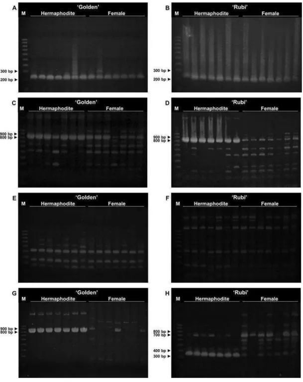
4. RESULTS
4.1. PCR assays with SCAR markers
Papaya SCAR markers were explored regarding their potential to
discriminate hermaphrodite from female individuals of the Brazilian cultivars
‘Golden’ and ‘Rubi’. A pair of primers used for amplifying a partial sequence
of the papain gene (gender-neutral) served as positive control of PCR
conditions. A fragment between 200 and 300 bp was amplified in all samples
of both cultivars (Figure 1a, b).
Each of the seven sex-specific SCAR markers investigated in this
study showed very similar banding patterns, containing fragments of varying
lengths, when comparing the two cultivars. Therefore, the considerations
made for each marker will serve for both cv. ‘Golden’ and ‘Rubi’, excepting
only one which showed PCR profiles slightly different between them (T12
marker). In this particular case, it could be noticed a more meaningful
production of 800 – 900-bp fragments in ‘Rubi’ hermaphrodite samples, while
there is a more homogeneous production of fragments within that range in all
‘Golden’ samples (Figure 1c, d).
Among the remaining SCAR markers, three of them (T1, W11 and
SDSP2) amplified different PCR products regardless the sex, while others
(NAPF-2, SDSP and SCARpm) generated at least one consistently
polymorphic band, clearly differentiating hermaphrodite from female samples.
Two representative PCR profiles show no difference on the sex-specific
banding behavior for the two sex forms of the two cultivars studied: SDSP2
amplification in ‘Golden’ samples (Figure 1e) and W11 amplification in ‘Rubi’
samples (Figure 1f).
Likewise in the amplification of T12 primers, NAPF-2 primers
produced hermaphrodite-specific 800 – 900-bp fragments in an expressive
quantity but interestingly in both cultivars (Figure 1g). Products of that same
length were also detected in some female samples, but in a very low
intensity. Another discrete hermaphrodite-specific band of
22
hermaphrodite-specific fragments of nearly 400 bp long and female-specific
fragments of slightly above 400 bp long. Besides, a low intensity
high-molecular-weight band was observed in female samples, which was absent
in hermaphrodites (data not shown). Amplification profile of the SCARpm
marker evidenced different sex-specific bands: hermaphrodite-specific 300 –
400-bp and female-specific 700 – 800-bp fragments, both in high intensity,
and three other female-specific bands (<700 bp long) in lower intensity
23
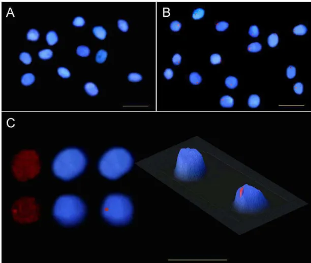
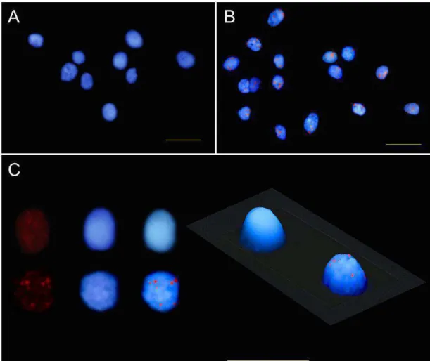
24 4.2. FISH
Based on the PCR amplification profiles of the SCAR molecular
markers which showed sex-specific polymorphism, the NAPF-2 marker was
selected for evaluating signal specificity in FISH analyses, considering its
highly contrasting banding pattern. For this, the PCR products obtained were
reamplified in a labeling reaction by testing four DNA polymerases. Although
the probes have been quantified by fluorometer and qualified by agarose-gel
electrophoresis, the labeling efficiency is in fact assessed on FISH analyses.
It is noteworthy that the protocol developed for obtaining nuclei
suspension from frozen leaf tissues was successful as intact and
well-individualized nuclei, with no cytoplasmic background noises, were gathered
in a great quantity per slide. These are important aspects for better reaching
of probes to nuclear DNA and further visualization of hybridization.
Thus, with adequate slide preparations, the labeling efficiency of each
DNA polymerase on probe construction could be evaluated by FISH. The
probe synthesized with Thermo SequenaseTM DNA Polymerase provided the best FISH result, giving strong and unambiguous fluorescence signals, and,
therefore, it was employed in the subsequent analyses.
FISH analyses in C. papaya by using NAPF-2 marker as probe
showed the same qualitative results in both cultivars ‘Golden’ and ‘Rubi’:
fluorescence signals present in hermaphrodite nuclei, and no detectable or
very low intensity fluorescence signal in female nuclei. In the hermaphrodite
slide of the cv. ‘Golden’, all the nuclei exhibited a single strong fluorescent
signal, most of them located at the nuclear peripheral region. Some smaller
and diffuse fluorescent spots, however, were also visualized in few nuclei. On
the other hand, many strong fluorescent signals spread on hermaphrodite
nuclei were evident in the cv. ‘Rubi’. The FISH results of the two sex types of
25
26
27
5. DISCUSSION
5.1. PCR assays with SCAR markers
Sex-linked DNA molecular markers have been developed for C.
papaya species over the years, and applied in early prediction of sexual type
(MING et al. 2007). This practice plays an important role on papaya fruit
production improvement, of great interest by farmers (MAGDALITA &
MERCADO 2003, CHAVES-BEDOYA & NUÑEZ 2007). Nevertheless,
considering that most of the markers come from arbitrary sequences, the
identification of markers conserved on papaya genome and specific for each
sex form regardless the genotype becomes a hard task. Moreover, sex
system in papaya is hypothesized to be determined by one genic locus with
three alleles, thus dominant markers like RAPD eventually would be linked to
the recessive allele, which is present in all sex types (OLIVEIRA et al. 2007).
So, it is necessary to investigate the potential sex-specific markers for each
papaya cultivar or variety of interest.
In this study, seven sex-specific SCAR markers were tested for
discriminating at molecular level hermaphrodite from female plants of two
commercially important Brazilian cultivars (male plants were not evaluated
because they are not cultivated for commercial purposes in Brazil), in
addition to the SCAR marker for papain gene used as positive control of PCR
conditions. In the latter case, the 200 – 300-bp fragment obtained after
amplification, in all samples, corroborates the expected fragment size of 221
bp (URASAKI et al. 2002b, RIMBERIA et al. 2005), indicating that the PCR
conditions adopted were suitable. Among the seven SCAR markers studied,
six of them (excluding T12 marker) showed very similar banding patterns
between the cultivars. This fact suggests a conservation of their sequences
in these two Brazilian cultivars. Conversely, PCR results for the T12 SCAR
marker demonstrate the presence of polymorphism intra- (between
hermaphrodite and female plants of cv. ‘Rubi’) and inter-cultivars. The
production of 800 – 900-bp fragments in ‘Rubi’ hermaphrodite samples was
28
Hawaiian cultivars. The RAPD-PCR fragment designated T12 had been
previously characterized and mapped on SEX1 locus (SONDUR et al. 1996).
In relation to the T1 SCAR marker, whose RAPD-PCR fragment was
also linked to the SEX1 locus (SONDUR et al. 1996), similar results to ours
were reported by DEPUTY et al. (2002). These authors observed a single
product of ~1300 bp in all plants, with no sex segregating pattern. However,
MAGDALITA & MERCADO (2003) found a 1300-bp PCR product in both
females and hermaphrodites, while male had no band. About W11 SCAR
marker, these three studies showed different results. While no segregating
band was detected for both Brazilian cultivars in the present study, a
polymorphic band (~800 bp) was produced in hermaphrodite and male plants
from Hawaiian cultivars (DEPUTY et al. 2002), and in hermaphrodites only
(MAGDALITA & MERCADO 2003).
Among the SCAR markers that generated sex polymorphism in our
analyses, the SDSP marker produced a hermaphrodite-specific fragment of
nearly 400 bp long, which is consistent with the amplification of a 369-bp
product in hermaphrodites and males from Colombian genotypes
(CHAVES-BEDOYA & NUÑEZ 2007), and the SCARpm marker produced a 300 –
400-bp fragment in hermaphrodites, also in line with a hermaphrodite and
male-specific 347-bp band obtained by URASAKI et al. (2002b). Also it can be
inferred that each of these sequences are conserved among their respective
cultivars analyzed. On the other hand, female-specific PCR products were
also detected for both SCAR markers, SDSP and SCARpm, but these
findings were not reported by CHAVES-BEDOYA & NUÑEZ (2007) neither
by URASAKI et al. (2002b), respectively.
NAPF-2 was considered by our analyses the best sex-specific SCAR
marker for the two cultivars, ‘Golden’ and ‘Rubi’, producing a consistently
polymorphic band of 800 – 900 bp long. PARASNIS et al. (2000), who
selected and tested this molecular marker, found a 831-bp fragment in male
plants, but absent in females. Only dioecious papaya varieties were tested so
that there were no hermaphrodite plants. The fact that the NAPF-2 marker
produced a ~800-bp fragment in hermaphrodite and male plants by these