Efficacy
of
curcumin
to
reduce
hepatic
damage
induced
by
alcohol
and
thermally
treated
oil
in
rats
Nasr
A.M.N.
El
‐
Deen
(1)&
Mohamed
Eid
(2)Summary
The authors investigated the effect of curcumin on markers of oxidative stress and liver damage in rats that chronically ingested alcohol and heated oil. Nine groups of ten Wistar male rats received combinations of curcumin 100 mg/kg body weight daily, ethanol 5 mg/kg, 15% dietary sunflower oil and 15% heated sunflower oil for 12 weeks. Serum and liver tissue were collected. Groups 4‐6, which had received compounds causing oxidative stress, showed increased serum aspartate aminotransferase, alanine aminotransferase, alkaline phosphatase, total bilirubin, cholesterol, triglycerides, low density lipoprotein, very low density lipoprotein and reduced high density lipoprotein, protein and albumin, compared with the controls. Reductions were observed in glutathione peroxidase and reductase gene expression, superoxide dismutase activity, glutathione peroxidase activity, glutathione reductase activity, reduced glutathione concentration and catalase enzyme activity. Groups 7, 8 and 9 which received curcumin with heated oil, ethanol or both, showed lower elevations in serum and oxidative damage markers compared with the corresponding non‐ curcumin treated groups. It can be concluded that curcumin reduces markers of liver damage in rats treated with heated sunflower oil or ethanol.
Keywords
Alcohol, Curcumin, Egypt, Liver, Oil, Rat, Stress.
Efficacia
della
curcumina
nella
riduzione
del
danno
epatico
in
ratti
trattati
con
alcol
e
olio
sottoposto
a
trattamento
termico
Riassunto
Gli autori hanno indagato l’effetto della curcumina
sui marcatori dello stress ossidativo e del danno
epatico in ratti trattati cronicamente con alcol e olio
riscaldato. Nove gruppi di dieci ratti Wistar maschi
hanno ricevuto giornalmente combinazioni di
curcumina (100 mg/kg di peso corporeo), etanolo
(5 mg/kg), olio di girasole dietetico (15%) e olio di
girasole riscaldato (15%) per 12 settimane. Sono
stati raccolti campioni di siero e tessuto epatico.
Rispetto al gruppo di controllo, i gruppi 4‐6 trattati
con composti responsabili dello stress ossidativo
hanno evidenziato aumento dei livelli sierici di
aspartato aminotransferasi, alanina amino‐
transferasi, fosfatasi alcalina, bilirubina totale,
colesterolo, trigliceridi, lipoproteina a bassa e
bassissima densità e riduzione dei livelli di
lipoproteina ad alta densità, proteine e albumina. Sono state osservate riduzioni, nellʹespressione
genica, del glutatione‐reduttasi e del glutatione‐
perossidasi, nellʹattività della superossido dismutasi,
del glutatione‐perossidasi e del glutatione‐reduttasi, nella concentrazione di glutatione e nell’attività
dell’enzima catalasi. I gruppi 7, 8 e 9 trattati con
curcumina e olio riscaldato, etanolo o entrambi,
hanno evidenziato aumenti inferiori dei marcatori
sierici e del danno ossidativo rispetto ai gruppi
corrispondenti non trattati con curcumina. È
possibile concludere che la curcumina riduce i
(1) Department of Clinical Pathology, Faculty of Veterinary Medicine, Zagazig University, 1 Elzeraa Street, Postal Code 44511, Zagazig City, Sharkia Province, Egypt
livelli dei marcatori del danno epatico nei ratti
trattati con olio di girasole riscaldato o etanolo.
Parole chiave
Alcol, Curcumina, Egitto, Fegato, Olio, Ratto, Stress.
Introduction
Oxidative stress occurs with an imbalance between pro‐oxidants and antioxidants (1). In a rat model, the administration of alcohol or thermally treated oil for 45 days caused excess generation of free radicals and oxidative injury to the liver (38). The mechanism for alcohol oxidative damage to the liver has been reported to be produced by the nuclear ethanol metabolising system which is capable of bioactivating ethanol to acetaldehyde and 1‐hydroxyethyl radicals. These reactive metabolites covalently bind to nuclear proteins and lipids, and provoke oxidative stress to nuclear components (11).
The newer vegetable oils which are termed ‘heart friendly’ are rich in polyunsaturated fatty acids. Deep frying of these oils has a deleterious effect on liver cells through the production of certain toxic products which damage hepatocytes (3, 50). The increased intake of polyunsaturated fatty acids increases the degree of unsaturation of biomembranes, making them more susceptible to lipid peroxidation (13). Thermally treated sunflower oil intake, with ethanol, has also been shown to aggravate hepatic toxicosis (4).
Turmeric, derived from the rhizome of the herb Curcuma longa, has been used for centuries as a dietary spice in Asia. Diferuloylmethane is an active principle present in curcumin which is the main active ingredient of turmeric; it exerts potent biological effects in vitro and in vivo (2, 43), particularly attenuating oxidative stress (15, 20). Curcumin has been used to treat various diseases, including hepatic disorders (32) and exhibits antioxidant, anti‐inflammatory, anti‐ viral, antibacterial, antifungal, and anticancer activities (2).
Material
and
methods
Animals
A total of 90 Albino Wistar strain male rats (100‐120 g body weight) were acquired from an animal farm in Helwan, Egypt. The animals were housed individually 15 days before the study in stainless steel wire‐mesh cages with free access to water and food (casein 180 g/kg, corn starch 460 g/kg, sucrose 220 g/kg, mineral mixture 50 g/kg, vitamin mixture 10 g/kg, cellulose 40 g/kg, sunflower oil 49 g/kg) according to Degrace et al. (10). All ingredients were obtained from Sigma‐Aldirsh in Cairo.
Experimental
design
The 90 rats were equally divided into nine groups. All treatments were administered for 12 weeks, as follows:
Group 1 received normal saline 5 ml/kg body weight daily via intragastric intubation Group 2 received curcumin (curcumin from
Curcuma longa) (Sigma Chemical, St Louis,
Missouri) 100 mg/kg body weight daily via intragastric intubation according to Reyes‐ Gordillo et al. (36)
Group 3 received 15% sunflower oil in the diet and normal saline 5 ml/kg bodyweight daily via intragastric intubation
Group 4 received 15% thermally treated sunflower oil in the diet (180°C for 30 min, twice) according to Rukkumani et al. (38) and normal saline 5 ml/kg body weight daily via intragastric intubation
Group 5 received ethanol (Hayman Limited, Witham, Essex,) 25% 5 ml/kg body weight daily via intragastric intubation according to Hussein et al. (21)
Group 6 received 15% thermally treated sun‐ flower oil in the diet and ethanol 5 ml/kg body weight daily via intragastric intubation Group 7 received curcumin 100 mg/kg
bodyweight daily via intragastric intubation and 15% thermally treated sunflower oil in the diet
Group 8 received curcumin 100 mg/kg bodyweight dissolved in ethanol 5 ml/kg body weight daily via intragastric intubation Group 9 received curcumin 100 mg/kg
bodyweight daily via intragastric intubation and 15% thermally treated sunflower oil in the diet.
Blood samples were collected by heart puncture (after anaesthetising the animal with ether in a cage/mask). Serum was obtained by centrifugation and the following analytes measured:
aspartate aminotransferase and alanine aminotransferase (34)
alkaline phosphatase (22) total bilirubin (28) total protein (17) albumin (53)
serum globulins were calculated as the difference between total protein and albumin cholesterol (37)
triglyceride (51)
high‐density lipoprotein cholesterol (52) low‐density lipoprotein cholesterol and very
low‐density lipoprotein cholesterol (14) Two liver samples were collected after anaesthetising the animal; these were immediately placed in liquid nitrogen. The first was used to determine glutathion peroxidase and glutathione reductase gene‐ expression according to Meadus (25). Liver homogenate was used to measure reduced glutathione concentrations according to Grunet and Phillips (19), glutathion peroxidase activity (23), glutathion reductase activity (5), superoxide dismutase activity (27) and catalase enzyme activity (44). Euthanasia was performed at the end of the experiment by freezing the rats while anaesthetised.
Samples were collected from all animals in each group and data were analysed using the MSTAT‐C computer program (42). The design used to analyse the data was one‐way analysis of variance (ANOVA) (F test) and comparison between means by least significant difference (LSD) values were considered statistically significant when p ≤0.05.
Results
Groups 2 and 3 revealed no alterations of all measured parameters. Groups 4, 5 and 6 showed a significant increase in serum aspartate transaminase (AST), alanine transaminase (ALT), alkaline phosphatase (ALP) and total bilirubin, together with a significant decrease in the serum concentrations of total protein and albumin, when compared with the control group. In addition, a significant decrease in the albumin/globulin ratio was reported in the group to which both ethanol and thermally treated oil had been administered (Table I). Groups 7, 8 and 9 which received curcumin in addition to liver oxidants, showed significantly less alterations in the various analytes when compared with the corresponding groups that had not been treated with curcumin.
Serum concentrations of cholesterol, triglycerides, low‐density lipoprotein and very low‐density lipoprotein had increased significantly, while a marked decrease in the high‐density lipoprotein was recorded in all groups except Groups 2 and 3 which had received curcumin and oil, respectively (Table II). Groups 7, 8 and 9 showed significantly less alterations in the various analytes when compared with the corresponding non‐curcumin‐treated groups and this improvement was more marked in groups treated with heated oil or ethanol than Group 9 which was treated by both.
Table I
Biochemical parameters measured in serum of the rats (mean values + standard error)
Group pa
ra m e te rs N o rm a l con trol
Curcumin Oil Heat
ed
oil
Ethanol Heat
ed
oil plus
ethanol Curcumin plus heated oil Curcumin plus ethanol Curcuminplus heated oil plus ethanol LSD
AST (U/l) 20.50(a)
±0.58 20.20(a) ±0.79 21.20(a) ±0.53 29.00(b) ±0.70 31.50(c) ±0.40 38.70(d) ±0.58 24.80(e) ±0.57 26.10(e) ±0.41 29.00(b) ±0.65 1.66
ALT (U/l) 15.20(a)
±0.51 15.20(a) ±0.41 16.20(a) ±0.63 22.70(b) ±0.84 25.40(c) ±0.91 29.00(d) ±0.70 19.90(e) ±0.59 20.50(e) ±0.64 23.30(b) ±0.67 1.88 Bilirubin (mg/dl) 0.58(e) ±0.04 0.57(e) ±0.04 0.60(e) ±0.03 0.87(c) ±0.05 0.90(c) ±0.04 1.02(d) ±0.03 0.72(b) ±0.05 0.75(b) ±0.05 0.87(c) ±0.03 0.1189
ALP (U/l) 71.20(f) ±1.35 70.20(f) ±1.60 73.00(f) ±0.89 96.50(b) ±1.65 101.90( c) ±0.71
114.60(d) ±1.05 78.60(a) ±1.93 79.00(a) ±2.76 91.20(e) ±1.14 4.48 Total protein (g/dl) 7.03(d) ±0.05 7.00(d) ±0.04 6.92(d) ±0.09 5.73(b) ±0.13 5.55(b) ±0.08 5.25(e) ±0.11
6.81(c, d) ±0.09 6.61(c) ±0.13 5.82(b) ±0.10 0.275 Albumin (g/dl) 4.748(d) ±0.07 4.72(d) ±0.05 4.64(d) ±0.06
3.588(b, e) ±0.08
3.392(e) ±0.07
3.12(a) ±0.04
4.548(c, d ) ±0.09
4.38(c) ±0.09 3.63(b) ±0.05 0.197 Globulin (g/dl) 2.28(d) ±0.04 2.28(d) ±0.05 2.28(d) ±0.06 2.14(d) ±0.05 2.16(d) ±0.05 2.13(d) ±0.07 2.26(d) ±0.03 2.23(d) ±0.06 2.19(d) ±0.05 NS
A/G ratio 2.08(c, d) ±0.06 2.07(d) ±0.06 2.05(d) ±0.05 1.68(c) ±0.02
1.58(b, c) ±0.05 1.47(b) ±0.03 2.01(d) ±0.05 1.98(d) ±0.05 1.66(c) ±0.03 0.13
Data are presented as mean ± SE, n = 10 and values within the same row which carrying different superscripts are significant at
p≤0.05 using the analysis of variance (ANOVA) test AST aspartate aminotransferase
U mean unit
ALT alanine aminotransferase ALP alkaline phosphatase A/G albumin globulin ratio LSD least significant difference NS not significant
Table II
Lipogram in the serum of the rats mean values + standard error)
Group pa
ra m e te rs N o rm a l con trol
Curcumin Oil Heat
ed
oil
Ethanol Heat
ed
oil plus
ethanol Curcumin a
n
d
heated oil Curcumin plus ethanol Curcumin plus heated oil plus ethanol LSD
Cholesterol (mg/dl) 105.40(a) ±1.08 103.90(a) ±2.81 107.00(a) ±2.45 132.00(b) ±0.77 124.60(c) ±0.97 141.90(d) ±0.98 115.80(e) ±1.89 114.30(e) ±2.18 122.10(c) ±1.81 5.25 Triglyceride (mg/dl) 81.70(f) ±0.86 81.10(f) ±1.64 82.30(f) ±1.48 116.90(b) ±0.70 109.50(c) ±0.79 123.60(d) ±1.17 92.70(a) ±1.94 88.90(a) ±3.86 103.20(e) ±0.80 5.05 HDL (mg/dl)
34.90(b, d) ±0.35 36.00(d) ±0.77 33.50(b) ±0.85 26.80(d) ±0.77 27.40(d) ±0.54 23.50(a) ±0.73 31.10(c) ±0.60 31.40(c) ±0.96 28.50(e) ±0.81 2.05
LDL (mg/dl) 54.16(a) ±1.08 51.68(a) ±2.98 57.04(a) ±2.31 81.82(b) ±1.08 75.30(c) ±1.31 93.68(d) ±1.27 66.16(e) ±1.77 65.12(e) ±2.15 72.96(c) ±1.50 5.128 VLDL (mg/dl) 16.34(f) ±0.17 16.22(f) ±0.33 16.46(f) ±0.30 23.38(b) ±0.15 21.90(c) ±0.16 24.72(d) ±0.23 18.54(a) ±0.39 17.78(a) ±0.77 20.64(e) ±0.29 1.011
Data are presented as mean ± SE, n = 10 and values within the same row which carrying different superscripts are significant at
p≤0.05 using the analysis of variance (ANOVA) test LSD least significant difference
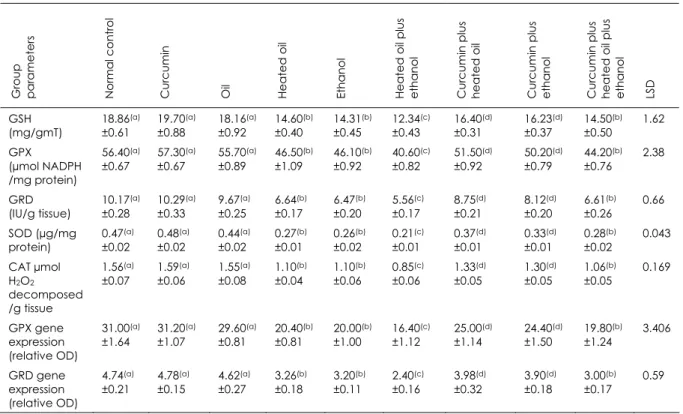
Table III
The antioxidants in hepatic tissue of rats (mean values + standard error)
Group pa
ra m e te rs N o rm a l con trol
Curcumin Oil Heat
ed
oil
Ethanol Heat
ed
oil plus
ethanol Curcumin plus heated oil Curcumin plus ethanol Curcumin plus heated oil plus ethanol LSD
GSH (mg/gmT) 18.86(a) ±0.61 19.70(a) ±0.88 18.16(a) ±0.92 14.60(b) ±0.40 14.31(b) ±0.45 12.34(c) ±0.43 16.40(d) ±0.31 16.23(d) ±0.37 14.50(b) ±0.50 1.62 GPX (µmol NADPH /mg protein) 56.40(a) ±0.67 57.30(a) ±0.67 55.70(a) ±0.89 46.50(b) ±1.09 46.10(b) ±0.92 40.60(c) ±0.82 51.50(d) ±0.92 50.20(d) ±0.79 44.20(b) ±0.76 2.38 GRD (IU/g tissue) 10.17(a) ±0.28 10.29(a) ±0.33 9.67(a) ±0.25 6.64(b) ±0.17 6.47(b) ±0.20 5.56(c) ±0.17 8.75(d) ±0.21 8.12(d) ±0.20 6.61(b) ±0.26 0.66 SOD (µg/mg protein) 0.47(a) ±0.02 0.48(a) ±0.02 0.44(a) ±0.02 0.27(b) ±0.01 0.26(b) ±0.02 0.21(c) ±0.01 0.37(d) ±0.01 0.33(d) ±0.01 0.28(b) ±0.02 0.043 CAT µmol H2O2
decomposed /g tissue 1.56(a) ±0.07 1.59(a) ±0.06 1.55(a) ±0.08 1.10(b) ±0.04 1.10(b) ±0.06 0.85(c) ±0.06 1.33(d) ±0.05 1.30(d) ±0.05 1.06(b) ±0.05 0.169 GPX gene expression (relative OD) 31.00(a) ±1.64 31.20(a) ±1.07 29.60(a) ±0.81 20.40(b) ±0.81 20.00(b) ±1.00 16.40(c) ±1.12 25.00(d) ±1.14 24.40(d) ±1.50 19.80(b) ±1.24 3.406 GRD gene expression (relative OD) 4.74(a) ±0.21 4.78(a) ±0.15 4.62(a) ±0.27 3.26(b) ±0.18 3.20(b) ±0.11 2.40(c) ±0.16 3.98(d) ±0.32 3.90(d) ±0.18 3.00(b) ±0.17 0.59
Data are presented as mean ± SE, n = 10 and values within the same row which carrying different superscripts are significant at p ≤0.05 using the analysis of variance (ANOVA) test
LSD least significant difference GSH reduced glutathione concentration GPX glutathione peroxidase activity
NADPH nicotinamide adenine dinucleotide phosphate GRD glutathione reductase activity
SOD superoxide dismutase activity CAT catalase enzyme activity
OD optical density
TBP Tata box binding protein Groups
1. Negative control 6. Heated oil plus ethanol
2. Curcumin 7. Curcumin plus heated oil
3. Oil 8. Curcumin plus ethanol
4. Heated oil 9. Curcumin plus heated oil plus ethanol
5. Ethanol
Figure 1
Polymerase chain reaction product of Tata box binding protein gene expression (internal control) 200 bp
100 bp
Groups 1 2 3 4 5 6 7 8 9
GPX glutathione peroxidase activity Groups
1. Negative control 6. Heated oil plus ethanol
2. Curcumin 7. Curcumin plus heated oil
3. Oil 8. Curcumin plus ethanol
4. Heated oil 9. Curcumin plus heated oil plus ethanol
5. Ethanol
Figure 2
Polymerase chain reaction product of glutathion peroxidase gene expression
GRD glutathione reductase activity Groups
1. Negative control 6. Heated oil plus ethanol
2. Curcumin 7. Curcumin plus heated oil
3. Oil 8. Curcumin plus ethanol
4. Heated oil 9. Curcumin plus heated oil plus ethanol
5. Ethanol
Figure 3
Polymerase chain reaction product of glutathion reductase gene expression
Discussion
In this study, the administration of liver oxidants thermally treated oil and ethanol were associated with increased serum levels of aminotransferases (AST and ALT), ALP and total bilirubin caused by hepatocellular damage (46). The liver is the main organ susceptible to damage by oral ethanol because it receives the portal blood directly from the
intestinal tract. Ethanol administration may cause injury to the liver through different mechanisms, including microsomal damage (24) and the release of metabolites from ethanol, such as malondialdehyde and acetaldehyde, which synergistically bind to proteins to form malondialdehyde‐ acetaldehyde modified proteins (48). These modified proteins induce pro‐inflammatory and pro‐fibrotic responses by liver endothelial
Groups 1 2 3 4 5 6 7 8 9
500 bp 100 bp
GRD GPX 198 bp
cells (47). Furthermore, the formation of 1‐hydroxyethyl free radicals from ethanol also damage the liver (33), induce cytochrome P450 2E1 (16) and sensitise Kupffer cells to endotoxin (lipopolysaccharide). These cells then produce tumour necrosis factor‐ which is critical for progression of alcoholic liver injury (12).
Heating oil rich in polyunsaturated fatty acids has a deleterious effect on liver cells through different pathways, including the production of toxic products which damage hepatocytes (3, 50). The increased intake of poly‐ unsaturated fatty acids increases the degree of unsaturation of biomembranes, making them more susceptible to lipid peroxidation (13), the generation of various cytotoxic aldehyde species (18) and the production of various lipid peroxidative as end‐products of heating (4).
Another explanation for the elevation in bilirubin may be the presence of ethanol that increases erythrocyte deformability (7), subsequently increasing the risk of haemolysis. Similarly, consumption of high levels of n‐3
polyunsaturated fatty acids may also lead to enhanced membrane lipid peroxidation by free
radicals (40). Previous studies on rats have reported similar findings (4, 41) with Pari and Karthikesan (31) also reporting increased cell wall permeability, damage and/or necrosis of hepatocytes.
A significant decrease was found in the total protein and albumin in groups that received thermally treated oil, ethanol or both. Similarly, the albumin globulin ratio significantly decreased in the group to which both thermally treated oil and ethanol had been administered. The hypoproteinaemia and decreased albumin globulin ratio were due to the hypoalbuminaemia which may be due to the loss of albumin through the gastro‐ intestinal tract (6
)
, increased excretion through damaged kidneys or disturbed production by the liver (31). Our findings were consistent with results obtained by Hussein et al. (21) who recorded hypoproteinemia and hypo‐ albuminaemia in rats after ingestion of ethanol. Similarly, Aruna et al. (4) found that the intake of thermally treated sunflower oil,together with alcohol, aggravated hepatic toxicity.
The lipogram revealed a significant increase in the levels of cholesterol, triglycerides, low density lipoprotein and very low density lipoprotein as well as a significant decrease in high density lipoprotein associated with the groups to which thermally treated oil, ethanol, or both had been administered. This is consistent with the findings of Aruna et al. and Rukkumani et al. (4, 39) who reported that levels of cholesterol, triglycerides, phos‐ pholipids and free fatty acids increased in the plasma of rats to which alcohol, thermally oxidised oil, or alcohol and thermally oxidised oil had been administered.
The antioxidant defence systems play an important role in protecting living organisms from the deleterious effects of reactive oxygen metabolites (38) produced by hepatic oxidant chemicals. Among these protective systems, superoxide dismutase, glutathione peroxidase, glutathione reductase and catalase play an important role. Our study showed that superoxide dismutase activity, glutathione peroxidase activity, glutathione reductase activity, reduced glutathione concentration and catalase enzymes significantly decreased following chronic ethanol and thermally treated oil ingestion in rats. The decreased glutathione peroxidase and glutathione reductase gene expression support our findings. Oxidative stress following the ingestion of thermally treated oil and ethanol caused increased use and weakened protective effect of the antioxidant enzymatic system in the rats in our study.
In our study, the administration of curcumin with thermally treated oil, alcohol, or both, caused a significant decrease in serum and gene markers of liver damage when compared to groups which had not been treated with curcumin. These findings support the ability of curcumin to counteract some of the oxidative stress produced by the ingestion of thermally treated oil, ethanol, or both, in rats.
Curcumin is believed to prevent necrosis factor ‐kappa activation and therefore suppress the secretion of pro‐inflammatory cytokines (35), inhibition of cyclooxygenase‐2, lipoxygenase and inducible nitric oxide synthase, which are important enzymes that mediate inflammatory processes (26). Previous studies have indicated that curcumin attenuates hepatic oxidative stress in rats (15, 20) and inhibits liver cirrhosis through multiple biological effects on hepatic stellate cells. The latter cells play a central role in the pathogenesis of hepatic fibrosis (29) and
may prevent and reverse cirrhosis by reducing transforming growth factor‐beta (TGF‐beta) expression (36) which affects development, homeostasis and tissue repair. Furthermore, the antioxidant effect of curcumin may occur through chelating metal ions which stimulate lipid peroxidation as iron (9, 45), scavenging of free radicals (49) and strong inhibitory properties towards cytochrome P450 and glutathione S‐transferase activities (8, 30).
Conclusions
It can be concluded that curcumin partially protects the liver by ameliorating the oxidative stress and activating the antioxidant defence systems.
References
1. Afify E.M., Saydate S.A., Nasr El Deen A.M.N. & Mahmoud A.E. 2007. Molecular and immunological studies on the effect of conjugated linoleic acid against the harmful effect of thermal treated oil in rats. Eg yp tia n J Bio c he m Mo le c Bio l, 25, 258-268.
2. Aggarwal B.B., Sundaram C., Malani N. & Ichikawa H. 2007. Curcumin: the Indian solid gold. Ad v Exp Me d Bio l, 595, 1-75.
3. Alexander J.C. 1981. Chemical and biological properties related to toxicity of heated fats. J To xic o l Enviro n He a lth, 7 (1), 125-138.
4. Aruna K., Rukkumani R., Varma P.S. & Menon V.P. 2005. Therapeutic role of cuminum cyminum on ethanol and thermally oxidized sunflower oil induced toxicity. Phyto the r Re s, 19 (5), 416-421.
5. Beutler E. 1975. Red cell metabolism. In A manual of biochemical methods, 2nd Ed. Grune and Stratton, New York, 69-70.
6. Bujanda L. 2004. The effects of alcohol consumption upon the gastrointestinal tract. Am J G a stro e nte ro l, 95 (12), 3374-3382.
7. Chmiel B.A., Olszowy Z.B., Turczynski B.B. & Kusmierski S.A. 1999. Effect of controlled ethanol intake on arterial blood pressure, heart rate and red blood cell deformability. C lin He mo rhe o l Mic ro c irc, 21,
325-328.
8. Choi H., Chun Y.S., Shin Y.J., Ye S.K., Kim M.S. & Park J.W. 2008. Curcumin attenuates cytochrome P450 induction in response to 2,3,7,8-tetrachlorodibenzo-p-dioxin by ROS-dependently degrading AhR and ARNT. C a nc e r Sc i, 99 (12), 2518-2524.
9. Dairam A., Fogel R., Daya S. & Limson J.L., 2008 Antioxidant and iron-binding properties of curcumin, capsaicin, and S-allylcysteine reduce oxidative stress in rat brain homogenate. J Ag ric Fo o d C he m,
56 (9), 3350-3356.
10. Degrace P., Demizieux L., Gresti J., Chardigny J.M., Sébédio J.L. & Clouet P. 2004. Hepatic steatosis is not due to impaired fatty acid oxidation capacities in C57 BL/6J mice fed the conjugated trans-10, cis-12-isomer of linoleicacid. J Nutr, 134, 861-867.
12. Enomoto N., Takei Y., Hirose M., Konno A., Shibuya T., Matsuyama S., Suzuki S., Kenichi K.I.T. & Sato N. 2003. Prevention of ethanol-induced liver injury in rats by an agonist of peroxisome proliferator-activated receptor-gamma, pioglitazone. J Pha rma c o l Exp The r, 306, 846-854.
13. Farrell S.O. & Jackson M.J. 1997. Dietary polyunsaturated fatty acids, vitamin E and hypoxia/reoxygenation-induced damage to cardiac tissue. C linic a C himic a Ac ta, 267, 197-211. 14. Friedewald W.T., Levy R.I. & Fredrickson D.S. 1972. Estimation of the concentration of low density
lipoprotein cholesterol in plasma without use of the preparative ultracentrifuge. C lin C he m, 18 (6), 499-502.
15. Glauert H.P., Tharappel J.C., Lu Z., Stemm D., Banerjee S., Chan L.S., Lee E.Y., Lehmler H.J., Robertson L.W. & Spear B.T. 2008. Role of oxidative stress in the promoting activities of PCBs. Enviro n To xic o l Pha rma c o l, 25 (2), 247-250.
16. Gouillon Z., Lucas D., Li J., Hagbjork A.L., Freuch B.A., Fu P. & Fang C. 2000. Inhibition of ethanol induced liver disease in the intragastric feeding rat model by chlomethiazole. Pro c So c Exp Bio Me d,
224, 302-308.
17. Grant G.H., Silverman L.M. & Christenson R.H. 1987. Amino acids and protein. In Fundamentals of clinical chemistry, 3rd Ed. (N.W. Tietz, ed.). WB Saunders Company, Philadelphia, 328-330.
18. Grootveld M., Atherton M.D, Sheerin A.N., Hawkes J., Blake D.R., Richens T.E., Silwood C.J.L, Lynch E. & Claxson A.W.D. 1998. In vivo absorption, metabolism, and urinary excretion of alpha, beta-unsaturated aldehydes in experimental animals. Relevance to the development of cardiovascular diseases by the dietary ingestion of thermally stressed polyunsaturate-rich culinary oils. J C lin Inve st,
101 (6), 1210-1218
19. Grunet R.R. & Phillips P.H. 1951. A modification of the nitroprussid method of analysis for glutathione. Arc h Bio c he m, 30, 217-225.
20. Gupta A., Vij G., Sharma S., Tirkey N., Rishi P. & Chopra K. 2009. Curcumin, a polyphenolic antioxidant, attenuates chronic fatigue syndrome in murine water immersion stress model. Immuno b io lo g y, 214 (1), 33-39.
21. Hussein J.S., Oraby F.S. & El-Shafey N. 2007. Antihepatotoxic effect of garlic and onion oils on ethanol-induced liver injury in rats. J Ap p l Sc i Re s, 3 (11), 1527-1533.
22. Kind P.R. & King E.G. 1954. Colorimetric determination of alkaline phosphatase activity. J C lin Pa tho l,
7, 322.
23. Lawrence R.A. & Burk R.F. 1976. Glutathione peroxidase activity in selenium-deficient rat liver. Bio he m Bio p hys Re s C o mmun, 71 (4), 952-958.
24. Lieber C.S. 1993. Biochemical factors in alcoholic liver disease. Se min Live r Dis, 13 (2), 136-153. 25. Meadus W.J. 2003. A semi-quantitative RT-PCR method to measure the in vivo effect of dietary
conjugated linoleic acid on protein muscle PPAR gene expression. Bio l Pro c O nline, 5 (1), 20-28. 26. Menon V.P. & Sudheer A.R. 2007. Antioxidant and anti-inflammatory properties of curcumin. Ad v
Exp Me d Bio l, 595, 105-125.
27. Minami M. & Yoshikawa H.A. 1979. Simplified assay method of superoxide dismutase activity for clinical use. C lin C him Ac ta, 92, 37-342.
28. Monnet L. 1963. Determination of bilirubin. Anna l Bio l C lin, 21, 717.
29. O’Connell M.A. & Rushworth S.A. 2008. Curcumin: potential for hepatic fibrosis therapy? Br J Pha rma c o l, 153 (3), 403-405.
30. Oetari S., Sudibyo M., Commandeur J.N.M., Samhoedi R. & Vermeulen N.P.E. 1996. Effects of curcumin on cytochrome P450 and glutathione S-transferase activities in rat liver. Bio c he m Pha rma c o l, 51 (1), 39-45.
31. Pari L. & Karthikesan K. 2007. Protective role of caffeic acid against alcohol-induced biochemical changes in rats. Fund a m C lin Pha rma c o l, 21 (4), 355-361.
32. Pari L., Tewas D. & Eckel J. 2008. Role of curcumin in health and disease. Arc h Physio l Bio c he m,
114 (2), 127-149.
33. Reinke L.A. & McCay P.B. 1997. Pin trapping of alcohol-initiated radicals in rat liver: influence of dietary fat. J Nutr, 127 (5 Suppl), 8995-9025.
35. Reyes-Gordillo K., Segovia J., Shibayama M., Vergara P., Moreno M.G. & Muriel P. 2007. Curcumin protects against acute liver damage in the rat by inhibiting NF-kappaB, proinflammatory cytokines production and oxidative stress. Bio c him Bio p hys Ac ta, 1770 (6), 989-996.
36. Reyes-Gordillo K., Segovia J., Shibayama M., Tsutsumi V., Vergara P., Moreno M.G. & Muriel P. 2008. Curcumin prevents and reverses cirrhosis induced by bile duct obstruction or CCl4 in rats: role of TGF-β modulation and oxidative stress. Fund a m C lin Pha rma c o l, 22 (4), 417-427.
37. Richmond W. 1973. Enzymatic determination of cholesterol. C lin C he m, 19, 1350-1356.
38. Rukkumani R., Aruna K., Varma P.S., Rajasekaran K.N. & Menon V.P. 2004. Comparative effects of curcumin and analog of curcumin on alcohol and PUFA induced stress. J Pha rm Pha rm Sc i, 7 (2), 274-283.
39. Rukkumani R., Aruna K., Varma P.S., Rajasekaran K.N. & Menon V.P. 2005. Comparative effects of curcumin and its analog on alcohol- and polyunsaturated fatty acid-induced alterations in circulatory lipid profiles. J Me d Fo o d, 8 (2), 256-260.
40. Saito M. & Nakatsugawa K. 1994. Increased susceptibility of liver to lipid peroxidation after ingestion of high fish oil diet. Int J Vit Nutr Re s, 64, 144-151.
41. Saravanan R., Viswanathan P. & Pugalendi K.V. 2006. Protective effect of ursolic acid on ethanol-mediated experimental liver damage in rats. Life Sc i, 78 (7), 713-718.
42. Software and Services (SAS) Institute Inc. 2002. SAS/STAT and user guide. SAS Institute Inc., Cary, North Carolina (support.sas.com/documentation/onlinedoc/stat/ accessed on 11 November 2009). 43. Sharma R.A., Gescher A.J. & Steward W.P. 2005. Curcumin: the story so far. Eur J C a nc e r, 41,
1955-1968.
44. Sinha K.A. 1972. Colorimetric assay of catalase. Ana l Bio c he m, 47, 389-394.
45. Sreejayan & Rao M.N. 1994. Curcuminoids as potent inhibitors of lipid peroxidation. J Pha rm Pha rma c o l, 46 (12), 1013-1016.
46. Stockham S.L. & Scott M.A. 2008. Fundamentals of veterinary clinical pathology, 2nd Ed. Blackwell Publishing Professional, Ames, Iowa, 550-559.
47. Thiele G.M., Duryee M.J., Wills M.S., Sorrell M.F., Freeman T.L., Tuma D.J. & Klassen L.W. 2004. malondialdehyde-acetaldehyde (MAA) modified proteins induce pro-inflammatory and pro-fibrotic responses by liver endothelial cells. C o mp He p a to l, 3 (Suppl 1), 525.
48. Tuma D.J., Thiele G.M., Xu D., Klassen L.W. & Sorrell M.F. I996. Acetaldehyde and malondialdehyde react together to generate distinct protein adducts in the liver during long-term ethanol administration. He p a to l, 23 (4), 872-880.
49. Unnikrishnan M.K. & Rao M.N.A. 1995. Curcumin inhibits nitrogen dioxide induced oxidation of hemoglobin. Mo le c C e ll Bio c he m, 146 (1), 35-37.
50. Varma P.S., Aruna K., Rukkumani R. & Menon V.P. 2004. Alcohol and thermally oxidized PUFA induced oxidative stress: role of N-acetyl cysteine. Ita l J Bio c he m, 53 (1), 10-15.
51. Wahlefeld A.W. & Bergmeyer H.W. 1974. Triglycerides determination after enzymatic hydrolysis. In Methods of enzymatic analysis. Berlachemie Zeinheim and Academic Press Inc., New York and London, 1831-1835.
52. Warnick G.R., Benderson V. & Albers, N. 1983. Estimation of HDL-cholesterol selected methods. C lin C he m, 1, 91-99.