Sleeping Beauty
Transposition of Chimeric
Antigen Receptors Targeting Receptor
Tyrosine Kinase-Like Orphan Receptor-1
(ROR1) into Diverse Memory T-Cell
Populations
Drew C. Deniger1,2¤, Jianqiang Yu3, M. Helen Huls1, Matthew J. Figliola1, Tiejuan Mi1,
Sourindra N. Maiti1, George F. Widhopf, 2nd3, Lenka V. Hurton1,2, Radhika Thokala1,2, Harjeet Singh1, Simon Olivares1, Richard E. Champlin4, William G. Wierda5, Thomas J. Kipps3, Laurence J. N. Cooper1,2*
1Pediatrics, Children’s Cancer Hospital, University of Texas MD Anderson Cancer Center, Houston, Texas, United States of America,2University of Texas Graduate School of Biomedical Sciences at Houston, Houston, Texas, United States of America,3Medicine, Moores Cancer Center, University of California San Diego, San Diego, California, United States of America,4Stem Cell Transplantation and Cellular Therapy, University of Texas MD Anderson Cancer Center, Houston, Texas, United States of America,5Leukemia, University of Texas MD Anderson Cancer Center, Houston, Texas, United States of America
¤ Current address: Surgery Branch, Center for Cancer Research, National Cancer Institute, National Institutes of Health, Bethesda, Maryland, United States of America
Abstract
T cells modified with chimeric antigen receptors (CARs) targeting CD19 demonstrated clinical activity against some B-cell malignancies. However, this is often accompanied by a loss of normal CD19+B cells and humoral immunity. Receptor tyrosine kinase-like or-phan receptor-1 (ROR1) is expressed on sub-populations of B-cell malignancies and solid tumors, but not by healthy B cells or normal post-partum tissues. Thus, adoptive transfer of T cells specific for ROR1 has potential to eliminate tumor cells and spare healthy tis-sues. To test this hypothesis, we developed CARs targeting ROR1 in order to generate T cells specific for malignant cells. TwoSleeping Beautytransposons were constructed with 2ndgeneration ROR1-specific CARs signaling through CD3ζand either CD28 (designated ROR1RCD28) or CD137 (designated ROR1RCD137) and were introduced into T cells. We selected for T cells expressing CAR through co-culture withγ-irradiated activating and propagating cells (AaPC), which co-expressed ROR1 and co-stimulatory molecules. Nu-meric expansion over one month of co-culture on AaPC in presence of soluble interleukin (IL)-2 and IL-21 occurred and resulted in a diverse memory phenotype of CAR+T cells as measured by non-enzymatic digital array (NanoString) and multi-panel flow cytometry. Such T cells produced interferon-γand had specific cytotoxic activity against ROR1+ tumors. Moreover, such cells could eliminate ROR1+tumor xenografts, especially T cells expressing ROR1RCD137. Clinical trials will investigate the ability of ROR1-a11111
OPEN ACCESS
Citation:Deniger DC, Yu J, Huls MH, Figliola MJ, Mi T, Maiti SN, et al. (2015)Sleeping Beauty
Transposition of Chimeric Antigen Receptors Targeting Receptor Tyrosine Kinase-Like Orphan Receptor-1 (ROR1) into Diverse Memory T-Cell Populations. PLoS ONE 10(6): e0128151. doi:10.1371/journal.pone.0128151
Academic Editor:Derya Unutmaz, Jackson Laboratory, UNITED STATES
Received:February 18, 2015
Accepted:April 22, 2015
Published:June 1, 2015
Copyright:This is an open access article, free of all copyright, and may be freely reproduced, distributed, transmitted, modified, built upon, or otherwise used by anyone for any lawful purpose. The work is made available under theCreative Commons CC0public domain dedication.
Data Availability Statement:All relevant data are within the paper and its Supporting Information files.
specific CAR+T cells to specifically eliminate tumor cells while maintaining normal B-cell repertoire.
Introduction
T cells can be rendered specific for tumor-associated antigens (TAAs) independent of their en-dogenous T-cell receptor (TCR) via gene transfer of chimeric antigen receptors (CARs) [1]. CARs are constructed from the genes encoding a single-chain variable fragment (scFv) of a TAA-specific monoclonal antibody (mAb), extracellular hinge or scaffold with transmembrane domain, and portions of CD3zand CD28 or CD137 (4-1BB) endodomains. Introduction of this chimeric gene generates T cells that proliferate, produce cytokines, and direct cytolysis of tumor cells in a TAA-dependent manner [2]. Infusion of T cells expressing CAR specific for CD19 with either CD3z/CD28 or CD3z/CD137 can induce complete tumor regressions in subsets of patients with B-lineage lymphomas, acute lymphoblastic leukemia (B-ALL), or chronic lymphocytic leukemia (CLL) [3–10]. In addition to the structure of the CAR, the subset of T cells that serves as a template for bioengineering can impact the anti-tumor effect. For in-stance, murine immunotherapy models have demonstrated that less differentiated T cells,i.e., naïve (TN), stem-cell memory (TSCM), or central memory (TCM) T cells, can clear tumor cells
more effectively than more differentiated T cells,i.e., effector memory (TEM) or
terminally-differentiated effector memory RA (TEMRA) T cells [11,12]. Thus, the specificity of the T cell
and the underlying memory phenotype can be important predictors for therapeutic efficacy. T cells that express CARs specific for CD19 cannot distinguish between neoplastic or nor-mal CD19-bearing B cells. Indeed, the initial clinical data has demonstrated that most patients benefiting from an anti-tumor response have concomitant B-cell depletion. Such B-cell aplasia requires that the patient receive timely intravenous infusions of normal immunoglobulin to al-leviate the threat for opportunistic infections [13]. Nevertheless, such treatment cannot elimi-nate the risk for serious infection, as one recipient of CD19-specific CAR+T cells died due to opportunistic infection [14]. Thus, targeting a TAA, which is not expressed on normal B cells or other adult tissues, would mitigate the risk for B-cell depletion and potentially improve outcome.
One TAA that may serve as an alternative to CD19-directed T-cell therapy is receptor tyro-sine kinase-like orphan receptor-1 (ROR1). ROR1 is principally expressed during embryonic development but its expression attenuates during fetal development and is negligible at term [15]. However, ROR1 is aberrantly expressed by some B-cell malignancies,e.g., lymphomas, CLL, and t(1;19) B-ALL, and by many solid-tumor malignancies,e.g., adrenal, bladder, breast, colon, lung, pancreas, prostate, ovary, skin, testes, uterus, and neuroblastoma [16–21]. ROR1 has also been discovered on ovarian cancer stem cells yielding the potential to eliminate tumor initiating cells by targeting ROR1 [22]. Monoclonal antibodies specific for ROR1 have reacted with some bone marrow adipocytes [23] and hematogones [24] (B-cell precursors dispensable for maintaining peripheral B-cell repertoire), but not on other normal tissues [18,20]. Non-human primates treated with autologous ROR1-specific T cells had no toxicity from treatment suggesting that targeting ROR1 is a safe approach for humans [25]. Thus, it appears T cells that express CARs specific for ROR1 would not deplete B-cells, but rather target neoplastic B cells or solid tumors.
Clinical trials have not yet evaluated the efficacy or safety of targeting ROR1 using adoptive immunotherapy. In preparation for the human application of genetically modified T cells, we
Mangurian, Jr., Fund for Leukemia Immunotherapy; Fund for Leukemia Immunotherapy; Institute of Personalized Cancer Therapy; Leukemia and Lymphoma Society SCOR; Lymphoma Research Foundation; Miller Foundation; Mr. Herb Simons; Mr. and Mrs. Joe H. Scales; Mr. Thomas Scott; MDACC Moon Shot; National Foundation for Cancer Research; Pediatric Cancer Research Foundation; Production Assistance for Cellular Therapies (PACT); TeamConnor; Thomas Scott; William Lawrence and Blanche Hughes Children's Foundation.
evaluated two 2ndgeneration CAR species specific for ROR1. Our approach is based on the methodology employed in our first-in-human clinical trials using CD19-specific T cells gener-ated by synchronous electro-transfer of CD19-specific CAR as aSleeping Beauty(SB) transpo-son and a hyperactive SB transposase [26,27]. Following transfection the T cells are co-cultured with irradiated activating and propagating cells (AaPC), which select for T cells that have stable expression of the CAR through direct interactions with AaPC bearing its cognate antigen,e.g., CD19 [28–31]. In this study we generated AaPC that express ROR1 and developed SB transposons to express CARs specific for ROR1 that signal via chimeric CD3z/CD28 (desig-nated ROR1RCD28) or chimeric CD3z/CD137 (designated ROR1RCD137). We show that T cells expressing either CAR can proliferate in response to cells bearing ROR1 and specifically kill ROR1+tumor cells. We found that ROR1RCD137+T cells had more effective anti-tumor ac-tivity than ROR1RCD28+T cells in immune-deficient mice engrafted with ROR1+tumor cells. A phase I clinical trial (NCT02194374) is open for the infusion of autologous ROR1RCD137 CAR+T cells in patients with CLL.
Materials and Methods
Ethics statement
All human samples acquired for this study were obtained after written informed consent was granted in accordance with protocols established and approved by the MD Anderson Cancer Center Internal Review Board (IRB). The identities of all samples were kept private. Animals treated in this study were handled in accordance with the strict guidelines established by the MD Anderson Cancer Center Institutional Animal Care and Use Committee (IACUC), which specifically approved this study (Protocol#03-06-04333). Mice were housed in pathogen-free conditions and were monitored daily for welfare-related assessments in accordance to IACUC guidelines. Moribund mice,e.g., ruffled fur, hunched posture, tumor size, were humanely eu-thanized with inhaled CO2as per direction of IACUC guidelines. No mice died without
eutha-nasia. All efforts were made to minimize animal suffering and inhaled isoflurane was administered for anesthesia as required.
Chimeric antigen receptors
Cloning of second generation CD19-specific CARs signaling through CD3zand CD28 (CD19RCD28) or CD137 (CD19RCD137) have been previously described [32,33]. Heavy and light chain immunoglobulin sequences from the 4A5 mAb hybridoma were codon optimized and synthesizedde novo(GeneArt; Invitrogen, Grand Island, NY) to create the“ROR1R” nu-cleotide sequence of (i) murine IgGκsignal peptide, (ii) VL, (iii) Whitlow linker
(GSTSGSG-KPGSGEGSTKG), (iv) VH, and (v) the first 73 amino acids of a modified human IgG4 stalk.
ROR1R was amplified by PCR with ROR1RCoOpF (GCTAGCCGCCACCATGGGCTGGTCCTG CATC) and ROR1Rrev (GCTCCTCCCGGGGCTTTGTCTTGGC) primers then sub-cloned into
pCR4-TOPO with TOPO TA Cloning Kit (Invitrogen) to generate ROR1R(CoOp)/pCR4-TOPO and sequence was verified with T7 and T13-0 primers (DNA Sequencing Core, MDACC). ROR1R(CoOp)/pCR4-TOPO and CD19RCD28mZ(CoOp)/pEK plasmids were digested with
digesting CD19R-CD28Tm-41BBCyt-Z(CoOp)/pSBSO-FRA withNheI,XmaI, andAntarctic Phosphataseand ROR1RCD28mZ(CoOp)/pSBSO-MCS withNheI,XmnI, andXmaIto generate ROR1RCD137/pSBSO-FRA plasmid. Identities of final ROR1R plasmids were distinguished from one another withBsrGIand from CD19R plasmids byPmlI(not present). The entire se-quence of both plasmids was verified by Sanger Sequencing (DNA Sequencing Core, MDACC).
Tumor cell tissue culture
EL4 cell line was acquired from American Type Culture Collection (Manassas, VA; cat# ATCC TIB-39). NALM-6 cell line was purchased from Deutsche Sammlung von Mikroorganismen und Zellkulturen (Germany; cat# ACC-128). Kasumi-2 was a gift from Jeffrey Tyner (Oregon Health & Science University) [34]. Clone#9 AaPC (previously referred to as artificial antigen presenting cells; aAPC) was generated though enforced co-expression of truncated CD19, CD64, CD86, and CD137L on K-562 cells genetically modified with lentiviral vectors and was a gift from Carl June (University of Pennsylvania) [35]. This AaPC was further modified using SB system to co-express IL15/IL15Rαfusion protein (designated membrane-bound IL-15; mIL15) and sub-cloned to generate clone#27. Clone#27 was then genetically modified using SB system to express ROR1, and single-cell clones were isolated by FACS based on co-staining of ROR1, CD137L, and IL15 during the sort. The clone#1 AaPC uniformly and stably co-express-ed CD19, CD32, CD64, CD86, CD137L, mIL15, and ROR1. These cell lines were maintainco-express-ed in complete media (RPMI, 10% FBS (Hyclone, Logan, UT), and 1x Glutamax-100). Identities of all cell lines were confirmed by STR DNA Fingerprinting at MDACC’s Cancer Center Sup-port Grant (CCSG) supSup-ported facility“Characterized Cell Line Core.”All tumor cells were free of mycoplasma and other microbial pathogens.
Numeric expansion of ROR1-specific CAR
+T cells
CAR+T cells were propagated based on modifying standard operating protocols as previously described [28,32]. Cryopreserved PBMC, obtained from healthy donors after informed con-sent, were thawed the day of the electroporation (designated day 0) and rested for 2 hours at 37°C. PBMC for electroporation were spun at 200g for 10 minutes and 2x10⁷cells were mixed with supercoiled DNA plasmids (5μg SB11 transposase and 15μg SB transposon) in Human T cell Nucleofector Solution (cat#VPA-1002, Lonza), added to a cuvette, and electroporated on the U-014 program of Amaxa Nucleofector II (Lonza). Electroporated cells were transferred to a 6-well plate containing phenol-free RPMI, 20% FBS, and 1x Glutamax-100. The following day, electroporated T cells were phenotyped and stimulated with AaPC clone#1 (irradiated using cesium source to 100 Gy) at 1:1 ratio of clone#1 to ROR1-specific CAR+T cells. Each co-culture was supplemented with IL-21 (cat# AF20021; Peprotech, Rocky Hill, NJ; 30 ng/mL) starting at initiation of culture and every 2–3 days thereafter and with IL-2 (Aldesleukin; Novartis, Switzerland; 50 IU/mL) added every 2–3 days starting at the second stimulation. CAR expression was evaluated weekly to determine the number of AaPC to add to co-cultures every 7 days. If contaminating NK cells reached>10% of the total population, they were
Abundance and identity of mRNA molecules by digital profiling
At designated times after co-culture on AaPC and cytokines, T cells were lysed with 160μL RLT Buffer (Qiagen, Valencia, CA) per 106cells and lysates were frozen at -80°C. RNA lysates were thawed and immediately analyzed using nCounter Analysis System (NanoString Technol-ogies, Seattle, WA) with“lymphocyte codeset array”(LCA). LCA data was normalized to both spike positive control RNA and housekeeping genes (ACTB,G6PD,OAZ1,POLR1B,POLR2A,
RPL27,RPS13, andTBP) as detailed previously [37] and normalized counts were reported. Limit-of-detection (LOD) was calculated from the negative control counts and reported as the mean plus two-times the standard deviation (mean+2xSD) and shown as dashed lines in graphs of mRNA data.
Flow cytometry and Immunohistochemistry
All mAbs were purchased from BD Biosciences (San Jose, CA), except for CCR7 mAb (eBioscience, San Diego, CA), IL-15 mAb (R&D Systems, Minneapolis, MN), Fc mAb used to detect CAR (Invitrogen), and 4A5 mAb used to detect ROR1 (Kipps TJ laboratory, UCSD). Staining was performed as described [38]. Samples for flow cytometry were acquired on FACS Calibur (BD Biosciences) and analyzed with FlowJo software (version 7.6.3). For immunohis-tochemistry, fresh frozen sections of malignant and normal pancreas were stained with 100μg/ ml of 4A5 mouse-anti-human ROR1 mAb or control mouse IgG2b mAb (isotype), and devel-oped using an anti-mouse secondary antibody conjugated with horse radish peroxidase. Tis-sues were visualized with diaminobenzidine (DAB) and counterstained with hematoxylin. Slides were evaluated by the study pathologist to identify the tissue or cell-type stained and in-tensity of staining.
In vitro
functional assays
In vitrospecific lysis was assessed using a standard 4-hour chromium release assay, as previ-ously described [32]. Expression of cytokines was evaluated by intracellular staining and flow cytometry. CAR+T cells were incubated with an equal volume and number of target cells for 6 hours at 37°C in the presence of Brefeldin-A (GolgiPlug; BD Biosciences) to block exocytosis and secretion of cytokines. Co-cultures were then (i) stained for surface markers,e.g., CD3 and CAR (Fc-specific antibody), (ii) fixed and permeabilized with BD Cytofix/Cytoperm (cat# 555028, BD Biosciences), (iii) stained for intracellular IFNγ, and (iv) analyzed by flow cytometry.
Mouse experiments
dosing and the following day. BLI from tumorffLucwas monitored twice per week. Log-rank (Mantel-Cox) test was used for statistical analysis between groups of mice (n = 5 per group).
Results
ROR1 is expressed on subsets of tumor cells, but not healthy pancreas
Before constructing CARs from 4A5, a mAb with specificity for human ROR1 [18–20,24], we sought to validate its staining of tumor cells. The murine T-cell lymphoma cell line EL4 did not express ROR1 but following genetic modification with a ROR1 transposon this cell line showed bright staining for ROR1 (Fig 1a). Consistent with other reports, all of the CLL samples that we tested were ROR1+as measured by 4A5 mAb staining (representative example is displayed). The t(1;19) B-ALL cell line Kasumi-2 expressed ROR1 whereas the non-t(1;19) B-ALL cell line NALM-6 did not. Eleven of twelve ovarian cancer cell lines tested positive for ROR1 (EFO27 and OC314 are shown as examples) and A2780 was the only ovarian cancer cell line we evalu-ated that did not express ROR1. Previous reports have identified ROR1 mRNA in the pancreas [39], so we tested 4A5 mAb staining against healthy and malignant pancreatic tissue using immunohistochemistry (Fig 1b). We were unable to detect ROR1 expression in healthy pancre-as but ROR1 wpancre-as widely and strongly expressed by pancreatic cancer tissue. Thus, this staining dataset corroborated our rationale to use the 4A5 mAb to construct CARs specific for ROR1 expressed on tumor cells.
Sleeping Beauty
transposition and co-culture on ROR1
+AaPC with
cytokines generates ROR1-specific T cells
SB transposition followed by co-culture with AaPC, IL-2, and IL-21 was employed to propagate T cells expressing a 2ndgeneration CD19-specific CAR that activates via CD3z/CD28 endodo-main (designated CD19RCD28) and these T cells have been infused into patients with B-cell malignancies [28–33,40]. This strategy was adapted to target ROR1 by replacing the
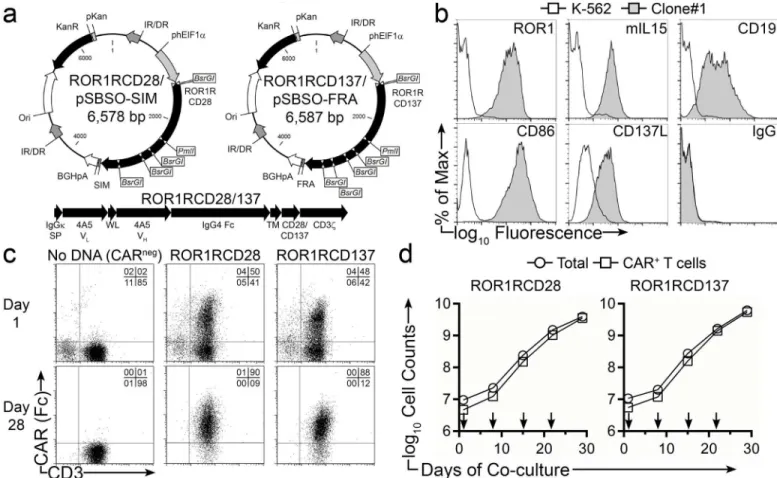
CD19-specific scFv sequence in the CAR with the scFv sequence derived from 4A5 mAb. Two SB transposons were constructed for side-by-side comparison of 2ndgeneration ROR1-specific CARs that differ in signaling via CD28 (ROR1RCD28) or CD137 (ROR1RCD137) (Fig 2a). The remaining portions of the CARs,e.g., murine IgGκsignal peptide, Whitlow linker, modi-fied human IgG4-Fc stalk, human CD28 transmembrane, and human CD3zintracellular
do-mains (with three ITAM activation signals) were identical between the two constructs. We used AaPC (designated clone#1) to achieve the selective expansion of CAR+T cells. These feed-er cells wfeed-ere dfeed-erived from K-562 cells genetically modified to co-express ROR1, CD19, CD86, CD137L, and a membrane-bound IL-15/IL-15-Receptor-α(IL15Rα) fusion protein (mIL15) (Fig 2b). Peripheral blood mononuclear cells (PBMC) from healthy donors were co-electropo-rated with DNA plasmids expressing SB11 transposase and either ROR1RCD28 or
ROR1RCD137 transposons. The following day, expression of CARs (from both integrated and episomal transgenes) was detected in ROR1RCD28+and ROR1RCD137+T cells at 41% ± 6% and 41% ± 8% (mean ± SD; n = 3), respectively, as evidenced by co-staining for Fc (the IgG4-Fc
ROR1RCD28 and ROR1RCD137, respectively (Fig 2cbottom). These two CAR-expressing T cell populations appear to proliferate at similar rates, as noted by the total numbers of cells counted at the end of culture (p = 0.66; Two-way ANOVA), and in number of CAR+T cells generated (p = 0.74). Total cell numbers closely coincided with CAR+T-cell counts for both ROR1RCD28 and ROR1RCD137, resulting in production of at least 109CAR+T cells (Fig 2d). In similar studies, CLL patient-derived PBMC, composed of>91% CD19+malignant cells,
were electroporated with SB11 and ROR1-specific CAR SB transposons. The following day, cultures were depleted of malignant cells with CD19 paramagnetic microbeads and stimulated weekly with clone#1 AaPC in the presence of IL-2 and IL-21, as was performed for healthy donor PBMC. This resulted in>160-fold expansion of CAR+T cells with 92% and 80%
ex-pressing ROR1RCD28 and ROR1RCD137, respectively. This indicates that autologous ROR1--specific T cells can be generated from recipients with advanced CLL. Thus, SB transposition and co-culture on AaPC clone#1 with IL-2/-21 generated clinically-appealing quantities of ROR1-specific T cells with high frequencies of CAR expression.
Electroporated and propagated CAR
+T cells have a transcriptional
profile consistent with diverse memory phenotypes and potential for
effector functions
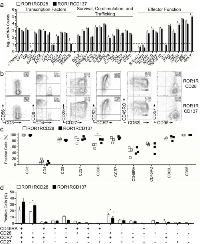
T-cell memory phenotype can be used to predict proliferation, survival, and effector potential of an infused CAR+T-cell product. We used non-enzymatic digital multiplex array of mRNA transcripts (NanoString) to assess the transcriptional profile of CAR+T cells following numeric expansion with AaPC and cytokines. Transcription factors associated with less differentiated T-cell phenotype,e.g.,CTNNB1(β-Catenin),GFI1(Growth Factor Independence-1),ID2 (In-hibitor of DNA Binding-2),BACH2(BTB and CNC Homology-2),KLF2(Kruppel-like Factor-2),FOXO1(Forkhead Box-O1), andLEF1(Lymphoid Enhancer Binding Factor-1), were ex-pressed by populations of ROR1-specific T cells concurrently with transcription factors associ-ated with T cells in later memory stages, includingBCL6(B-cell Lymohoma-6),PRDM1
Fig 1. Evaluation of ROR1 expression on tumor cells, pancreatic cancer, and healthy pancreas.(a) Flow cytometry plots of samples stained with 4A5 mouse-anti-human ROR1 mAb (filled histograms) or matched isotype control (open histograms). Cell types tested were: EL4 murine T-cell lymphoma cell line, EL4 cells genetically modified to express ROR1 (EL4-ROR1), PBMC from patient with CLL diagnosis, non-t(1;19) B-ALL cell line NALM-6, t(1;19) B-ALL cell line Kasumi-2, and ovarian cancer cell lines A2780, EFO27, and OC314. (b) Fresh frozen sections of malignant and normal pancreas were stained with 4A5 mAb or isotype mAb, and developed using an anti-mouse secondary antibody conjugated with horse radish peroxidase. Tissues were visualized with diaminobenzidine and counterstained with hematoxylin. Slides were evaluated by the study pathologist to identify the tissue or cell-type stained and intensity of staining.
(BLIMP-1), andTBX21(T-bet), suggesting that the outgrowth of CAR+T cells was heteroge-neous in memory gene regulation (Fig 3aleft). Quantities of transcription factorsID3 (Inhibi-tor of DNA Binding-3; reciprocal and upstream toID2),TCF7(T-cell Factor-1; TCF1; reciprocal to and upstream ofLEF1), andEOMES(Eomesodermin; similar in function to T-bet) were at or below the limit-of-detection, indicating that coordination through some of the gene regulation cascades had occurred during or prior to expansion on AaPC. Thus, CAR+T cells had a continuum of memory transcription factor expression. Genes associated with intrin-sic memory T-cell survival,e.g.,BCL2(B-cell Lymphoma-2) andBCLXL(BCL2-related protein long isoform), and receptors for cytokines responsible for extrinsic memory T-cell survival and proliferation,e.g.,IL2RA(IL-2-Receptor-α; CD25),IL2RB(IL-2-Receptor-β; CD122),IL2RG
(IL-2-Receptor-γ; CD132),IL7R(IL-7-Receptor-α; CD127), andIL15RA(IL-15-Receptor-α),
Fig 2. Sustained numeric expansion of ROR1-specific CAR+T cells upon clone#1 AaPC and cytokines.(a) DNA plasmid vector maps for
ROR1RCD28/pSBSO-SIM and ROR1RCD137/pSBSO-FRA. Abbreviations are IR/DR:Sleeping BeautyInverted Repeat/Direct Repeat, phElF1α: Human Elongation Factor-1-αregion hybrid promoter, ROR1RCD28: Human codon-optimized ROR1-specific scFv:Fc:CD28:CD3ζCAR, ROR1RCD137: Human codon-optimized ROR1-specific scFv:Fc:CD137:CD3ζCAR, SIM:“SIM”PCR tracking oligonucleotides, FRA:“FRA”PCR tracking oligonucleotides, BGHpA; bovine growth hormone polyadenylation sequence, Ori: minimalE.coliorigin of replication, KanR: Bacterial selection gene encoding Kanamycin resistance, pKan: prokaryotic Kanamycin promoter. Digestion withBsrGIenzyme can distinguish the two plasmids, which have high degrees of similarity, and withPmlI enzyme can distinguish them from CD19RCD28 plasmid, which does not havePmlIsite. The entire plasmid sequences were verified by Sanger-based sequencing techniques. (b) Parental K-562 (open histograms) and clone#1 AaPC (shaded histograms) were stained for CD19, CD86, CD137L, ROR1, and IL15 (membrane-bound IL-15; mIL15) where isotype (IgG) was used a negative control. (c) Expression CAR in ROR1RCD28 (middle) and ROR1RCD137 (right) T cells the day following electroporation (top panels) and after 28 days of co-culture on clone#1 AaPC (bottom panels) where“no DNA”T cells (left) were used as negative controls. T cells were marked by CD3 staining and CAR+cells were detected with Fc-specific antibody. Quadrant frequencies are
displayed in upper right corners. (d) Proliferation kinetics of total cells (circles) and CAR+T cells (squares) on clone#1 AaPC over 28 days of co-culture.
ROR1RCD28 displayed on the left and ROR1RCD137 shown on the right. Arrows represent addition ofγ-irradiated clone#1 AaPC. Data are representative of 3 donors expanded in 3 independent experiments. Expansion data can be found inS1 Dataset, Fig 2 tab.
were detected in CAR+T cells suggesting that persistence and survival of these cells following adoptive transfer is plausible (Fig 3amiddle). Memory-associated T-cell trafficking molecules,
e.g.,SELL(L-Selectin; CD62L),CCR7(CCR7),CXCR3(CXCR3), andCCR5(CCR5), and lym-phocyte co-stimulatory molecules,e.g.,TNFRSF7(CD27) andCD28(CD28), were detected in ROR1RCD28+and ROR1RCD137+T cells, indicating that the CAR+T cells may have been de-rived from or developed into T-cell memory cells. Importantly, genes associated with exhaus-tion and terminal differentiaexhaus-tion, includingB3GAT1(Beta-1,3-Glucuronyltransferase-1; CD57) andKLRG1(KLRG1) were not expressed above the limit-of-detection by the CAR+T cells (Fig 3aright). Genes encoding surface receptors leading to killer function,e.g.,NCAM1
(Neural cell adhesion molecule-1; CD56) andNKG2D(NKG2D), cytokines with pro-inflam-matory properties,e.g.,IFNG(Interferon-γ) andTNF(Tumor Necrosis Factor), and molecules responsible for directing cell death and/or cytolysis,e.g.,TNFSF6(Fas-ligand),TNFSF10
(TRAIL),GZMA(Granzyme-A),GZMB(Granzyme-B),PRF1(Perforin-1), andGNLY (Gran-ulysin), were expressed by CAR+T cells suggesting that these cells could have effector function in addition to their potential for memory formation. In aggregate, CAR+T cells which emerge after recurrent additions of AaPC and IL-2/IL-21 appear to contain sub-populations with de-sirable gene expression predictive of therapeutic efficacy after adoptive transfer.
CAR
+T cells express surface markers consistent with T-cell memory
Flow cytometry was used to validate the NanoString panel, examine the surface expression of canonical T-cell markers, and determine the frequencies of memory populations with multi-parameter staining. CAR+T-cell populations expressed CD56 (NCAM1), although CD3negCD56+NK cells were absent from final cultures (Fig 3b, first panels). A preponderance of CD8+T cells was present in the cultures relative to CD4+T cells (Fig 3b, second panels), and
>99% of cells expressed CD3. High frequencies of CAR+T cells were found co-expressing
CD28 and CD27 co-stimulatory molecules (Fig 3b, third panels), which are characteristics of T-cell memory. CD27, in particular, has been correlated to durable regressions resulting from CD8+T-cell treatments [41]. The stemness of CD8+memory cells has been linked to expres-sion of L-selectin [12], which was detected in CAR+T cell’s mRNA (SELL) and as protein (CD62L), and was concurrently expressed with the associated lymphoid homing marker CCR7 (Fig 3b, fourth panels). Thus, such CAR+T cells with long-lived potential could migrate to sec-ondary lymphoid structures harboring ROR1+leukemias. Few CD45RO+CD62Lnegcells were detected, which are generally associated with TEMor late-stage effector memory cells, and
could be contrasted to the larger abundance of CD45ROnegCD62L+cells usually associated with less differentiated T cells (Fig 3b, fifth panels). Virtually all cells expressed CD95, a marker for activation and differentiation beyond TNlineages, which was expected on T cells culturedex
vivo, but the same T cells did not express CD57, a marker of exhaustion (Fig 3b, sixth panels). In aggregate, the staining of these markers was consistent amongst donors (Fig 3c). Multi-parame-ter flow cytometry revealed that most electroporated/propagated T cells belonged to less-differ-entiated memory phenotype primarily composed of TSCM(CD45RA+CD27+CD28+CCR7+) and
TCM(CD45RAnegCD27+CD28+CCR7+) (Fig 3d) [42,43]. There were appreciable frequencies of
CD62L and CCR7, CD45RO and CD62L, or CD95 and CD57 in cells gated for CAR expression based staining with Fc-specific antibody. ROR1RCD28+T cells are shown in the top panels and ROR1RCD137 T cells are displayed in the bottom panels. One of 3 representative donors is displayed and quadrant frequencies are shown in the upper right corners. (c) Cumulative frequencies of cells staining positive for each memory marker within an extended memory phenotype panel. (d) Multi-parameter flow cytometry was used to determine frequencies of cells staining positive for combinations of CD45RA, CD27, CD28, and CCR7. For (c) and (d) ROR1RCD28 are in open shapes/bars and ROR1RCD137 are in closed shapes/bars and lines displayed in (c) are means (n = 3) and in (d) are mean±SD (n = 3). Student’s two-tailed t-tests were used for statistical analysis between the two groups.*p<0.05 Expression and phenotype data can be found inS1 Dataset, Fig 3 tab.
CD45RA+CD27negCD28+CCR7+and CD45RAnegCD27negCD28+CCR7+, which we were not able to define as a specific memory subset, and almost no TEMRAcells (CD45RA+CD27neg CD28-negCCR7neg). In aggregate, the surface phenotype of ROR1-specific CAR+T cells corroborated
the mRNA digital barcoding profiling data (NanoString) and indicated that these cells have de-sirable characteristics for fighting ROR1+malignancies.
ROR1-specific CAR
+T cells produce IFN
γ
in response to ROR1
+tumor
cells
We monitored for the production of IFNγby intracellular cytokine staining of healthy donor CAR+T cells following 6 hour co-culture with target cells. Both ROR1RCD28+and
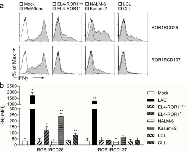
ROR1RCD137+T cells produced significant quantities of IFNγwhen treated with PMA/Iono-mycin (non-specific mitogenic stimulus; positive control) relative to mock-activated (media only; negative control) cultures (Fig 4). ROR1RCD28+T cells stained brightly for IFNγwhen co-cultured with ROR1+tumor targets EL4-ROR1, Kasumi-2, and CLL cells, but did not pro-duce IFNγ, as evidenced by similar staining of unchallenged cultures (mock; media only) to co-cultures with ROR1negcells from EBV-transformed healthy donor B-cell lymphoblastoid cell lines (LCL), NALM-6, and parental EL4 cells (Fig 4atop). IFNγproduction by ROR1RCD137+ T cells slightly increased only in response to Kasumi-2 cells, demonstrating that there were dif-ferences in the effector cytokine production of the two CAR populations to certain tumor cells (Fig 4abottom). In summary, effector function of CAR+T cells, as manifested by IFNγ expres-sion, was restricted to ROR1+tumor cells and ROR1RCD28+T cells produced more IFNγthan did ROR1RCD137+T cells in response to ROR1.
ROR1-specific killing by CAR
+T cells
in vitro
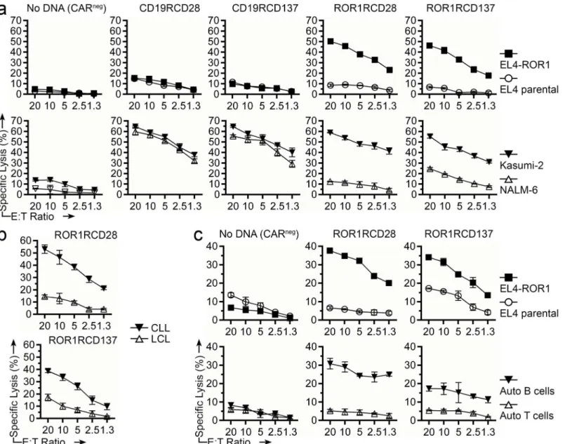
The killing of tumor cells by genetically modified T cells is another measurement of re-directed specificity. Four-hour chromium release assay was used to assess the specific lysis of tumor-cell lines and primary tumor cells. ROR1RCD28+and ROR1RCD137+T cells derived from healthy donor PBMC efficiently lysed EL4-ROR1+and Kasumi-2 but showed minimal lysis of EL4 pa-rental (ROR1neg) and NALM-6 cells (Fig 5a). Furthermore, autologous CD19RCD28+and CD19RCD137+T cells displayed minimal lysis of EL4 and EL4-ROR1 but lysed both Kasumi-2 and NALM-6 (both CD19+) suggesting that the ROR1RCD28+and ROR1RCD137+T cells were more discriminant in their targeting of these B-ALL cell lines (Fig 5abottom). Autologous CARnegT cells (No DNA) showed no lysis of these 4 cell lines indicating that the CAR responses were specific. ROR1RCD28+and ROR1RCD137+T cells generated from healthy donor PBMC also lysed allogeneic CLL patient B cells (ROR1+), but spared allogeneic LCL (ROR1neg) derived from healthy donor B-cells (Fig 5b). Similarly, ROR1RCD28+and ROR1RCD137+T cells gener-ated from PBMC of a patient with CLL were able to lyse EL4-ROR1 and autologous tumor cells (ROR1+; isolated with CD19-specific magnetic beads), while autologous T cells and parental EL4 (both ROR1neg) cells were spared (Fig 5c). In summary, ROR1-specific CAR+T cells dem-onstrated effectivein vitrospecific lysis of ROR1+tumor cells in both autologous and allogeneic settings.
In vivo
clearance of leukemia by ROR1-specific CAR
+T cells
firefly luciferase(ffLuc) [44] for serial non-invasive bioluminescence imaging (BLI) of tumor burdenin vivo. NOD.scid.γc-/-(NSG) mice were used because they lack functional adaptive im-mune systems and can therefore accept human tumor xenografts. Healthy, 6–12 week old fe-male NSG mice were engrafted with Kasumi-2-ffLuc-mKate and distributed into 3 treatment groups (n = 5 per group): (i) no treatment, (ii) ROR1RCD28+T-cell treated, and (iii)
ROR1RCD137+T-cell treated, where T cells were given at days 1, 8, and 15 post-tumor cell en-graftment (Fig 6). Untreated mice had consistent log10-fold increases in bioluminescence flux
from their tumors (Fig 6ablack circles and6btop) and succumbed to disease after an average of 27 days following engraftment (Fig 6cblack circles). ROR1RCD28+T cells were able to di-minish tumor burden compared to untreated mice as measured by tumor BLI flux (Fig 6ablue squares and6bmiddle) and prolonged survival compared to untreated mice (Fig 6cblue squares). ROR1RCD137+T cells eliminated tumor burden above both untreated mice and ROR1RCD28+T cell-treated mice (Fig 6cred triangles and6bbottom), and were able to in-crease survival compared to both untreated mice and ROR1RCD28+T cell-treated (Fig 6cred
Fig 4. IFNγproduction by ROR1-specific CAR+T cells in response to ROR1+targets.At day 29 of co-culture with AaPC/IL2/IL-21, CAR+T cells were
co-cultured for 6 hours at 37°C with tumor targets or non-specific mitogenic stimuli (PMA/Iono) then analyzed for expression of IFNγin CAR+T cells (gated
based on Fc expression). Brefeldin-A (GolgiPlug) was added to T cells to block IFNγsecretion. (a) Representative flow cytometry plots where ROR1RCD28 cultures are on the top and ROR1RCD137 cultures are on the bottom. Percentage of max (y-axes) normalized total cell numbers in each sample for consistency between samples so that IFNγfluorescent intensity (x-axes) could be compared between conditions. (b) Cumulative mean fluorescence intensities (MFI) of IFNγstaining from co-cultures where mean±SD (n = 3 donors) is shown. Student’s paired, 1-tailed t-test for statistical analysis between target co-culture and T cells alone (Mock; media only).*p<0.05 and**p<0.01 IFNγdata can be found inS1 Dataset, Fig 4 tab.
triangles). In summary, ROR1-specific CAR+T cells can efficiently treat ROR1+leukemia in mice which supports testing of these T cells in the clinic as an investigational therapy for pa-tients with ROR1+malignancies.
Discussion
This work reveals the pre-clinical basis to support our current“first-in-human”Phase I clinical trial of CAR+T cells for investigational therapy of ROR1+malignancies. We demonstrate that
Fig 5. Specific cytolysis of ROR1+tumor cells by CAR+T cells.Four-hour chromium release assay was used to assess specific lysis by T cells at decreasing effector to target (E:T) ratios. (a) No DNA (CARneg), CD19RCD28+, CD19RCD137+, ROR1RCD28+, or ROR1RCD137+T cells generated from
healthy donor PBMC were challenged with EL4-ROR1+cell line (closed squares), EL4 parental (ROR1neg) cell line (open circles), Kasumi-2 cell line (closed
inverted triangles), or NALM-6 cell line (open triangles). (b) CAR+T cells generated from healthy donor PBMC were challenged with allogeneic CLL cells
(closed inverted triangles), healthy donor allogeneic B-cell LCL (open triangles). (c) No DNA (CARneg), ROR1RCD28+, or ROR1RCD137+T cells generated from CLL patient PBMC were challenged with EL4-ROR1+cell line (closed squares), EL4 parental (ROR1neg) cell line (open circles), autologous B cells
(closed inverted triangles), or autologous T cells (open triangles). Data are mean±SD of triplicate measurements in CRA and are representative of four donors from four independent experiments. Lysis data can be found inS1 Dataset, Fig 5 tab.
SB transposition achieved stable CAR expression in T cells and, in concert with co-culture on clone#1 AaPC, resulted in heterogeneous outgrowth of T-cell memory populations with ROR1-restricted anti-tumor activity. Currently, this process takes 28 days to achieve clinical numbers of T cells. Potentially shortening the production time for cell growth may (i) simplify the overall process, (ii) preserve more of the minimally differentiated subsets, and (iii) could take advantage ofin vivoproliferation of CAR+T cells thereby augmenting therapeutic efficacy. As SB transposition was used to express ROR1-specific CARs in naïve or minimally-differenti-ated T-cell populations present in quiescent PBMC, as has been done with other SB-based CAR studies [31,38], the prospect of giving T cells earlier in the proliferative cycle is of great interest to our group. Homeostatic cytokines,e.g., IL-15, can promote the survival of less differ-entiated T cells, and trans-presentation of IL-15 by IL15Rαhas higher signaling potency than IL-15 alone [45,46]. Thus, we chose to introduce mIL15 (IL-15/IL-15Rαfusion protein) rather than IL-15 tethered to a hinge/Fc stalk, which is the moiety expressed on AaPC used to gener-ate CAR+T cells expressing CD19RCD28 [47], on the clone#1 AaPC. The AaPC feeder plat-form, which is amenable to genetic introduction of TAAs (e.g., ROR1) and molecules that promote T-cell survival (e.g., mIL15), in concert with SB transposition can support the propa-gation of populations of T cells predicted to have sustained engraftment,i.e., TSCMand
CD62L+TCMpopulations [12,48], while enforcing CAR expression on T cells through
recur-sive antigen exposure; therefore, they are attractive platforms for the generation of ROR1-spe-cific T cells for cancer immunotherapy.
The choice of signaling motif within a 2ndgeneration CAR endodomain may impact the therapeutic activity of infused T cells. CD19-specific CARs activating T cells via chimeric CD28 or CD137 endodomains led to objective clinical responses [3–7]. We designed CARs that used the same extracellular structure in order to reveal differences arising between the endodomains. The most notable differences were that ROR1RCD137+T cells displayed
Fig 6.In vivotumor clearance by ROR1-specific CAR+T cells.NSG mice were intravenously (i.v.) injected with Kasumi-2-ffLuc-mKate cells and were treated with three i.v. doses of T cells to assess the ability of ROR1-specific T cells to manage disease. High-dose IL-2 was added intraperitoneally the day of injection and the following day. (a) Non-invasive BLI reported as flux was serially measured in untreated (black circles), ROR1RCD28+T cell-treated (blue
squares), and ROR1RCD137+T cell-treated (red triangles) mice. Data are mean±SEM (n = 5). Two-way ANOVA was used for statistical analysis. (b) Representative BLI images at day +23 post-tumor cell engraftment. (c) Overall survival of mice.*p<0.05,**p<0.01,***p<0.001 BLI and survival data can be found in Supporting Information, Fig 6 tab.
enhancedin vivotumor clearance (Fig 6) despite showing reduced IFNγproductionin vitro
relative to ROR1RCD28+T cells (Fig 4). The decrease in IFNγ-response with CARs signaling through CD137 relative to those signaling through CD28 is consistent with independent stud-ies comparing ROR1-specific CARs [25,39,49,50]. These studies introduced CARs derived from different ROR1-specific antibodies (2A2 and R12) into TCMcells with lentivirus and
showed that CD137 signaling resulted in reduced cytokine production and increased anti-tumor activityin vivo[50]. CARs with short extracellular spacers, especially those without Fc stalks that could be bound by cells expressing CD64 (high-affinity Fc receptor), were optimal for other ROR1 CARs (2A2 and R12 moieties) [49]. Future studies from our group will evalu-ate whether truncation or replacement of the Fc stalk in CARs constructed from 4A5 mAb, in-troduced into T cells with SB, and cultured on AaPC will produce similar benefits. An inverse correlation betweenin vitroeffector function (cytokine production and cytolysis) andin vivo
tumor clearance by CD8+tumor-specific T cells has also been established for adoptive immu-notherapy of melanoma, further supporting the notion thatex vivoculture conditions and memory phenotype of infused T cells are useful parameters to correlate with therapeutic effica-cy [11]. A side-by-side comparison of the two CARs using a competitive repopulation experi-ment in a clinical trial is likely needed to reveal which CAR design is superior for a given tumor and perhaps even a particular patient.
A major advantage of targeting ROR1 over the current T-cell therapies targeting CD19 is that recipients would not deplete B cells and develop hypogammaglobulinemia, thereby miti-gating the risk for impaired humoral immunity [32,51]. ROR1 was originally identified on the surface of CLL cells with absent expression on normal tissues, including hematopoietic cells [18,24]. Subsequently, ROR1 was described on some acute leukemias and solid tumors [19,20,
34]. The 4A5 ROR1-specific mAb (used in this study to construct the CAR) did not detect ROR1 in healthy tissues by either immunoblot or immunohistochemistry (Fig 1b) [18,20]. The only healthy cells to be stained with 4A5 mAb are hematogones, which are rare pre-B-cells that do not appear to be obligate precursors to mature B cells [24]. There has been some discor-dance between the mRNA transcript data and protein staining data, which may be explained by off targeting of ROR2 gene, which is expressed after birth in many tissues, and is highly ho-mologous to ROR1. Given that ROR1-specific T cells propagated in this study have potential for long-term engraftment due to their memory T-cell phenotype, it is plausible that patients treated with these genetically modified T cells could experience prolonged anti-tumor effects with potential for elimination of minimal disease or relapsed ROR1+malignancies. As a control for adverse events, conditional suicide genes,e.g., inducible Caspase9, could be co-expressed with CAR in order to eliminate T cellsin vivoas needed [52].
In summary, our data support the clinical application of ROR1-specific CAR+T cells and a Phase I clinical trial is open to patients with CLL as of July 2014 (NCT02194374). Clinical-grade DNA plasmids coding for ROR1RCD137 and SB11 have been produced and a master-cell bank of AaPC clone#1 has been generated. Furthermore, regulatory documents and mate-rials are in place to undertake the clinical trial. This will be the first time ROR1 on CLL has been targeted by T cells; therefore, the primary endpoint will be to determine toxicity and max-imum tolerated T-cell dose. Future trials can then assess the clinical impact of targeting ROR1 on solid tumors.
Supporting Information
multiparameter memory phenotype of T cells (bottom). (Fig 4tab) MFI of IFNγstaining of CAR+T cells following 6 hour co-culture with target cells. (Fig 5tab) 4-hour chromium release assay of T cells co-cultured with target cells. (Fig 6tab) BLI flux kinetics of Kasumi2-ffLuc-mKate cells following challenge with CAR+T cells (top) and days of mouse euthanasia (bottom). (XLSX)
Acknowledgments
We thank Dr. Carl June and colleagues (University of Pennsylvania) for help generating K-562-derived AaPC clone #9 and Dr. Perry Hackett (University of Minnesota) for his assistance with the SB system. The flow cytometry core lab (MDACC) assisted with FACS. We thank Dr. Eric Tran (NCI Surgery Branch) for editing this document. Drew C. Deniger was an American Legion Auxiliary Fellow in Cancer Research (UT-GSBS-Houston), Andrew Sowell-Wade Hug-gins Scholar in Cancer Research (Cancer Answers Foundation), and a Teal Pre-doctoral Schol-ar (DepSchol-artment of Defense OvSchol-arian Cancer ReseSchol-arch Program) during this study.
Author Contributions
Conceived and designed the experiments: DCD REC WGW TJK LJNC. Performed the experi-ments: DCD JY MHH MJF TM SNM GFW RT. Analyzed the data: DCD MHH MJF TM JY GFW. Contributed reagents/materials/analysis tools: DCD JY LVH GFW HS SO WGW TJK. Wrote the paper: DCD WGW TKJ LJNC.
References
1. Jena B, Dotti G, Cooper LJ. Redirecting T-cell specificity by introducing a tumor-specific chimeric anti-gen receptor. Blood. 2010; 116(7):1035–44. Epub 2010/05/05. doi: blood-2010-01-043737 [pii]doi:10. 1182/blood-2010-01-043737PMID:20439624; PubMed Central PMCID: PMC2938125.
2. June CH. Principles of adoptive T cell cancer therapy. J Clin Invest. 2007; 117(5):1204–12. Epub 2007/ 05/04. doi:10.1172/JCI31446PMID:17476350; PubMed Central PMCID: PMC1857246.
3. Grupp SA, Kalos M, Barrett D, Aplenc R, Porter DL, Rheingold SR, et al. Chimeric antigen receptor-modified T cells for acute lymphoid leukemia. N Engl J Med. 2013; 368(16):1509–18. Epub 2013/03/27. doi:10.1056/NEJMoa1215134PMID:23527958.
4. Brentjens RJ, Riviere I, Park JH, Davila ML, Wang X, Stefanski J, et al. Safety and persistence of adop-tively transferred autologous CD19-targeted T cells in patients with relapsed or chemotherapy refracto-ry B-cell leukemias. Blood. 2011; 118(18):4817–28. Epub 2011/08/19. doi: 10.1182/blood-2011-04-348540PMID:21849486; PubMed Central PMCID: PMC3208293.
5. Kochenderfer JN, Dudley ME, Feldman SA, Wilson WH, Spaner DE, Maric I, et al. B-cell depletion and remissions of malignancy along with cytokine-associated toxicity in a clinical trial of anti-CD19 chime-ric-antigen-receptor-transduced T cells. Blood. 2012; 119(12):2709–20. Epub 2011/12/14. doi:10. 1182/blood-2011-10-384388PMID:22160384; PubMed Central PMCID: PMC3327450.
6. Kalos M, Levine BL, Porter DL, Katz S, Grupp SA, Bagg A, et al. T cells with chimeric antigen receptors have potent antitumor effects and can establish memory in patients with advanced leukemia. Sci Transl Med. 2011;3(95: ):95ra73. Epub 2011/08/13. doi: 3/95/95ra73 [pii]doi:10.1126/scitranslmed.3002842
PMID:21832238.
7. Porter DL, Levine BL, Kalos M, Bagg A, June CH. Chimeric antigen receptor-modified T cells in chronic lymphoid leukemia. N Engl J Med. 2011; 365(8):725–33. Epub 2011/08/13. doi:10.1056/
NEJMoa1103849PMID:21830940.
8. Cruz CR, Micklethwaite KP, Savoldo B, Ramos CA, Lam S, Ku S, et al. Infusion of donor-derived CD19-redirected virus-specific T cells for B-cell malignancies relapsed after allogeneic stem cell trans-plant: a phase 1 study. Blood. 2013; 122(17):2965–73. Epub 2013/09/14. doi: 10.1182/blood-2013-06-506741PMID:24030379; PubMed Central PMCID: PMC3811171.
10. Lee DW, Kochenderfer JN, Stetler-Stevenson M, Cui YK, Delbrook C, Feldman SA, et al. T cells ex-pressing CD19 chimeric antigen receptors for acute lymphoblastic leukaemia in children and young adults: a phase 1 dose-escalation trial. Lancet. 2015; 385(9967):517–28. doi:10.1016/S0140-6736(14) 61403-3PMID:25319501.
11. Gattinoni L, Klebanoff CA, Palmer DC, Wrzesinski C, Kerstann K, Yu Z, et al. Acquisition of full effector function in vitro paradoxically impairs the in vivo antitumor efficacy of adoptively transferred CD8+ T cells. J Clin Invest. 2005; 115(6):1616–26. Epub 2005/06/03. doi:10.1172/JCI24480PMID:15931392; PubMed Central PMCID: PMC1137001.
12. Graef P, Buchholz VR, Stemberger C, Flossdorf M, Henkel L, Schiemann M, et al. Serial transfer of sin-gle-cell-derived immunocompetence reveals stemness of CD8(+) central memory T cells. Immunity. 2014; 41(1):116–26. Epub 2014/07/19. doi:10.1016/j.immuni.2014.05.018PMID:25035956. 13. Jolles S, Sewell WA, Misbah SA. Clinical uses of intravenous immunoglobulin. Clin Exp Immunol.
2005; 142(1):1–11. doi:10.1111/j.1365-2249.2005.02834.xPMID:16178850; PubMed Central PMCID: PMC1809480.
14. Brentjens R, Yeh R, Bernal Y, Riviere I, Sadelain M. Treatment of chronic lymphocytic leukemia with genetically targeted autologous T cells: case report of an unforeseen adverse event in a phase I clinical trial. Mol Ther. 2010; 18(4):666–8. Epub 2010/04/02. doi:10.1038/mt.2010.31PMID:20357779; PubMed Central PMCID: PMC2862525.
15. Nomi M, Oishi I, Kani S, Suzuki H, Matsuda T, Yoda A, et al. Loss of mRor1 enhances the heart and skeletal abnormalities in mRor2-deficient mice: redundant and pleiotropic functions of mRor1 and mRor2 receptor tyrosine kinases. Mol Cell Biol. 2001; 21(24):8329–35. Epub 2001/11/20. doi:10.1128/ MCB.21.24.8329–8335.2001PMID:11713269; PubMed Central PMCID: PMC99997.
16. Baskar S, Kwong KY, Hofer T, Levy JM, Kennedy MG, Lee E, et al. Unique cell surface expression of receptor tyrosine kinase ROR1 in human B-cell chronic lymphocytic leukemia. Clin Cancer Res. 2008; 14(2):396–404. Epub 2008/01/29. doi:10.1158/1078-0432.CCR-07-1823PMID:18223214.
17. Daneshmanesh AH, Mikaelsson E, Jeddi-Tehrani M, Bayat AA, Ghods R, Ostadkarampour M, et al. Ror1, a cell surface receptor tyrosine kinase is expressed in chronic lymphocytic leukemia and may serve as a putative target for therapy. Int J Cancer. 2008; 123(5):1190–5. Epub 2008/06/12. doi:10. 1002/ijc.23587PMID:18546292.
18. Fukuda T, Chen L, Endo T, Tang L, Lu D, Castro JE, et al. Antisera induced by infusions of autologous Ad-CD154-leukemia B cells identify ROR1 as an oncofetal antigen and receptor for Wnt5a. Proc Natl Acad Sci U S A. 2008; 105(8):3047–52. Epub 2008/02/22. doi:10.1073/pnas.0712148105PMID:
18287027; PubMed Central PMCID: PMC2268582.
19. Zhang S, Chen L, Cui B, Chuang HY, Yu J, Wang-Rodriguez J, et al. ROR1 is expressed in human breast cancer and associated with enhanced tumor-cell growth. PLoS One. 2012; 7(3):e31127. Epub 2012/03/10. doi:10.1371/journal.pone.0031127PMID:22403610; PubMed Central PMCID: PMC3293865.
20. Zhang S, Chen L, Wang-Rodriguez J, Zhang L, Cui B, Frankel W, et al. The onco-embryonic antigen ROR1 is expressed by a variety of human cancers. Am J Pathol. 2012; 181(6):1903–10. Epub 2012/10/ 09. doi:10.1016/j.ajpath.2012.08.024PMID:23041612; PubMed Central PMCID: PMC3509760. 21. Rebagay G, Yan S, Liu C, Cheung NK. ROR1 and ROR2 in Human 1Malignancies: Potentials for
Tar-geted Therapy. Front Oncol. 2012; 2:34. Epub 2012/06/02. doi:10.3389/fonc.2012.00034PMID:
22655270; PubMed Central PMCID: PMC3356025.
22. Zhang S, Cui B, Lai H, Liu G, Ghia EM, Widhopf GF 2nd, et al. Ovarian cancer stem cells express ROR1, which can be targeted for anti-cancer-stem-cell therapy. Proc Natl Acad Sci U S A. 2014; 111 (48):17266–71. doi:10.1073/pnas.1419599111PMID:25411317; PubMed Central PMCID: PMC4260559.
23. Dave H, Anver MR, Butcher DO, Brown P, Khan J, Wayne AS, et al. Restricted cell surface expression of receptor tyrosine kinase ROR1 in pediatric B-lineage acute lymphoblastic leukemia suggests target-ability with therapeutic monoclonal antibodies. PLoS One. 2012; 7(12):e52655. Epub 2013/01/04. doi:
10.1371/journal.pone.0052655PMID:23285131; PubMed Central PMCID: PMC3527582.
24. Broome HE, Rassenti LZ, Wang HY, Meyer LM, Kipps TJ. ROR1 is expressed on hematogones (non-neoplastic human B-lymphocyte precursors) and a minority of precursor-B acute lymphoblastic leuke-mia. Leuk Res. 2011; 35(10):1390–4. Epub 2011/08/05. doi:10.1016/j.leukres.2011.06.021PMID:
21813176; PubMed Central PMCID: PMC3163753.
26. Hackett PB, Largaespada DA, Cooper LJ. A transposon and transposase system for human applica-tion. Mol Ther. 2010; 18(4):674–83. Epub 2010/01/28. doi: mt20102 [pii]doi:10.1038/mt.2010.2PMID:
20104209; PubMed Central PMCID: PMC2862530.
27. Hackett PB, Largaespada DA, Switzer KC, Cooper LJ. Evaluating risks of insertional mutagenesis by DNA transposons in gene therapy. Transl Res. 2013; 161(4):265–83. Epub 2013/01/15. doi:10.1016/j. trsl.2012.12.005PMID:23313630; PubMed Central PMCID: PMC3602164.
28. Huls MH, Figliola MJ, Dawson MJ, Olivares S, Kebriaei P, Shpall EJ, et al. Clinical application of Sleep-ing Beauty and artificial antigen presentSleep-ing cells to genetically modify T cells from peripheral and umbili-cal cord blood. J Vis Exp. 2013;(72: ):e50070. Epub 2013/02/15. doi:10.3791/50070PMID:23407473; PubMed Central PMCID: PMC3596954.
29. Maiti SN, Huls H, Singh H, Dawson M, Figliola M, Olivares S, et al. Sleeping beauty system to redirect T-cell specificity for human applications. J Immunother. 2013; 36(2):112–23. Epub 2013/02/05. doi:10. 1097/CJI.0b013e3182811ce9PMID:23377665; PubMed Central PMCID: PMC3568214.
30. Singh H, Figliola MJ, Dawson MJ, Olivares S, Zhang L, Yang G, et al. Manufacture of Clinical-Grade CD19-Specific T Cells Stably Expressing Chimeric Antigen Receptor Using Sleeping Beauty System and Artificial Antigen Presenting Cells. PLoS One. 2013; 8(5):e64138. Epub 2013/06/07. doi:10.1371/ journal.pone.0064138PMID:23741305; PubMed Central PMCID: PMC3669363.
31. Singh H, Huls H, Kebriaei P, Cooper LJ. A new approach to gene therapy using Sleeping Beauty to ge-netically modify clinical-grade T cells to target CD19. Immunol Rev. 2014; 257(1):181–90. Epub 2013/ 12/18. doi:10.1111/imr.12137PMID:24329797.
32. Singh H, Figliola MJ, Dawson MJ, Huls H, Olivares S, Switzer K, et al. Reprogramming CD19-Specific T Cells with IL-21 Signaling Can Improve Adoptive Immunotherapy of B-Lineage Malignancies. Cancer Res. 2011; 71(10):3516–27. Epub 2011/05/12. doi: 0008-5472.CAN-10-3843 [pii]doi: 10.1158/0008-5472.CAN-10-3843PMID:21558388; PubMed Central PMCID: PMC3096697.
33. Singh H, Manuri PR, Olivares S, Dara N, Dawson MJ, Huls H, et al. Redirecting specificity of T-cell pop-ulations for CD19 using the Sleeping Beauty system. Cancer Res. 2008; 68(8):2961–71. Epub 2008/ 04/17. doi: 68/8/2961 [pii]doi:10.1158/0008-5472.CAN-07-5600PMID:18413766; PubMed Central PMCID: PMC2424272.
34. Bicocca VT, Chang BH, Masouleh BK, Muschen M, Loriaux MM, Druker BJ, et al. Crosstalk between ROR1 and the Pre-B cell receptor promotes survival of t(1;19) acute lymphoblastic leukemia. Cancer Cell. 2012; 22(5):656–67. Epub 2012/11/17. doi:10.1016/j.ccr.2012.08.027PMID:23153538; PubMed Central PMCID: PMC3500515.
35. Suhoski MM, Golovina TN, Aqui NA, Tai VC, Varela-Rohena A, Milone MC, et al. Engineering artificial antigen-presenting cells to express a diverse array of co-stimulatory molecules. Mol Ther. 2007; 15 (5):981–8. Epub 2007/03/22. doi:10.1038/mt.sj.6300134PMID:17375070.
36. O'Connor CM, Sheppard S, Hartline CA, Huls H, Johnson M, Palla SL, et al. Adoptive T-cell therapy im-proves treatment of canine non-Hodgkin lymphoma post chemotherapy. Sci Rep. 2012; 2:249. Epub 2012/02/23. doi:10.1038/srep00249PMID:22355761; PubMed Central PMCID: PMC3278154. 37. Deniger DC, Maiti SN, Mi T, Switzer KC, Ramachandran V, Hurton LV, et al. Activating and propagating
polyclonal gamma delta T cells with broad specificity for malignancies. Clin Cancer Res. 2014; 20 (22):5708–19. doi:10.1158/1078-0432.CCR-13-3451PMID:24833662; PubMed Central PMCID: PMC4233015.
38. Deniger DC, Switzer K, Mi T, Maiti S, Hurton L, Singh H, et al. Bispecific T-cells expressing polyclonal repertoire of endogenous gammadelta T-cell receptors and introduced CD19-specific chimeric antigen receptor. Mol Ther. 2013; 21(3):638–47. Epub 2013/01/09. doi:10.1038/mt.2012.267PMID:
23295945; PubMed Central PMCID: PMC3589159.
39. Hudecek M, Schmitt TM, Baskar S, Lupo-Stanghellini MT, Nishida T, Yamamoto TN, et al. The B-cell tumor-associated antigen ROR1 can be targeted with T cells modified to express a ROR1-specific chi-meric antigen receptor. Blood. 2010; 116(22):4532–41. Epub 2010/08/13. doi: 10.1182/blood-2010-05-283309PMID:20702778; PubMed Central PMCID: PMC2996114.
40. Kebriaei P, Huls H, Jena B, Munsell M, Jackson R, Lee DA, et al. Infusing CD19-directed T cells to aug-ment disease control in patients undergoing autologous hematopoietic stem-cell transplantation for ad-vanced B-lymphoid malignancies. Hum Gene Ther. 2012; 23(5):444–50. Epub 2011/11/24. doi:10. 1089/hum.2011.167PMID:22107246; PubMed Central PMCID: PMC3360496.
41. Rosenberg SA, Yang JC, Sherry RM, Kammula US, Hughes MS, Phan GQ, et al. Durable complete re-sponses in heavily pretreated patients with metastatic melanoma using T-cell transfer immunotherapy. Clin Cancer Res. 2011; 17(13):4550–7. Epub 2011/04/19. doi:10.1158/1078-0432.CCR-11-0116
42. Di Mitri D, Azevedo RI, Henson SM, Libri V, Riddell NE, Macaulay R, et al. Reversible senescence in human CD4+CD45RA+CD27- memory T cells. J Immunol. 2011; 187(5):2093–100. Epub 2011/07/27. doi:10.4049/jimmunol.1100978PMID:21788446.
43. Klebanoff CA, Gattinoni L, Restifo NP. CD8+ T-cell memory in tumor immunology and immunotherapy. Immunol Rev. 2006; 211:214–24. Epub 2006/07/11. doi:10.1111/j.0105-2896.2006.00391.xPMID:
16824130; PubMed Central PMCID: PMC1501075.
44. Rabinovich BA, Ye Y, Etto T, Chen JQ, Levitsky HI, Overwijk WW, et al. Visualizing fewer than 10 mouse T cells with an enhanced firefly luciferase in immunocompetent mouse models of cancer. Proc Natl Acad Sci U S A. 2008; 105(38):14342–6. Epub 2008/09/17. doi:10.1073/pnas.0804105105PMID:
18794521; PubMed Central PMCID: PMC2567214.
45. Stonier SW, Schluns KS. Trans-presentation: a novel mechanism regulating IL-15 delivery and re-sponses. Immunol Lett. 2010; 127(2):85–92. Epub 2009/10/13. doi:10.1016/j.imlet.2009.09.009PMID:
19818367; PubMed Central PMCID: PMC2808451.
46. Stonier SW, Ma LJ, Castillo EF, Schluns KS. Dendritic cells drive memory CD8 T-cell homeostasis via IL-15 transpresentation. Blood. 2008; 112(12):4546–54. Epub 2008/09/25. doi: 10.1182/blood-2008-05-156307PMID:18812469; PubMed Central PMCID: PMC2597127.
47. Manuri PV, Wilson MH, Maiti SN, Mi T, Singh H, Olivares S, et al. piggyBac transposon/transposase system to generate CD19-specific T cells for the treatment of B-lineage malignancies. Hum Gene Ther. 2010; 21(4):427–37. Epub 2009/11/13. doi:10.1089/hum.2009.114PMID:19905893; PubMed Central PMCID: PMC2938363.
48. Gattinoni L, Lugli E, Ji Y, Pos Z, Paulos CM, Quigley MF, et al. A human memory T cell subset with stem cell-like properties. Nat Med. 2011; 17(10):1290–7. Epub 2011/09/20. doi:10.1038/nm.2446
PMID:21926977; PubMed Central PMCID: PMC3192229.
49. Hudecek M, Sommermeyer D, Kosasih PL, Silva-Benedict A, Liu L, Rader C, et al. The nonsignaling extracellular spacer domain of chimeric antigen receptors is decisive for in vivo antitumor activity. Can-cer Immunol Res. 2015; 3(2):125–35. doi:10.1158/2326-6066.CIR-14-0127PMID:25212991. 50. Hudecek M, Lupo-Stanghellini MT, Kosasih PL, Sommermeyer D, Jensen MC, Rader C, et al. Receptor
affinity and extracellular domain modifications affect tumor recognition by ROR1-specific chimeric anti-gen receptor T cells. Clin Cancer Res. 2013; 19(12):3153–64. doi:10.1158/1078-0432.CCR-13-0330
PMID:23620405; PubMed Central PMCID: PMC3804130.
51. Kochenderfer JN, Wilson WH, Janik JE, Dudley ME, Stetler-Stevenson M, Feldman SA, et al. Eradica-tion of B-lineage cells and regression of lymphoma in a patient treated with autologous T cells genetical-ly engineered to recognize CD19. Blood. 2010; 116(20):4099–102. Epub 2010/07/30. doi: blood-2010-04-281931 [pii]doi:10.1182/blood-2010-04-281931PMID:20668228; PubMed Central PMCID: PMC2993617.