Negatively Regulates T Helper 17-Mediated Immunity to
Listeria monocytogenes
Infection
Yeonseok Chung1,2,3*, Tomohide Yamazaki1, Byung-Seok Kim1, Yongliang Zhang1¤a,
Joseph M. Reynolds1¤b, Gustavo J. Martinez1,3, Seon Hee Chang1, Hoyong Lim2, Mark Birkenbach4, Chen Dong1,3*
1Department of Immunology and Center for Inflammation and Cancer, The University of Texas MD Anderson Cancer Center, Houston, Texas, United States of America,
2Center for Immunology and Autoimmune Diseases, Institute of Molecular Medicine, The University of Texas Medical School at Houston, Houston, Texas, United States of America, 3The Graduate School of Biomedical Sciences, The University of Texas Health Science Center at Houston, Houston, Texas, United States of America,
4Department of Medicine, Section of Rheumatology, Temple University, Philadelphia, Pennsylvania, United States of America
Abstract
Although the protective functions by T helper 17 (Th17) cytokines against extracellular bacterial and fungal infection have been well documented, their importance against intracellular bacterial infection remains unclear. Here, we investigated the contribution of Th17 responses to host defense against intracellular bacteriaListeria monocytogenesand found that Th17 cell generation was suppressed in this model. Unexpectedly, mice lacking both p35 and EBI3 clearedL. monocytogenesas efficiently as wild-type mice, whereas p35-deficient mice failed to do so. Furthermore, both innate cells and pathogen-specific T cells from double-deficient mice produced significantly higher IL-17 and IL-22 compared to wild-type mice. The bacterial burden in the liver of double-deficient mice treated with anti-IL-17 was significantly increased compared to those receiving a control Ab. Transfer of Th17 cells specific for listeriolysin O as well as administration of IL-17 and IL-22 significantly suppressed bacterial growth in p35-deficient mice, indicating the critical contribution of Th17 responses to host defense against the intracellular pathogen in the absence of IL-12 and proper Th1 responses. Our findings unveil a novel immune evasion mechanism whereby the intracellular bacteria exploit IL-27EBI3 to suppress Th17-mediated protective immunity.
Citation:Chung Y, Yamazaki T, Kim B-S, Zhang Y, Reynolds JM, et al. (2013) Epstein Barr Virus-Induced 3 (EBI3) Together with IL-12 Negatively Regulates T Helper 17-Mediated Immunity toListeria monocytogenesInfection. PLoS Pathog 9(9): e1003628. doi:10.1371/journal.ppat.1003628
Editor:Sarah Gaffen, University of Pittsburgh, United States of America
ReceivedMarch 24, 2013;AcceptedAugust 2, 2013;PublishedSeptember 19, 2013
Copyright:ß2013 Chung et al. This is an open-access article distributed under the terms of the Creative Commons Attribution License, which permits
unrestricted use, distribution, and reproduction in any medium, provided the original author and source are credited.
Funding:This study was funded in part by grants from the National Institutes of Health and Cancer Prevention and Research Institute of Texas (CD). The Flow Cytometry Core Facility is supported by The University of Texas MD. Anderson Cancer Center Support Grant CA16672 (NIH). The funders had no role in study design, data collection and analysis, decision to publish, or preparation of the manuscript.
Competing Interests:The authors have declared that no competing interests exist.
* E-mail: Yeonseok.Chung@uth.tmc.edu (YC); cdong@mdanderson.org (CD)
¤a Current address: Department of Microbiology, National University of Singapore, Singapore.
¤b Current address: Department of Microbiology and Immunology, Rosalind Franklin University of Medicine and Science, North Chicago, Illinois, United States of America.
Introduction
The generation of pathogen-specific T cell responses is essential for the clearance of infectious agents. This involves the differen-tiation of naı¨ve T cells into distinct pathogen-specific helper T cell lineages in a process that largely depends on the cytokine milieu created by innate immune cells upon their activation. Among these innate cytokines, the IL-12 family plays a pivotal role during the differentiation of helper T cells by promoting or inhibiting the lineage program of Th1 or Th17 cells. IL-12 and Th1 responses mediate protective immunity against intracellular pathogens such asMycobacterium tuberculosis, Francisella tularemia, andListeria monocy-togenes [1,2]. Conversely, the production of IL-23 and the generation of Th17 responses are thought to mediate host defense against extracellular bacteria such asStaphylococcus aureus, Klebsiella pneumoniae, andCitrobacter rodentum[3,4,5,6], as well as fungi such as
Candida albicansandPneumocystis carnii[7,8]. The function of Th17 cells following intracellular bacterial infection is less clear.
The IL-12 gene family consists of p35, p40, p19, p28 and Epstein-Barr virus-induced 3 (EBI3). Different combination of two gene-products from this family results in the production of four cytokines: IL-12 (p35/p40), IL-23 (p19/p40), IL-27 (p28/EBI3) and IL-35 (p35/EBI3) [9,10]. IL-12, IL-23 and IL-27 are produced by antigen-presenting cells such as dendritic cells (DC) and macrophages, whereas IL-35 is primarily produced by regulatory T cells [9,10]. IL-12 is essential for promoting IFNc production by innate cells such as NK and NKT cells following viral and bacterial infections. The IL-12 family also impacts adaptive T cell responses where IL-12 promotes Th1 generation and IL-23 promotes Th17 cells. IL-27 is thought to mediate the early phase of Th1 responses [11]. For instance, mice deficient in IL-27Ra exhibit reduced Th1 responses following infection with intracellular pathogens such asListeria monocytogenesandLeishmania major [12,13]. In contrast, others have shown that the IL-27 receptor signal is not required for Th1 polarization but rather inhibits IFNcproduction by CD4+
Toxoplasma gondii infection [14]. IL-27 has also been shown to suppress Th17 differentiation and Th17-mediated tissue inflam-mation [15,16], probably by inducing the expression of PD-L1 on T cells [17]. More recently, it has been demonstrated that IL-27 drives the differentiation of IL-10 producing CD4+
T cells [18,19,20], suggesting anti-inflammatory function of this cytokine. Thus, IL-12 family of cytokines are involved in complex and often opposing roles in the development of helper T cell responses during infection and inflammation.
Listeria monocytogenes (Lm) is a Gram-positive, intracellular bacterium that can cause meningitis and encephalitis in im-mune-compromised individuals as well as reproductive issue in pregnant women [21]. The host defense against Lm involves a complex network of innate and adaptive immune cells. Following infection, Lm promptly triggers a series of innate immune cell activation where IFNc produced mainly by natural killer (NK) cells contributes to initial resistance then triggers the induction of TNF-a and iNOS-producing dendritic cells (Tip-DC) that can control bacterial growth in vivo. In addition, neutrophils and macrophages are recruited and mediate killing of the intracellular pathogen. Finally, pathogen specific CD4+
T cells and CD8+
T cells are generated and mediate efficient bacterial clearance and recall responses to the pathogen [21]. cd T cells may also be involved in an innate capacity as mice deficient incd T cells are more susceptible to the Lm infection [22]. In this regard, a recent study showed that IL-23 mediated activation of IL-17-producing cd T cells can contribute the resistance against Lm infection [23,24].
The importance of Th17 responses in the host defense against extracellular pathogens has been well described, however, whether Th17 cells and Th17 cytokines play a role against intracellular pathogen remains unclear. In addition, no study to date has fully addressed the relative contribution of IL-12 family cytokines following intracellular bacterial infection. To address these issues, we investigated anti-Listeria immunity in mice deficient in IL-12p35, IL-27EBI3, or both. Unexpectedly, our findings uncovered a dominant negative regulatory role of IL-27EBI3 in the protective immunity to Lm, especially in the absence of IL-12p35. The
function of EBI3 was, at least in part, mediated by inhibiting the production of Th17 cytokines.
Results
Innate and helper T cell responses againstListeria monocytogenesinfection
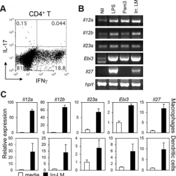
Systemic infection with Lm is known to induce pathogen-specific Th1 cells. To examine if pathogen-pathogen-specific Th17 cells are also generated during infection, we intravenously infected C57BL/ 6 mice with Lm expressing ovalbumin (Lm-Ova) [25], and examined the expression of IL-17 and IFNcby splenic CD4+T
cells after restimulation with an Lm-specific, MHC II-restricted peptide (listeriolysin O (LLO)190–201). As expected, intravenous infection with live Lm-Ova induced a high percentage of IFNc -producing CD4+T cells (Figure 1A). By contrast, very few CD4+T
cells expressed IL-17 in the spleens of the infected mice. Among the IL-12 family cytokines, IL-23 mediates Th17 immunity while IL-12 and IL-27 induce Th1 and suppress Th17 responses. To determine if the Lm dominant Th1 responses were due to a preferential induction of IL-12 and IL-27, we examined the induction of IL-12 family genes in dendritic cells and macrophages stimulated with lethally irradiated Lm. Importantly, irradiation induces the inactivation of Lm without affecting adjuvanticity and immunogenicity [26]. Stimulation of bone marrow-derived dendritic cells or macrophages with irradiated Lm induced the expression of Il12a (encoding IL-12p35), Il12b
(encoding IL-12/IL23p40), Il23a (encoding IL-23p19), Ebi3
Figure 1. CD4+ T cell responses and the induction of IL-12 family genes after infection withL. monotytogenes. A, C57BL/6 mice were intravenously infected with 2.56104Lm-Ova on day 0, and
splenocytes were obtained on day 7. CD4+
T cells expressing IFNcand IL-17 were measured by intracellular staining after stimulation with LLO190–201. B and C, Bone marrow-derived DC or macrophages were
stimulated with LPS, Pam3CSK4, or irradiated Lm-Ova for four hours.
Cells were harvested and analyzed for the mRNA expression of IL-12 family genes by using RT-PCR (B, bone marrow-derived DC), or quantitative real-time PCR analysis (C). Values are mean 6SD. Data shown represent two independent experiments.
doi:10.1371/journal.ppat.1003628.g001 Author Summary
(encoding IL-27EBI3) andIl27(encoding IL-27p28) as efficiently as LPS stimulation (Figure 1B & C). Together, these data demonstrate that while all genes in the IL-12 family were induced upon Lm encounter, only Th1 immunity was induced after systemic infection with Lm-Ovain vivo.
IncreasedListeria-specific Th17 responses in p352/2 EBI32/2mice
We next sought to address whether the lack of pathogen-specific Th17 immunity in wild-type mice after Lm-Ova infection was due to 12 and 27. To analyze the relative contribution of IL-12p35 and IL-27EBI3, we first crossed p352/2mice with EBI32/2 to generate p352/2EBI32/2mice. Wild-type, p352/2, EBI32/2, or p352/2EBI32/2mice were then systemically infected with Lm-Ova via the intravenous route. Seven days later, we restimulated splenocytes from the infected mice with LLO190–201 to measure pathogen-specific CD4+
T cell responses. As expected, we observed high percentages of IFNc-producing CD4+
T cells (,20%), while few CD4+
T cells produced IL-17 in the wild-type mice (,0.5%) (Figure 2A & B). Compared with wild-type mice, the production of IFNc by LLO-specific CD4+ T cells was greatly diminished in
p352/2mice. Notably, although the IL-27 may be an inducer of Th1 responses [12,13], we did not observe any defect in the percentage of IFNc-producing CD4+
T cells in EBI32/2 mice (Figure 2A & B). Instead, we observed that the frequency of IL-17-producing CD4+
T cells in the EBI3-deficient mice was significantly higher than those of wild-type mice, likely due to the increased population producing both IFNc and IL-17 among CD4+T cells
(Figure 2A & B). Notably, compared with p352/2and EBI32/2mice, p352/2EBI32/2mice exhibited a significantly increased frequency of IL-17+
IFNc2 CD4+
T cells (Figure 2A & B). Consequently, the production of IL-17 and IL-22 by Lm-specific CD4+
T cells was far higher in the p352/2EBI32/2 mice compared to wild-type mice (Figure 2C). p352/2and EBI32/2mice both showed a slight increase in the frequency of IL-17+CD4+T cells, however, the amounts of
IL-17 produced after antigen restimulation were far less than that of p352/2EBI32/2 mice. Thus, p352/2EBI32/2 mice exhibited diminished Th1 and enhanced Th17 responses to Lm-Ova infection, indicating that IL-27EBI3 and IL-12p35 cooperatively suppress the generation of pathogen-specific Th17 cells after infection.
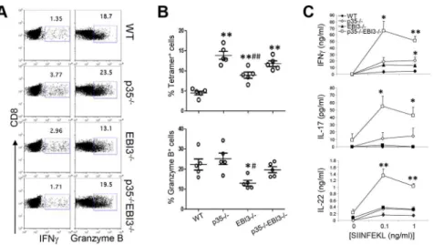
Activation of CD8+T cells and innate cells in the absence of IL-12p35 and/or IL-27EBI3 followingL. monocytogenes infection
To measure the pathogen-specific CD8+T cell responses to
Lm-Ova, we restimulated splenocytes from infected mice with SIINFEKL peptide. CD8+
T cells derived from p352/2 mice and EBI32/2 mice exhibited similar or higher percentages of IFNc compared to wild-type T cells (Figure 3A). Moreover, the percentages of Ova-specific MHC I tetramer-positive CD8+
T cells were significantly higher in p352/2, EBI32/2, and p352/ 2EBI32/2 mice compared to wild-type mice (Figure 3B). The
frequencies of CD8+ T cells expressing granzyme B were
comparable among wild-type, p352/2, and p352/2EBI32/2 mice while decreased in EBI32/2mice (Figure 3A & B). Hence, the generation of pathogen-specific CD8+
T cells is largely independent of p35 and EBI3. These results are consistent, in part, with a previous study showing that IL-12 is not required for IFNc production but rather inhibits the generation of memory CD8+T cells [27]. By contrast, we observed that the amounts of
IL-17 and IL-22 produced by CD8+
T cells were remarkably higher in p352/2EBI32/2 mice than those in the wild-type (Figure 3C). Hence, in the absence of IL-12p35 and IL-27EBI3,
systemic Lm-Ova infection triggers increased production of IL-17 and IL-22 by pathogen-specific CD8+
T cells.
To further examine the regulation of host defensive immunity by the cytokines of IL-12 family, we analyzed the activation of innate immune cells during the early phase of Lm infection. IL-12 triggers IFNc production in NK cells and NKT cells which is critical for the activation of innate cells and the prevention of Lm propagation [28]. Consistent with this notion, we observed a significant reduction of IFNc-producing NKT cells and NK cells in p352/2mice as well as in p352/2EBI32/2mice infected with Lm-Ova (Figure 4A). The percentages of IFNc-producing NKT cells and NK cells in EBI32/2 mice were comparable to those from wild-type mice, indicating that there is no significant role of EBI3 in the induction of IFNcfrom NK and NKT cells after Lm-Ova infection. Ly6C+CD11bhi
dendritic cells, also known as Tip-DC, suppress the dissemination of Lm [28,29]. We observed comparable percentages of the Ly6C+CD11bhi
DC in p352/2, EBI32/2as well as p352/2EBI32/2mice with that of wild-type mice (Figure 4B). Therefore, the induction of Tip-DC was likely normal in mice lacking p35, EBI3, or both in this experimental setting.
NK, NKT, andcdT cells represent additional sources of innate Th1 and Th17 cytokines that could be potentially released following Lm infection. To investigate the contributions of the cellular subsets, we measured the production of IFNc, IL-17, and IL-22 from splenocytes obtained three days after Lm-Ova infection. As depicted in Figure 4C, p352/2 mice as well as p352/2 EBI32/2 mice showed significantly diminished IFNc Figure 2. Increased pathogen-specific Th17 responses in the absence of IL-12p35 and IL-27EBI3.C57BL/6 (WT) or the indicated strains of mice (n = 5 per group) were intravenously infected with 2.56104Lm-Ova on day 0. Seven days later, lymphoid cells from the
spleen were obtained and CD4+
T cells expressing IFNcand IL-17 were measured by intracellular staining after stimulation with LLO190–201(A
and B). The lymphoid cells from the spleen were stimulated with LLO190–201peptide for three days, and the concentrations of IFNc, IL-17
and IL-22 in the supernatant were measured by ELISA(C). Bars in B are mean 6 SEM. Values in C are mean 6 SEM. Data shown are representative of two independent experiments. *,p,0.05; **,p,0.01 in comparison with WT group.#,p,0.05;##,p,0.01 in comparison with p352/2group.
while EBI32/2 mice showed comparable IFNc production. In contrast, the amounts of IL-17 in the supernatant were higher in EBI32/2 and p352/2 EBI32/2 mice compared with those of wild-type mice. The IL-22 production was higher in p352/2and p352/2 EBI32/2 mice. Collectively, these data suggest that the regulation of Th1 and Th17 cytokines by innate immune cells is also under the control of multiple IL-12 family cytokines.
p352/2EBI32/2mice are resistant toL. monocytogenes infection
We next addressed the differential roles of the IL-12 family cytokines in host defense against Lm infection. Wild-type, p352/2, EBI32/2, or p352/2EBI32/2 mice were intravenously infected with Lm-Ova and bacterial burden in the livers and spleens were measured three days later. As expected, p352/2 mice showed higher bacterial burden in the livers compared to wild-type
Figure 3. Pathogen-specific CD8+T cell responses in the absence of IL-12p35 and IL-27EBI3.
C57BL/6 (WT) or the indicated strains of mice (n = 5 per group) were intravenously infected with 2.56104Lm-Ova on day 0. Seven days later, lymphoid cells from the spleen were obtained and
CD8+
T cells expressing IFNcand granzyme B were measured by intracellular staining after stimulation with SIINFEKL (A and B). CD8+ T cells expressing T cell receptors specific to Ova were analyzed by staining with SIINFEKL-loaded Kbtetramer (B). The lymphoid cells from the spleen were
stimulated with SIINFEKL peptide for three days, and the concentrations of IFNc, IL-17 and IL-22 in the supernatant were measured by ELISA(C). Bars in B are mean6SEM. Values inCare mean6SEM. Data shown are representative of two independent experiments. *,p,0.05; **,p,0.01 in comparison with WT group.#,p,0.05;##,p,0.01 in comparison with p352/2group.
doi:10.1371/journal.ppat.1003628.g003
Figure 4. Enhanced production of Th17 cytokines in the p352/2EBI32/2 mice during the early phase of infection with L. monocytogenes.C57BL/6 (WT) or the indicated strains of mice (n = 3 per group) were intravenously infected with 2.56104Lm-Ova on day 0. Two to
three days later, lymphoid cells were analyzed for the production of IFNcby NK and NKT cells (A), or for the frequency of CD11b+Ly6C+Tip DC (B). The production of IFNc, IL-17, IL-17F and IL-22 by the splenocytes obtained three days after the infection was measured (C). Values are mean6SD. Data shown are representative of two independent experiments. *,p,0.05; **,p,0.01 in comparison with WT group.
controls (Figure 5A). We observed significantly less bacterial burden in the livers of EBI32/2mice compared with those from wild-type, indicating that EBI3 is not required for the host defense against the infection. To our surprise, the bacterial burden in the livers of p352/2EBI32/2mice was significantly lower than that of p352/2mice, to levels comparable to EBI32/2mice (Figure 5A). Within the spleens, p352/2EBI32/2mice exhibited significantly lower bacterial burden compared to p352/2mice; however, there was no evident difference in bacterial burden between wild-type and EBI32/2or p352/2EBI32/2mice (Figure S1A).
We also measured bacterial burden 7 days after infection and found that p352/2 mice failed to control bacterial growth with significantly higher levels of bacteria in the livers compared to wild-type animals (Figure 5B). However, EBI32/2 as well as p352/2 EBI32/2 mice showed comparable levels of bacteria in the livers compared to wild-type mice (Figure 5B). We also observed similar pattern of bacterial burdens in the spleens of these mice (Figure S1B). Therefore, the bacterial resistance observed at day 3 largely remained intact by day 7 post infection. Collectively, these findings demonstrate that EBI3-deficiency conferred resis-tance to Lm-Ova infection in the absence of IL12p35, indicative of possible antagonistic function of IL-12p35 and IL-27EBI3 in host defense to the intracellular bacterial infection. Furthermore, in the absence of IL-12p35, IL-27EBI3 likely exerts strong immunosup-pressive activity and thus mediates immune evasion of the Lmin vivo.
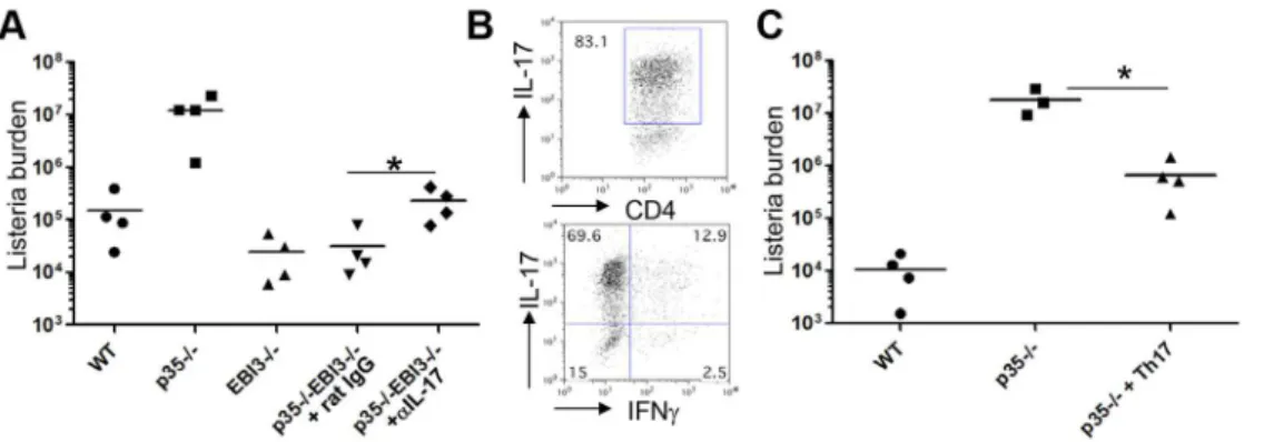
IL-17 and IL-22 mediate anti-Listeriaimmunity in the absence of IL-12p35
The enhanced production of IL-17 and IL-22 we observed in p352/2EBI32/2 mice by both innate and adaptive immune compartments led us to hypothesize that the induction of the Th17 cytokines might be responsible for the observed resistance of p352/2EBI32/2mice against Lm-Ova infection. To test this hypothesis, we infected p352/2EBI32/2 mice with Lm-Ova and then injected anti-IL-17 or control Ab. Notably, the bacterial burden in the livers of the mice receiving anti-IL-17 showed a modest but significant increase (8 times higher) compared with that of the control Ab group; however the burden was still substantially lower than that observed in p352/2mice (Figure 6A). This result demonstrates that the upregulated production of IL-17, at least in part, contributed to the observed resistance of p352/2EBI32/2 mice to Lm-Ova infection.
Based on our findings, we hypothesized that IL-17-producing cells suppress the growth of Lm, especially in the absence of IL-12p35. To address this point, we investigated if Lm-specific Th17 cells are sufficient to limit the growth of Lm in the absence of IL-12-mediated innate and adaptive immunity. To obtain Lm-specific Th17 cells, we first isolated lymphoid cells from IL-17Frfp mice [30] after immunization with LLO190–201emulsified in CFA and then restimulated them with peptide in the presence of IL-23, IL-1b and anti-IFNcto specifically expand the Th17 population [31]. After 5 days culture, we sorted RFP+CD4+cells (Figure 6B;
.80% IL-17+and
,15% IFNc+), and transferred them i.v. into p352/2mice. Wild-type and p352/2mice receiving no cells were used as controls. All mice were then infected with Lm-Ova, and the bacterial burden in the liver was measured 7 days post infection. As shown in Figure 6C, the p352/2mice receiving the RFP+CD4+T cells showed significantly less bacterial load in the
liver compared to p352/2 mice receiving no cells (26.8 times lower), although the bacterial burden in the former group was still higher than that of the wild-type mice.
These results demonstrated that Lm-specific Th17 cells are protective against Lm-Ova in the absence of IL-12p35; however, it is possible that small population of IFNc-producers among the RFP+donor T cells (
,15%) mediated this protection. To rule out this possibility and to further determine the protective immunity mediated by IL-17 and IL-22, we next examined if administration of recombinant IL-17 or IL-22 mediates host defense against Lm-Ova in the absence of IL-12p35. As depicted in Figure 7, p352/2 mice treated with IL-17 or IL-22 alone showed a slightly lower, but not statistically significant, bacterial load in the liver than saline-treated mice. Notably, administration of both cytokines induced a significantly lower bacterial burden in the liver than saline-, IL-17- or IL-22-treated p352/2mice (29.5 times less than saline-treated mice). Administration of IL-17 and IL-22, however, did not fully restore the resistance of p352/2 mice, since the bacterial load was still higher than that of wild-type mice (Figure 7 and Figure S2). The inhibition of bacterial growth by exogenous IL-17 or IL-22 was more evident in the bacterial load in the spleens (Figure S2). Taken together, these results indicate that the Th17 cytokines IL-17 and IL-22 act synergistically to induce protective anti-Listeria immunity in the absence of IL-12p35.
Figure 5. p352/2EBI32/2mice are resistant to the infection withL. monocytogenes.C57BL/6 (WT) or the indicated strains of mice (n = 4–5 per group) were intravenously infected with 2.56104Lm-Ova on day 0. Three (A) or seven (B) days later, the bacterial burden in the livers of the
infected mice was analyzed by measuring colony-forming unit. Bars are mean values. Data shown are representative of three independent experiments. *, p,0.05 and **, p,0.01 in comparison between two indicated groups.
Discussion
In this study, we comparatively analyzed the contribution of IL-12p35 and IL-27EBI3 to the host defense against the intracellular pathogen Lm. We demonstrate that, although p352/2mice failed to control bacterial growth, mice deficient in both p35 and EBI3 had no such defect in controlling bacterial growth. Our study also revealed that IL-17 is involved in the protective immunity in
p352/2EBI32/2mice. Furthermore, administration of Th17 cells as well as recombinant IL-17 and IL-22 significantly suppressed bacterial growth in p352/2mice. These findings strongly suggest that Lm utilizes IL-27EBI3 to escape Th17-mediated immune surveillance in IL-12p35-deficient mice. Thus, the present study unveils a previously unappreciated immune escape mechanism of intracellular bacteria through IL-27EBI3, and that Th17 responses play an important role in intracellular bacterial infection, especially in the absence of IL-12 and Th1-mediated immunity.
NK cells, NKT cells and Tip-DC are well known innate effector cells that suppress bacterial growth during the early phase of Lm infection [28,29]. IL-12 is required for the induction of IFNcfrom NK and NKT cells which then mediates the recruitment of Tip-DC. Comparative analysis between p352/2and p352/2EBI32/2 mice showed no apparent difference in the activation of NK and NKT cells and the frequency of Tip-DC. In addition, the percentages of effector CD8+
T cells expressing granzyme B were similar between p352/2 and p352/2EBI32/2 mice. Moreover, although 27 has been reported to drive the differentiation of IL-10 producing CD4+
T cells [18,19,20], we observed comparable expression of theIl10transcript between wild-type and EBI32/2 mice after Lm-Ova infection (data not shown). Therefore, we conclude that the increased resistance to Lm in p352/2EBI32/2 mice is not due to the enhanced activity of these innate immune cells nor CD8+
T cells.
Accumulating evidence suggests that some of the Th1 cells recruited to inflamed tissues are actually derived from Th17 cells [32,33]. However, we observed that very few LLO-specific IFNc -producing CD4+
T cells in wild-type mice after Lm infection co-expressed IL-17. In addition, LLO-specific, IFNc-producing CD4+
T cells in IL-17FCre6Rosa26eYFP mice after Lm infection were.99% YFP-negative (data not shown), indicating that Th1 cells do not originate from Th17 cells in this model.
Notably, we observed increased production of IL-17 and IL-22 by innate immune cells, presumablycdT cells [24,34], as well as Lm-Ova-specific CD4+T and CD8+T cells in p352/2EBI32/2 mice. IL-22 is a Th17 cytokine that induces a series of anti-microbial peptides upon infection [4,5,35,36]. The mechanism of protection by these Th17 cytokines, however, significantly differs
Figure 6. A role for IL-17 and Th17 cells on the resistance of p352/2EBI32/2mice againstL. monocytogenesinfection.A, C57BL/6 (WT) or the indicated strains of mice (n = 4 per group) were intravenously infected with 2.56104Lm-Ova on day 0. The mice were i.p. injected with 100mg
of anti-IL-17 or rat IgG on day 0, 2, 4. Seven days after the infection, bacterial burden in the livers of the infected mice was determined by measuring colony-forming unit. *, p,0.05 in comparison with rat IgG-treated group.B and C, IL-17Frfpreporter mice were s.c. immunized with LLO peptide emulsified in CFA. Seven days later, lymphoid cells from spleen and draining lymph nodes of the immunized mice were isolated and stimulated with the LLO peptide in the presence of IL-23 (50 ng/ml), IL-1b(10 ng/ml) and anti-IFNcfor 5 days. CD4+RFP+cells were isolated by flow cytometry, and the expression of IL-17 and IFNcwas measured by intracellular staining (B). The sorted Th17 cells (56105cells per transfer) were i.v. transferred into
p352/2mice, followed by i.v. infection with 2.5
6104Lm-Ova. WT or p352/2mice without the cell transfer were used as controls. Seven days after
infection, bacterial burden in the liver was measured (C). Data shown are representative of two independent experiments. *, p,0.05 in comparison with p352/2mice without Th17 cell transfer.
doi:10.1371/journal.ppat.1003628.g006
Figure 7. IL-17 and IL-22 cooperatively promote protective immunity againstL. monocytogenesinfection in p352/2mice.A, C57BL/6 (WT) or groups of p352/2 mice (n = 5 per group) were intravenously infected with 2.56104 Lm-Ova on day 0. Some of the
p352/2mice were i.p. injected with 1mg of recombinant IL-17, IL-22, or both on day 0, 2, 4. Seven days after the infection, bacterial burden in the livers of the infected mice was determined by measuring colony-forming unit. Bars are mean values. *, p,0.05 in comparison between two indicated groups.
from that of IFNc due to the distribution of receptors and differential downstream targets. IFNcmediates protective immu-nity by multiple mechanisms including the induction of iNOS and autophagy [21,37,38], whereas IL-17 does so possibly through neutrophil recruitment and by enhancing cross-presentation of bacterial antigens [24,34]. In the present study, the amounts of IL-17 and IL-22 produced by innate cells and the pathogen-specific T cells were significantly increased in p352/2EBI32/2 mice. Furthermore, exogenous IL-17 and IL-22 synergistically induced protective immunity in p35-deficient mice, while each cytokine individually could only invoke marginal protection. Supporting this notion, it has been documented that IL-17 and IL-22 synergistically induce the expression of antimicrobial peptides [36]. Conversely, IL-22 has been shown to be dispensable for the clearance of Lm in p35-sufficient mice [39]. Our present work combined with other reports then suggests that, in the absence of IL-12-mediated protective immunity, Th17 cytokines IL-17 and IL-22 cooperatively inhibit the growth of Lm and are negatively regulated by EBI3. Importantly, since the bacterial burden in p352/2 mice treated with exogenous IL-17 and IL-22 was still higher than that of wild-type mice, undefined alternative protective mechanism may still exist.
One can assume that the difference between p352/2mice and p352/2EBI32/2mice in anti-Lm immunity could be due to the effect of IL-35, which is composed of p35 and EBI3 [40]. Given that p352/2 mice cannot produce IL-12 and IL-35, and that p352/2EBI32/2 mice cannot produce IL-12, IL-35 and IL-27, the only cytokine that is lacking in the latter mice compared with the former mice is IL-27. Recent studies have shown that the other subunit of IL-27, IL-27p28, can be secreted in the absence of EBI3 to act as an antagonist of gp130 [15,41,42] or alternatively form a heterodimer with Cytokine-Like Factor 1 (p28/CLF) to promote NK and T cell activity [43]. Hence, EBI3-deficiency may lead to the production of p28 and p28/CLF, which may exert biological activities independently of IL-27. The role of p28 subunit of IL-27 during host defense in the present study is not clear. Future studies with p28-deficient mice will be important for a complete understanding on the mechanism by which EBI3 regulates protective immunity to intracellular pathogens.
IL-27 has been shown to trigger preliminary Th1 responses, where mice deficient in the IL-27 receptor (WSX-12/2; TCCR2/2) are more susceptible toLeishmania major[12] and Lm infection [13] due to decreased Th1 responses. On the contrary, WSX-12/2 mice generate more IFNc-producing CD4+T cells than wild-type mice
after infection withToxoplasma gondii[14], indicating that IL-27 signal is not necessary for the generation of Th1 immunity to the infection. Therefore the effect of IL-27 on pathogen-specific Th1 response is likely dependent on the infectious agents. It is not clear why IL-27EBI32/2 mice in the present study did not recapitulate the phenotype of IL-27R2/2mice in a previous study [13]. It is possible that the route of infection (intravenous versus subcutaneous) results in distinct immune responses to Lm. Alternatively, it is possible that the phenotype of EBI32/2mice described in this study may in fact be IL-27 independent and instead mediated through IL-IL-27p28 [42,44,45]. Interestingly, fundamental differences have also been reported between WSX-12/2and EBI32/2mice. For instance, WSX-12/2 mice exhibited enhanced liver inflammation, whereas EBI32/2mice showed reduced liver inflammation in the same Con A-induced hepatitis animal model [46,47]. Moreover, while T cells from WSX-12/2mice produce less IFNc, T cells from EBI32/2mice produce higher IFNcand less IL-4 than wild-type T cells [12,13,48]. Further study is needed to demonstrate the mechanism of these differences in
the regulation of infectious and inflammatory diseases between the EBI3 and IL-27 receptor signaling pathways.
Collectively our findings demonstrate that the immune system produces IL-12 to suppress bacterial growth upon infection while Lm utilizes another host immune component, EBI3, to escape immune surveillance. Increased susceptibility to intracellular pathogens in patients with deficiency in IL-12 or its receptor has been demonstrated [49,50]. Based on our findings, blockade of EBI3 may provide a new therapeutic approach for the treatment of infectious diseases, particularly in patients with defective IL-12 immunity.
Materials and Methods
Ethics statement
All the animal experiments were performed in accordance with the Guide for the Care and Use of Laboratory Animals of the National Institutes of Health and with the permission of the American Association for the Assessment and Accreditation of Laboratory Animal Care. The protocol was reviewed and approved by the Institutional Animal Care and Use Committee of MD Anderson Cancer Center (identification number: 10-04-09833) and University of Texas Health Science Center at Houston (identification number: HSC-AWC-12-008).
Mice
C57BL/6 and IL-12p352/2 mice were purchased from the Jackson Laboratory. IL-27EBI32/2 mice were generated as described previously [48]. Double-deficient mice (p352/2EBI32/2) were obtained by crossing IL-12p352/2and IL-27EBI32/2mice. IL-17Frfp-reporter mice were generated as described previously [30]. All mice were kept under specific pathogens-free condition. The animal experiments were performed at the age of 6–12 weeks.
Stimulation of bone marrow-derived dendritic cells (BM-DC) and macrophages (BM-M)
Bone marrow cells from femurs and tibia of C57BL/6 mice were cultured with 10% FBS supplemented RPMI containing GM-CSF or M-CSF for 6 days. For irradiation, log-phase cultured Lm-Ova were exposed to 300 K rad of c-irradiation. After extensive washing, BM-DC and BM-M cells were incubated with the irradiated Lm-Ova at the ratio of 1:10. As controls, LPS (100 ng/ml) and Pam3CysSK4 (Pam; 1mg/ml) were added in the culture. Four hours after the stimulation, cells were harvested and resuspended in Trizol for mRNA expression analysis.
Infection with Listeria monocytogenes
Antibodies used for flow cytometric analysis
The following antibodies were used for cell surface and intracellular staining; PerCPCy5-5- or FICT-labeled anti-TCRb (H57-597), PerCPCy5-5-labeled anti-CD4 (GK1.5), Alexa 488-labeled anti-CD8 (5H10-1), APC-488-labeled anti-CD11b (M1/70) from Biolegnd; PE- or Alexa 488-labeled anti-IFNc (XMG1.2), PE-labeled anti-IL-17 (clone TC11-18H10), Alexa 647-labeled GranzymeB (GB11) FITC- or PerCPCy5.5-labeled anti-NK1.1 (PK136), PerCPCy5-5-labeled anti-Ly6C (AL21) from BD Biosciences. For intracellular staining, cells were incubated with permeabilization buffer (BD Biosciences), and then further stained with intracellular staining Abs described above. These cells were analyzed by using LSRII flow cytometer (BD Bioscience) and Flowjo software.
Real-time PCR analysis for mRNA expression
Total RNA was prepared from splenocytes with TriZol reagent (Invitrogen). Complementary DNA (cDNA) was synthesized with Superscript reverse transcriptase and oligo(dT) primers (Invitro-gen), and gene expression was examined with a Bio-Rad iCycler Optical System with iQ SYBR green real-time PCR kit (Bio-Rad Laboratories). The data were normalized toActb reference. The following primer pairs were used:ActB: F-GAC GGC CAG GTC ATC ACT ATT G and R-AGG AAG GCT GGA AAA GAG CC;Ifng: F-GAT GCA TTC ATG AGT ATT GCC AAG T and R-GTG GAC CAC TCG GAT GAG CTC;Il17: F-CTG GAG GAT AAC ACT GTG AGA GT and R-TGC TGA ATG GCG ACG GAG TTC; Il17f: F-CTG GAG GAT AAC ACT GTG AGA GT-39 and R-TGC TGA ATG GCG ACG GAG TTC;
Il22: F-CAT GCA GGA GGT GGT ACC TT and R-CAG ACG CAA GCA TTT CTC AG;Il10: F-ATA ACT GCA CCC ACT TCC CAG TC and R-CCC AAG TAA CCC TTA AAG TCC TGC;Ebi3: F-TCC CCG AGG TGC AAC TGT TCT CC and R-GGT CCT GAG CTG ACA CCT GG. Primers for p35, p40, p19 were described previously [53].
Adoptive transfer study To obtain IL-17-producing CD4+
T cells specific for Lm-Ova, we s.c. immunized IL-17Frfp-reporter mice with LLO peptide in CFA. A week later, lymphoid cells from the draining lymph nodes and spleen were pooled and restimulated with the same peptide in the presence of IL-23 (50 ng/ml) and IL-1b(10 ng/ml) plus anti-IFNc (5mg/ml; XMG1.2) for five days. The cells were stained with APC-labeled anti-CD4, and APC-positive and RFP-positive
cells were sorted by using FACS-Influx (BD Biosciences). 2.56105 sorted cells/mouse were intravenously injected into IL-12p352/2 mice followed by Lm-Ova inoculation and analysis of bacterial burden, as described above.
Statistical analysis
The Student t test was used to assess the statistical values. P
values were determined, and error bars represent standard error of the mean (SEM) or standard deviation (SD).
Supporting Information
Figure S1 Bacterial load in the spleens of p352/2 and
EBI32/2 mice after infection with L. monocytogenes.
C57BL/6 (WT) or the indicated strains of mice (n = 4–5 per group) were intravenously infected with 2.56104Lm-Ova on day 0. Three (A) or seven (B) days later, the bacterial burden in the spleens of the infected mice was analyzed by measuring colony-forming unit. Bars are mean values. Data shown are representative of three independent experiments. *, p,0.05 and **, p,0.01 in comparison between two indicated groups.
(PDF)
Figure S2 Bacterial load in the spleens of p352/2mice
treated with IL-17 or IL-22.A, C57BL/6 (WT) or groups of
p352/2 mice (n = 5 per group) were intravenously infected with 2.56104Lm-Ova on day 0. Some of the p352/2mice were i.p. injected with 1mg of recombinant IL-17, IL-22, or both on day 0, 2, 4. Seven days after the infection, bacterial burden in the spleens of the infected mice was determined by measuring colony-forming unit. Bars are mean values. *, p,0.05 in comparison between two indicated groups.
(PDF)
Acknowledgments
We thank Dr. Hao Shen (University of Pennsylvania) for Lm-Ova, the FACS Core Facility at the MD Anderson Cancer Center for assistance with cell sorting.
Author Contributions
Conceived and designed the experiments: YC CD. Performed the experiments: YC TY BSK YZ JMR GJM SHC HL. Analyzed the data: YC BSK YZ CD. Contributed reagents/materials/analysis tools: MB. Wrote the paper: YC CD.
References
1. Romani L, Puccetti P, Bistoni F (1997) Interleukin-12 in infectious diseases. Clin Microbiol Rev 10: 611–636.
2. Trinchieri G (1998) Interleukin-12: a cytokine at the interface of inflammation and immunity. Adv Immunol 70: 83–243.
3. Ye P, Rodriguez FH, Kanaly S, Stocking KL, Schurr J, et al. (2001) Requirement of interleukin 17 receptor signaling for lung CXC chemokine and granulocyte colony-stimulating factor expression, neutrophil recruitment, and host defense. J Exp Med 194: 519–527.
4. Aujla SJ, Chan YR, Zheng M, Fei M, Askew DJ, et al. (2008) IL-22 mediates mucosal host defense against Gram-negative bacterial pneumonia. Nat Med 14: 275–281. 5. Zheng Y, Valdez PA, Danilenko DM, Hu Y, Sa SM, et al. (2008) Interleukin-22
mediates early host defense against attaching and effacing bacterial pathogens. Nat Med 14: 282–289.
6. Ishigame H, Kakuta S, Nagai T, Kadoki M, Nambu A, et al. (2009) Differential roles of interleukin-17A and -17F in host defense against mucoepithelial bacterial infection and allergic responses. Immunity 30: 108–119.
7. Rudner XL, Happel KI, Young EA, Shellito JE (2007) Interleukin-23 (IL-23)-IL-17 cytokine axis in murine Pneumocystis carinii infection. Infect Immun 75: 3055–3061.
8. Conti HR, Shen F, Nayyar N, Stocum E, Sun JN, et al. (2009) Th17 cells and IL-17 receptor signaling are essential for mucosal host defense against oral candidiasis. J Exp Med 206: 299–311.
9. Goriely S, Neurath MF, Goldman M (2008) How microorganisms tip the balance between interleukin-12 family members. Nat Rev Immunol 8: 81–86. 10. Kastelein RA, Hunter CA, Cua DJ (2007) Discovery and biology of IL-23 and
IL-27: related but functionally distinct regulators of inflammation. Annu Rev Immunol 25: 221–242.
11. Yoshida H, Miyazaki Y (2008) Regulation of immune responses by interleukin-27. Immunol Rev 226: 234–247.
12. Yoshida H, Hamano S, Senaldi G, Covey T, Faggioni R, et al. (2001) WSX-1 is required for the initiation of Th1 responses and resistance to L. major infection. Immunity 15: 569–578.
13. Chen Q, Ghilardi N, Wang H, Baker T, Xie MH, et al. (2000) Development of Th1-type immune responses requires the type I cytokine receptor TCCR. Nature 407: 916–920.
14. Villarino A, Hibbert L, Lieberman L, Wilson E, Mak T, et al. (2003) The IL-27R (WSX-1) is required to suppress T cell hyperactivity during infection. Immunity 19: 645–655.
17. Hirahara K, Ghoreschi K, Yang XP, Takahashi H, Laurence A, et al. (2012) Interleukin-27 Priming of T Cells Controls IL-17 Production In trans via Induction of the Ligand PD-L1. Immunity 36: 1017–1030.
18. Awasthi A, Carrier Y, Peron JP, Bettelli E, Kamanaka M, et al. (2007) A dominant function for interleukin 27 in generating interleukin 10-producing anti-inflammatory T cells. Nat Immunol 8: 1380–1389.
19. Fitzgerald DC, Zhang GX, El-Behi M, Fonseca-Kelly Z, Li H, et al. (2007) Suppression of autoimmune inflammation of the central nervous system by interleukin 10 secreted by interleukin 27-stimulated T cells. Nat Immunol 8: 1372–1379.
20. Stumhofer JS, Silver JS, Laurence A, Porrett PM, Harris TH, et al. (2007) Interleukins 27 and 6 induce STAT3-mediated T cell production of interleukin 10. Nat Immunol 8: 1363–1371.
21. Pamer EG (2004) Immune responses to Listeria monocytogenes. Nat Rev Immunol 4: 812–823.
22. Hiromatsu K, Yoshikai Y, Matsuzaki G, Ohga S, Muramori K, et al. (1992) A protective role of gamma/delta T cells in primary infection with Listeria monocytogenes in mice. J Exp Med 175: 49–56.
23. Hamada S, Umemura M, Shiono T, Tanaka K, Yahagi A, et al. (2008) IL-17A produced by gammadelta T cells plays a critical role in innate immunity against listeria monocytogenes infection in the liver. J Immunol 181: 3456–3463. 24. Meeks KD, Sieve AN, Kolls JK, Ghilardi N, Berg RE (2009) IL-23 is required
for protection against systemic infection with Listeria monocytogenes. J Immunol 183: 8026–8034.
25. Pope C, Kim SK, Marzo A, Masopust D, Williams K, et al. (2001) Organ-specific regulation of the CD8 T cell response to Listeria monocytogenes infection. Journal of immunology 166: 3402–3409.
26. Datta SK, Okamoto S, Hayashi T, Shin SS, Mihajlov I, et al. (2006) Vaccination with irradiated Listeria induces protective T cell immunity. Immunity 25: 143– 152.
27. Pearce EL, Shen H (2007) Generation of CD8 T cell memory is regulated by IL-12. J Immunol 179: 2074–2081.
28. Kang SJ, Liang HE, Reizis B, Locksley RM (2008) Regulation of hierarchical clustering and activation of innate immune cells by dendritic cells. Immunity 29: 819–833.
29. Serbina NV, Salazar-Mather TP, Biron CA, Kuziel WA, Pamer EG (2003) TNF/iNOS-producing dendritic cells mediate innate immune defense against bacterial infection. Immunity 19: 59–70.
30. Yang XO, Nurieva R, Martinez GJ, Kang HS, Chung Y, et al. (2008) Molecular antagonism and plasticity of regulatory and inflammatory T cell programs. Immunity 29: 44–56.
31. Chung Y, Chang SH, Martinez GJ, Yang XO, Nurieva R, et al. (2009) Critical regulation of early Th17 cell differentiation by interleukin-1 signaling. Immunity 30: 576–587.
32. Martin-Orozco N, Chung Y, Chang SH, Wang YH, Dong C (2009) Th17 cells promote pancreatic inflammation but only induce diabetes efficiently in lymphopenic hosts after conversion into Th1 cells. European journal of immunology 39: 216–224.
33. Hirota K, Duarte JH, Veldhoen M, Hornsby E, Li Y, et al. (2011) Fate mapping of IL-17-producing T cells in inflammatory responses. Nature immunology 12: 255–263.
34. Xu S, Han Y, Xu X, Bao Y, Zhang M, et al. (2010) IL-17A-producing gammadeltaT cells promote CTL responses against Listeria monocytogenes infection by enhancing dendritic cell cross-presentation. Journal of immunology 185: 5879–5887.
35. Chung Y, Yang X, Chang SH, Ma L, Tian Q, et al. (2006) Expression and regulation of IL-22 in the IL-17-producing CD4+T lymphocytes. Cell Res 16: 902–907.
36. Liang SC, Tan XY, Luxenberg DP, Karim R, Dunussi-Joannopoulos K, et al. (2006) Interleukin (IL)-22 and IL-17 are coexpressed by Th17 cells and cooperatively enhance expression of antimicrobial peptides. J Exp Med 203: 2271–2279.
37. Gutierrez MG, Master SS, Singh SB, Taylor GA, Colombo MI, et al. (2004) Autophagy is a defense mechanism inhibiting BCG and Mycobacterium tuberculosis survival in infected macrophages. Cell 119: 753–766.
38. Chang YP, Chen CL, Chen SO, Lin YS, Tsai CC, et al. (2011) Autophagy facilitates an IFN-gamma response and signal transduction. Microbes and infection/Institut Pasteur 13: 888–894.
39. Graham AC, Carr KD, Sieve AN, Indramohan M, Break TJ, et al. (2011) IL-22 production is regulated by IL-23 during Listeria monocytogenes infection but is not required for bacterial clearance or tissue protection. PloS one 6: e17171. 40. Collison LW, Vignali DA (2008) Interleukin-35: odd one out or part of the
family? Immunol Rev 226: 248–262.
41. Pflanz S, Timans JC, Cheung J, Rosales R, Kanzler H, et al. (2002) IL-27, a heterodimeric cytokine composed of EBI3 and p28 protein, induces proliferation of naive CD4(+) T cells. Immunity 16: 779–790.
42. Stumhofer JS, Tait ED, Quinn WJ, . (2010) A role for IL-27p28 as an antagonist of gp130-mediated signaling. Nat Immunol 11: 1119–1126.
43. Crabe S, Guay-Giroux A, Tormo AJ, Duluc D, Lissilaa R, et al. (2009) The IL-27 p28 subunit binds cytokine-like factor 1 to form a cytokine regulating NK and T cell activities requiring IL-6R for signaling. Journal of immunology 183: 7692–7702.
44. Crabe S, Guay-Giroux A, Tormo AJ, Duluc D, Lissilaa R, et al. (2009) The IL-27 p28 subunit binds cytokine-like factor 1 to form a cytokine regulating NK and T cell activities requiring IL-6R for signaling. J Immunol 183: 7692–7702. 45. Shimozato O, Sato A, Kawamura K, Chiyo M, Ma G, et al. (2009) The secreted
form of p28 subunit of interleukin (IL)-27 inhibits biological functions of IL-27 and suppresses anti-allogeneic immune responses. Immunology 128: e816–825. 46. Yamanaka A, Hamano S, Miyazaki Y, Ishii K, Takeda A, et al. (2004) Hyperproduction of proinflammatory cytokines by WSX-1-deficient NKT cells in concanavalin A-induced hepatitis. Journal of immunology 172: 3590–3596. 47. Siebler J, Wirtz S, Frenzel C, Schuchmann M, Lohse AW, et al. (2008) Cutting
edge: a key pathogenic role of IL-27 in T cell- mediated hepatitis. Journal of immunology 180: 30–33.
48. Nieuwenhuis EE, Neurath MF, Corazza N, Iijima H, Trgovcich J, et al. (2002) Disruption of T helper 2-immune responses in Epstein-Barr virus-induced gene 3-deficient mice. Proc Natl Acad Sci U S A 99: 16951–16956.
49. Filipe-Santos O, Bustamante J, Chapgier A, Vogt G, de Beaucoudrey L, et al. (2006) Inborn errors of IL-12/23- and IFN-gamma-mediated immunity: molecular, cellular, and clinical features. Semin Immunol 18: 347–361. 50. Picard C, Fieschi C, Altare F, Al-Jumaah S, Al-Hajjar S, et al. (2002) Inherited
interleukin-12 deficiency: IL12B genotype and clinical phenotype of 13 patients from six kindreds. Am J Hum Genet 70: 336–348.
51. Sun JC, Bevan MJ (2003) Defective CD8 T cell memory following acute infection without CD4 T cell help. Science 300: 339–342.