Overexpression of ROR
γ
t Enhances
Pulmonary Inflammation after Infection with
Mycobacterium Avium
Masashi Matsuyama1, Yukio Ishii1*, Hirofumi Sakurai1, Satoshi Ano1, Yuko Morishima1, Keigyou Yoh2, Satoru Takahashi3,4,5,6, Kenji Ogawa7, Nobuyuki Hizawa1
1Department of Respiratory Medicine, Division of Clinical Medicine, University of Tsukuba, Tsukuba, Japan, 2Department of Nephrology, Division of Clinical Medicine, University of Tsukuba, Tsukuba, Japan, 3Department of Anatomy and Embryology, University of Tsukuba, Tsukuba, Japan,4International Institute for Integrative Sleep Medicine, Life Science Center, University of Tsukuba, Tsukuba, Japan,5Tsukuba Advanced Research Alliance, University of Tsukuba, Tsukuba, Japan,6Laboratory Animal Resource Center, University of Tsukuba, Tsukuba, Japan,7Department of Clinical Research, National Hospital Organization, Higashinagoya National Hospital, Nagoya, Japan
*ishii-y@md.tsukuba.ac.jp
Abstract
Mycobacterium aviumcomplex (MAC) is the most common cause of nontuberculous myco-bacterial disease in humans. The role of Th17 immunity in the pathogenesis of intracellular bacteria, such as MAC, is not currently understood. Transcription factor RAR-related orphan receptor gamma t (RORγt) is known as the master regulator for Th17 cell develop-ment. Here, we investigated the role of RORγt in host responses against MAC infection. Wild-type (WT) mice andRORγt-overexpressing mice were infected with MAC via intratra-cheal inoculation. Systemic MAC growth was not different between WT mice andRORγt -overexpressing mice. However, neutrophilic pulmonary inflammation following MAC infection was enhanced inRORγt-overexpressing mice compared with that in WT mice. The cytokine expression shifted toward a Th17 phenotype in the lungs ofRORγt-overexpressing mice following MAC infection; the levels of IL-6 and IL-17 were significantly higher in the lung of these mice than in WT mice. In addition to the increase in IL-17 single-positive T cells, T cells producing both IL-17 and interferon-γwere elevated in the lung ofRORγt -over-expressing mice following MAC infection. These findings suggest that RORγt overexpres-sion-mediated Th17 bias contributes to local inflammation rather than systemic responses, by regulating neutrophil recruitment into the sites of infection during MAC infection.
Introduction
Nontuberculous mycobacteria (NTM) are an important cause of morbidity and mortality in pulmonary infectious diseases. The prevalence of NTM is increasing worldwide, especially in
industrialized countries [1].Mycobacterium aviumcomplex (MAC) is the most common NTM
that causes disease in humans [2]. Pulmonary MAC disease is divided into two forms: the
OPEN ACCESS
Citation:Matsuyama M, Ishii Y, Sakurai H, Ano S, Morishima Y, Yoh K, et al. (2016) Overexpression of RORγt Enhances Pulmonary Inflammation after Infection withMycobacterium Avium. PLoS ONE 11 (1): e0147064. doi:10.1371/journal.pone.0147064
Editor:Samithamby Jeyaseelan, Louisiana State University, UNITED STATES
Received:September 8, 2015
Accepted:December 27, 2015
Published:January 19, 2016
Copyright:© 2016 Matsuyama et al. This is an open access article distributed under the terms of the
Creative Commons Attribution License, which permits unrestricted use, distribution, and reproduction in any medium, provided the original author and source are credited.
Data Availability Statement:All relevant data are within the paper.
Funding:This work was supported by the Grants-in-Aid for Scientific Research (C) (KAKENHI) Grant Number 24591152, Japan Society for the Promotion of Science (http://www.jsps.go.jp/english/index.html) to YI. The funder had no role in study design, data collection and analysis, decision to publish, or preparation of the manuscript.
primary form usually develops in nonsmoking post-menopausal women without known ante-cedent pulmonary disease, while the secondary form usually develops in patients with
underly-ing pulmonary diseases, includunderly-ing old tuberculosis and bronchiectasis [3]. The factors
predisposing to pulmonary MAC infection are not well understood, but some host factors may regulate susceptibility to pulmonary MAC disease.
T cell immunity is thought to be an important host factor regulates MAC susceptibility because disseminated MAC disease is often developed in patients with acquired immunodefi-ciency syndrome (HIV/AIDS). Among the T cells, type 1 T (Th1) cell-mediated immune responses play a central role in providing protection against intracellular pathogens, including
MAC. This is because the Th1 cytokine interferon-gamma (IFN-γ) activates nitric oxide
pro-duction in macrophages, which subsequently enhances mycobactericidal activities [4]. It is
thought that susceptibility to mycobacteria could be explained by an immune dominance of either a Th1 or Th2 phenotype because Th2 cytokines interleukin (IL)-4 and IL-13 inhibit
Th1-mediated mycobactericidal activity [5]. However, the majority of studies failed to reveal
the presence of mycobacteria-specific Th2 cells [6,7].
The transcription factor T-box expressed in T cells (T-bet) is known as the critical regulator
of Th1 differentiation and Th1 cytokine production [8]. We recently demonstrated that
T-bet-overexpressing mice were resistant to pulmonary MAC infection and their cytokine expression
was shifted toward Th1 phenotype [9]. However, T-bet-deficient mice were susceptible to
MAC and had a higher expression of Th17 cytokines, such as IL-6 and IL-17. These findings suggest that the Th1/Th17 balance is a more critical determinant for host resistance to MAC infection than the Th1/Th2 balance. It is generally accepted that Th17 cells participate in host defense against fungi and extracellular bacteria [10]. As for their role in defending against intracellular pathogens, Ross and colleague have demonstrated that Th17 responses
protec-tively contribute to host immunity against infection withBordetella pertussis[11]. However,
the role of Th17 immunity in host defense against mycobacteria is not fully understood. The transcription factor RAR-related orphan receptor gamma t (RORγt), a member of the nuclear receptor superfamily, was recently described as a master regulator for Th17 differentiation under the influence of several cytokines, such as transforming growth factor (TGF)-β, 6,
IL-1β, and IL-23 in mice and humans [12–14]. We recently generated transgenic mouse
overex-pressing RORγt specifically in lymphocytes (RORγt-tgmouse) [15,16]. Using these mice, we
investigated the role of RORγt in the susceptibility to MAC disease.
Materials and Methods
Ethics Statement
All animal procedures were performed in accordance with the University of Tsukuba guide-lines for proper conduct of animal experiments. All animal studies were approved by the
Insti-tutional Review Board of the University of Tsukuba (permit number: 13–093).
Mycobacteria
A clinically isolatedM.avium subsp.hominissuisstrain obtained from a non-HIV-infected
Mice and Infection
Wild-type (WT) C57BL/6 mice were purchased from Charles River (Yokohama, Japan).
RORγt-tgmise under the control of the CD2 promoter were generated as previously described,
and we confirmed that transgenicRORγt-tgmRNA is expressed in the T cells but not in the B
cells or CD11b-positive macrophages in these mice [15]. Female mice (8 to 12 weeks-old) were
used in all experiments. Mice were anesthetized with isoflurane and intubated orotracheally
with a 22-gauge intravenous catheter, followed by the administration of 1x10⁷CFU ofM.
aviumin 50μl sterile saline. Control mice were treated with 50μl saline.
Histology
Mice were euthanized 2 months after MAC infection. Lungs were removed and fixed with 10% neutral buffered formalin at 25 cm H2O pressure for 48 hours, washed with phosphate-buff-ered saline (PBS), processed, and embedded in paraffin. The paraffin blocks were sectioned into 2-μm sections for histopathological analysis. The sections were then deparaffinized, hydrated, and stained with hematoxylin and eosin (H&E) as well as with a Ziehl-Neelsen reagent to identify the bacilli. Inflammation in lung sections was semi-quantitatively graded for severity by scanning multiple random fields in three sections of lung tissue per mouse: 0 = no lesion, 1 = minimal lesion(s) (1–10% of the involved area), 2 = mild lesion(s) (11–30% of the
involved area), 3 = moderate lesion(s) (31–50% of the involved area), 4 = marked lesion(s)
(50–80% of the involved area), 5 = severe lesion(s) (>80% of the involved area) [17].
Bronchoalveolar Lavage (BAL)
The trachea of deeply anesthetized mice by pentobarbital was exposed and an 18 gauge teflon tube was inserted into the trachea. The lungs were then lavaged with six sequential 1-ml ali-quots of saline. BAL fluids were collected into 15-ml conical centrifuge tube (Thermo Scien-tific) and centrifuged at 1500 rpm for 5 minutes. The cell pellets were resuspended with PBS containing 0.1 mM EDTA. The cells were then counted using a hemocytometer, and differen-tial cell counts were obtained after staining with Diff-Quick (Polysciences, Inc.).
Quantitative reverse transcription-Polymerase Chain Reaction
(qRT-PCR)
Total RNA was extracted from lungs tissues using RNeasy Mini Kit (Qiagen Inc) according to the manufacturer's instructions. qRT-PCR was performed using a sequence detector (ABI7700; Applied Biosystems) according to the manufacturer's instructions. The PCR primers used in
this study are listed inTable 1. The gene expression levels for each amplicon were calculated
using the∆∆CT method and normalized against glyceraldehydes 3-phosphate dehydrogenase
(GAPDH) mRNA expression.
Fluorescence-Activated Cell Sorting (FACS)
The lungs were removed 2 months after infection and digested with 75 U/ml collagenase (type 1; Sigma) at 37°C for 90 minutes. Isolated cells were filtered through a 20-μm nylon mesh and
then stained with anti-CD4, anti-CD8, anti-CD3 and anti-TCRβantibodies (Biolegend) to
detect T cell subsets and analyzed by flow cytometry. T cell cytokine production was
deter-mined by flow cytometric intracellular cytokine analysis as described previously [18]. Briefly,
cells were suspended at 106/ml in RPMI 1640 containing 10% fetal calf serum, incubated with
phosphomolybdic acid (50 ng/ml; Sigma) and ionomycin (500 ng/ml; Sigma) for 2 hours, and
washed in PBS and fixed with 2% formaldehyde in PBS for 15 minutes at room temperature. The fixed cells were washed in PBS supplemented with 0.5% bovine serum albumin (BSA) and 0.02% sodium azide (PBS/BSA/azide). For intracellular cytokine detection, the cells were per-meabilized with 0.5% saponin (Sigma) in PBS/BSA/azide, stained with PE-conjugated
anti-mouse IFN-γ(Biolegend), PE-conjugated anti-mouse T-bet (eBioscience), PE-conjugated or
APC-conjugated anti-mouse IL-17A (BD PharMingen), or APC-conjugated anti-mouse RORγT (eBioscience).
Statistical Analysis
Data are expressed as the mean ± SEM. Data comparisons among the experimental groups were performed using a one-way ANOVA followed by post-hoc tests. Survival data were
ana-lyzed by a Kaplan-Meier and log-rank test. Values ofp<0.05 were considered to be
statisti-cally significant.
Results
ROR
γ
t Overexpression Exhibits Limited Effects on Systemic MAC
Infection
To assess the influence of RORγt on MAC susceptibility, we first evaluated the survival of WT
mice andRORγt-tgmice 9 months after MAC infection. More than 70% of mice in both groups
survived throughout the observation period. Notably, the survival rate was not different
between WT mice and theRORγt-tgmice (Fig 1A).
We next evaluated the mycobacterial burden in WT mice andRORγt-tgmice following
MAC infection. Intratracheal administration of 1x10⁷CFU ofM.aviumcaused systemic
infec-tion in both genotypes. Two months after MAC infecinfec-tion, mycobacterial growth was detected in the lungs, spleens, and livers of both genotypes. Measurement of organ CFU revealed that
mycobacterial counts in these tissues were not different between WT mice andRORγt-tgmice
Table 1. Primers used for RT-PCR.
Primer target Sequence
GAPDH 5’-CCGCATCTTCTTGTGCAGTG-3’(forward),
5’-CGTTGATGGCAACAATCTCC-3’(reverse)
IFN-γ 5’-CACGGCACAGTCATTGAAAG-3’(forward),
5’-TCTGGCTCTGCAGGATTTTC-3’(reverse)
IL-17 5’-AAAGCTCAGCGTGTCCAAAC-3’(forward),
5’-TGGAACGGTTGAGGTAGTCTG-3’(reverse)
IL-4 5’- ACGGAGATGGATGTGCCAAAC-3’(forward),
5’- AGCACCTTGGAAGCCCTACAGA-3’(reverse)
IL-6 5’-TAGTCCTTCCTACCCCAATTTCC-3’(forward),
5’-TTGGTCCTTAGCCACTCCTTC-3’(reverse)
IL-10 5’- GCTCTTACTGACTGGCATGAG-3’(forward),
5’- CGCAGCTCTAGGAGCATGTG-3’(reverse)
IL-23p19 5’- ATGCTGGATTGCAGAGCAGTA -3’(forward),
5’- ACGGGGCACATTATTTTTAGTCT -3’(reverse)
IL-12p40 5’- TGGTTTGCCATCGTTTTGCTG -3’(forward),
5’- ACAGGTGAGGTTCACTGTTTCT -3’(reverse)
TNF-α 5’-CCCTCACACTCAGATCATCTTCT-3’(forward),
5’-GCTACGACGTGGGCTACAG-3’(reverse)
(Fig 1B). These results indicate that RORγt-mediated host responses do not influence the growth in organs or its systemic spread.
ROR
γ
t Overexpression Enhanced MAC-Induced Pulmonary
Inflammation
We histopathologically evaluated the MAC-induced pulmonary inflammation in both WT
mice andRORγt-tgmice. Inflammatory cell infiltration was observed in peribronchial regions
of WT mice 2 months after MAC infection (Fig 2A). InRORγt-tgmice, the inflammatory cell
infiltration, especially the neutrophil infiltration, was more severe than in the WT mice, and it
Fig 1. Responses to MAC in WT and RORγt-tg mice.(A) Survival of WT mice (filled circles) andRORγt-tgmice (open circles) after intratracheal inoculation of 1 x 10⁷CFU of MAC or saline. n = 20 in each group. (B) Mycobacteria outgrowth in the lungs, spleens, and livers of WT andROR
γt-tgmice 2 months after intratracheal inoculation of 1 x 10⁷CFU of MAC. The results are expressed as CFU per organ. The experiments were performed in duplicate with eight mice in each group. Data are expressed as the mean±SEM.
extended to the perivascular and alveolar regions (Fig 2A). No abnormal findings were
observed in saline-administrated controls (Fig 2A). We then evaluated the distribution of
mycobacteria in the lung tissues of both WT mice andRORγt-tgmice. Two months after MAC
infection, acid-fast bacilli were most prominently observed in macrophages in the alveolar
region of both genotypes (Fig 2B). The bacilli were also detected in granuloma-like lesions,
where macrophages accumulated, located in peribronchial inflamed sites ofRORγt-tgmice
(Fig 2B). The lung inflammation was semi-quantitatively evaluated in both genotypes by using a scoring method. After MAC infection, the lung histological inflammation score was
signifi-cantly higher inRORγt-tgmice than in WT mice (Fig 2C). Further, we quantitatively evaluated
the degree of pulmonary inflammation in this infection model with both WT mice and
RORγt-tgmice by assessing the number of cells recovered from the BAL. The number of
BAL-recovered inflammatory cells was increased following MAC infection in both genotypes. Additionally, the numbers of neutrophils and lymphocytes were significantly higher in the
BAL fluids ofRORγt-tgmice than those in the BAL fluids of WT mice (Fig 2D). These results
indicate that RORγt overexpression enhances MAC-induced pulmonary inflammation, partic-ularly neutrophilic inflammation.
ROR
γ
t Overexpression Induces Th17 Cytokines in Lung Tissues after
MAC Infection
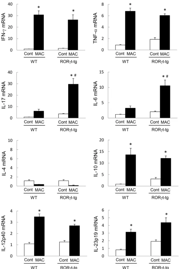
Because RORγt is known as the critical regulator of Th17 cell differentiation and Th17 cytokine
expression, we next assessed the cytokine expression in the lungs of WT mice andRORγt-tg
mice 2 months post-MAC infection. Although the expression of IFN-γand tumor necrosis
fac-tor-α(TNF-α) increased after MAC infection in the lungs of both WT mice andRORγt-tg
mice, the expression level was not different between the genotypes (Fig 3). The levels of lung
IL-17 and IL-6 expression also increased in both genotypes after MAC infection. However, in MAC-infected mice, the levels of IL-17 and IL-6 expression were significantly higher in
RORγt-tgmice than in WT mice (Fig 3). IL-4 expression was not induced in the lungs of any
mice following MAC infection (Fig 3). IL-10, IL-12, and IL-23 expressions were induced
signif-icantly in the lungs of WT mice andRORγt-tgmice after MAC infection (Fig 3), but the
expres-sion levels of these cytokines were not different between the genotypes. These results indicate
that Th1 cytokines are induced in the lungs of both WT mice andRORγt-tgmice after MAC
infection. In contrast, Th17 cytokines are specifically and strongly induced in the lung of
RORγt-tgmice.
ROR
γ
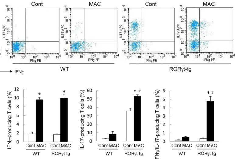
t Overexpression Enhances IL-17 Production in Lung T Cells
We assessed the production of IFN-γand IL-17 in CD4-positive T cells obtained from the
lungs of WT mice andRORγt-tgmice to clarify the contribution of CD4-positive T cells to Th1
and Th17 cytokine production. The number of IFN-γ-producing CD4-positive T cells
increased in the lungs of both genotypes after MAC infection (Fig 4). In the MAC-infected
Fig 2. MAC-induced pulmonary inflammation in WT and RORγt-tg mice.(A) Representative microphotographs of lungs from WT andRORγt-tgmice 2 months after the intratracheal inoculation of 1 x 10⁷CFU of MAC or saline (Cont). Magnification, x100.Insetsshow the inflammatory lesions at higher
magnifications. (B) Representative photographs of Ziehl-Neelsen staining of an alveolar region from WT mice (left panel), and alveolar (center panel) and peribronchial (right panel) regions ofRORγt-tgmice 2 months after the intratracheal inoculation of 1 x 10⁷CFU of MAC. Arrows indicate acid-fast bacilli. Magnification, x400. (C) Semi-quantitative scoring of inflammation in the lungs of WT andRORγt-tgmice 2 months after intratracheal inoculation of 1 x 10⁷ CFU of MAC or saline (Cont). (D) The number of total cells, neutrophils, macrophages, and lymphocytes in BAL fluids from WT andRORγt-tgmice 2 months after the intratracheal inoculation of 1 x 10⁷CFU of MAC or saline (Cont). All experiments were performed in duplicate with four mice in each group.
*Significant difference between MAC and Cont group (p<0.05). #Significant difference between genotypes after MAC infection (p<0.05). Data are expressed as the mean±SEM.
mice, the amount of IFN-γ-producing CD4 positive T cells was not different between the geno-types. The proportion of IL-17-producing CD4-positive T cells was significantly higher in the
lung ofRORγt-tgmice than in the lungs of WT mice, regardless of MAC infection (Fig 4). In
RORγt-tgmice, the proportion of IL-17-producing cells was significantly elevated after MAC
infection, compared with that in uninfectedRORγt-tgmice (Fig 4). The proportion of IFN-γ/
IL-17-coproducing CD4-positive T cells increased significantly in the lungs ofRORγt-tgmice
after MAC infection, compared with that in the lungs of uninfectedRORγt-tgmice (Fig 4).
To clarify the distribution of the T cell subsets, we then assessed the proportion of
T-bet-positive T cells and RORγt-positive T cells in the lungs of WT mice andRORγt-tgmice. Similar
to the cytokine production results, the number of T-bet-positive T cells increased in the lungs
each mRNA was analyzed by qRT-PCR, and the y-axis of each graph represents the relative expression of the respective genes calculated using theΔΔCT method and normalized against GAPDH mRNA. Experiments were performed in duplicate with five mice in each group.*Significant difference between MAC and Cont group (p<0.05). #Significant difference between genotypes after MAC infection (p<0.05). Data are expressed as the mean±SEM.
doi:10.1371/journal.pone.0147064.g003
Fig 4. IL-17-producing T cells in the lungs of WT and RORγt-tg mice after MAC infection.The proportion of IL-17- and IFN-γ-producing cells in CD4-positive T cells obtained from the lungs of WT andRORγt-tgmice 2 months after intratracheal inoculation of 1 x 10⁷CFU of MAC or saline (Cont). The
IFN-γ- and IL-17-positive cells were detected by FACS using PE-conjugated anti-mouse IFN-γand APC-conjugated anti-mouse IL-17 antibodies.
Representative plots (upper panel) and mean value among triplicate samples (lower panel) are shown.*Significant difference between MAC and Cont group (p<0.05). #Significant difference between genotypes after MAC infection (p<0.05). Data are expressed as the mean±SEM.
of both genotypes after MAC infection (Fig 5A). In the MAC-infected mice, the proportion of
T-bet-positive lung T cells was not different between the genotypes. The proportion of RORγ
t-positive lung T cells was significantly higher inRORγt-tgmice than in WT mice, regardless of
MAC infection (Fig 5A). InRORγt-tgmice, the proportion of RORγt-positive lung T cells was
significantly elevated after MAC infection, relative to the proportion in uninfectedRORγt-tg
mice (Fig 5A). The proportion of T-bet/RORγt-double positive lung T cells increased
signifi-cantly in the lung ofRORγt-tgmice after MAC infection, compared with the proportion in
uninfectedRORγt-tgmice (Fig 5A). These results indicate that the balance of the immune
response in the lung is shifted toward a Th17 phenotype inRORγt-tgmice after MAC
infection.
To clarify the contribution of CD8-positive T cells to the Th17 bias that we observed in this infection model, the IL-17 production was evaluated in the CD4-positive T cells and
CD8-posi-tive T cells obtained from the lungs of WT mice andRORγt-tgmice 2 months following MAC
infection. As stated above, IL-17-producing CD4-positive cells increased markedly after MAC
infection (Fig 5B). However, a low level of IL-17-producing CD8-positive cells was observed,
and there was no significant difference in the level of these cells between the WT mice and
RORγt-tgmice after MAC infection (Fig 5B). These results indicate that CD8-positive T cells
are not strongly involved in the generation of a Th17 bias in MAC-infectedRORγt-tgmice.
Discussion
In response to antigen, such as those from microorganisms, naïve CD4-positive T cells can be differentiated into different T cell subpopulations, including Th1, Th2, Th17, and regulatory T (Treg) cells depending on the cytokine milieu to which they are exposed. RORγt is a transcrip-tion factor belonging to a large family of hormone nuclear receptors and is known as a lineage-specific transcription factor for the development of Th17 cells [12–14,19,20]. RORγt is
expressed in Th17 cells and directs the transcriptional activation of theIL-17gene, which is
responsible for the lineage-specific cytokine of the Th17 cells [14,21,22]. In the present study,
higher levels IL-17 expression were observed in the lung CD4-positive T cells ofRORγt-tgmice
than in those of WT control mice, under both uninfected and MAC-infected conditions. We
also demonstrated that the amount of RORγt-expressing T cells was higher in the lungs of
RORγt-tgmice than in the lungs of WT mice. These findings suggest that the lung Th balance
is shifted toward Th17 phenotype in our transgenic mice that overexpress RORγt. It is unclear
why all the T cells inRORγt-tgmice do not express RORγt because the expression of the
RORγt transgene is under the control of the CD2 promoter. Potential explanations for our
findings are the detection limit of FACS or an inactivation of RORγt through interaction with other lineage-specific transcription factors during T cell development.
It is generally accepted that Th17 immunity plays a central role in the protection against fungi and extracellular bacteria [10,23]. However, the role of Th17 in regulating intracellular pathogens, such as mycobacteria, is not fully understood. Initial studies suggested that the
IL-17/Th17 pathway was not essential for protection against mycobacteria, such asM.tuberculosis
andM.bovis[24]. However, recent studies demonstrated that the IL-17/Th17 pathway may play a role in anti-mycobacterial immunity by recruiting neutrophils to the site of infection at the early stage of tuberculosis [25,26], or by accelerating the accumulation of Th1 cells and
enhancing Th1 anti-mycobacterial responses [26–28]. IL-17 also plays a role in the formation
and maintenance of granulomas in mycobacteria-infected lungs [29,30]. Re-exposure of
tuber-culosis-infected mice to high levels of tuberculosis antigen promotes further Th17 responses that cause extensive lung damage, which is associated with elevated neutrophil recruitment
inRORγt-tgmice compared with the corresponding values in WT control mice, even though
the concentration of IL-17 was significantly elevated in response to MAC infection. Corre-spondingly, we previously demonstrated that IL-17 neutralization did not exacerbate the bacte-rial burden in Th17-biased T-bet-deficient mice [9]. Taken together, it is likely that an increase in IL-17/Th17 responses does not directly link to the enhancement of anti-mycobacterial activ-ity in our MAC infection model.
In the present study, we demonstrated that neutrophilic pulmonary inflammation was
enhanced in the lungs ofRORγt-tgmice following MAC infection. IL-17 is considered to be an
important mediator for neutrophilic inflammation that acts by inducing the production of
GM-CSF, which activates neutrophil differentiation [32], and the production of neutrophil
attractant CXC chemokines [22,33,34]. Therefore, an enhancement of neutrophilic pulmonary
inflammation may be associated with an increase in the IL-17 level inRORγt-tgmice following
MAC infection. The findings in our previous study, that neutralization of IL-17 clearly attenu-ated MAC-induced neutrophil recruitment in Th17-biased T-bet-deficient mice, support the
hypothesis [9]. In patients without immunodeficiencies, neutrophils were the main cellular
constituents in BAL fluids during pulmonary MAC infection [35]. Neutrophilic pulmonary
inflammation with decreased CD4-positive lymphocytes reflected disease progression in these
patients [36]. In our MAC infection model, neutrophils were not essential for mycobacterium
killing because organ MAC CFUs were not different betweenRORγt-tgand WT mice. Thus,
enhanced neutrophil recruitment derived from Th17 deviation might have pathological, rather than protective, effects during MAC infection in our model.
It is generally accepted that lineage-specific transcription factors can inhibit the differentia-tion of other Th subsets. In fact, we previously demonstrated that T-bet suppresses IL-17 pro-duction and Th17 cell differentiation by controlling the nitric oxide level after MAC infection
[9]. However, in the present study, lung IFN-γlevel was not suppressed inRORγt-tgmice
fol-lowing MAC infection. We also found that CD4-positive T cells producing both IL-17 and
IFN-γincreased inRORγt-tgmice following MAC infection. Previous studies have
demon-strated that, in addition to cells producing either IL-17 or IFN-γ, elevated numbers of IL-17/
IFN-γdouble-positive T cells were observed in both human and mouse inflamed tissues
[37,38]. It was also reported that CD4-positive cells that express both IFN-γand IL-17 were
observed in the peripheral blood and pleural fluid from patients with tuberculosis [39]. This
suggests a complex process of transcription factor-regulated T cell differentiation during
infec-tion. Bonifaceet al. have demonstrated that the development of IL-17/IFN-γdouble-positive T
cells is under the influence of RORγt and that these cells may belong to the Th17 lineage but
they are distinct from Th1 lineage [40]. The expression analysis of the transcription factor
revealed that, equal with the increase in IL-17/IFN-γdouble-positive T cells, T cells
co-express-ing RORγt and T-bet increased followco-express-ing MAC infection.
It has been reported that Th17 cells may represent a heterogeneous population with distinct trafficking profiles and differing abilities. Ghoreschiet al. have demonstrated that Th17 cells
express both RORγt and T-bet when these cells are generated in the absence of TGF-β[41].
RORγt and T-bet double-positive Th17 cells were presentin vivoin lesional tissue in
Fig 5. RORγt-expressing T cells in the lungs of WT and RORγt-tg mice after MAC infection. (A)The proportion of CD4-positive T cells expressing RORγt and/or T-bet in the lungs of WT andRORγt-tgmice 2 months after an intratracheal inoculation of 1 x 10⁷CFU of MAC or saline (Cont). The T-bet- and
RORγt-positive cells were detected by FACS using PE-conjugated anti-T-bet and APC-conjugated anti-RORγt antibodies. Representative plots (upper panel) and mean value among triplicate samples (lower panel) are shown.(B)The proportion of IL-17-producing cells in CD4-positive (upper panels) and CD8-positive T cells (lower panels) obtained from the lungs of WT andRORγt-tgmice 2 months after an intratracheal inoculation of 1 x 10⁷CFU of MAC. The IL-17-, CD4-, and CD8-positive cells were detected by FACS using PE-conjugated anti-IL-17, FITC-conjugated anti-CD4, and PerCP-conjugated anti-CD8 antibodies. Representative plots (left panel) and mean value among duplicate samples (lower panel) are shown.*Significant difference between the MAC and Cont group (p<0.05). #Significant difference between genotypes after MAC infection (p<0.05). Data are expressed as the mean±SEM.
experimental allergic encephalomyelitis [41] and multiple sclerosis [38]. These cells might be more relevant to the pathogenesis of diseases that develop both Th1- and Th17-mediated pathology. The role of RORγt and T-bet double-positive T cells in the pathogenesis of MAC disease should be elucidated in the future.
In summary, lymphocyte-restricted overexpression of RORγt induced a Th17 bias in the
lung tissue following MAC infection. This RORγt-mediated Th17 bias did not affect the
sys-temic growth of MAC, whereas it enhanced the neutrophilic pulmonary inflammation follow-ing MAC infection. Excessive Th17 responses might produce pathological effects, rather than
provide protection, during MAC infection. The lung histopathology inRORγt-tgmice
resem-bled the histopathology in patients with pulmonary MAC disease. Therefore, we will examine the appearance of Th17 cells and the IL-17 level in the BAL fluids and lung tissues of patients with pulmonary MAC disease in our next study.
Author Contributions
Conceived and designed the experiments: MM YI. Performed the experiments: MM SA YM HS. Analyzed the data: MM YI. Contributed reagents/materials/analysis tools: KY ST KO. Wrote the paper: MM YI NH.
References
1. Marras TK., Chedore P, Ying AM, Jamieson F. Isolation prevalence of pulmonary non-tuberculous mycobacteria in Ontario, 1997–2003. Thorax 2007; 62: 661–666. PMID:17311842
2. Cassidy P. Maureen PM., Hedberg K, Saulson A, McNelly E, Winthrop KL. Nontuberculous mycobacte-rial disease prevalence and risk factors: A changing epidemiology. Clin Infect Dis 2009; 49: e124–
e129. doi:10.1086/648443PMID:19911942
3. Plotinsky RN., Talbot EA., von Reyn CF. Proposed definitions for epidemiologic and clinical studies of Mycobacterium avium complex pulmonary disease. PLoS One 2013; 8: e77385. doi:10.1371/journal. pone.0077385PMID:24265675
4. Trinchieri G. Cytokines acting on or secreted by macrophages during intracellular infection (10, IL-12, IFN gamma). Curr Opin Immunol 1997; 9: 17–23. PMID:9039773
5. Rook GA. Th2 cytokines in susceptibility to tuberculosis. Curr Mol Med 2007; 7: 327–337. PMID:
17504117
6. North RJ. Mice incapable of making IL-4 or IL-10 display normal resistance to infection with Mycobacte-rium tuberculosis. Clin Exp Immunol 1998; 113: 55–58. PMID:9697983
7. Jung YJ, LaCourse R, Ryan L, North RJ. Evidence inconsistent with a negative influence of T helper 2 cells on protection afforded by a dominant T helper 1 response against Mycobacterium tuberculosis lung infection in mice. Infect Immun 2002; 70: 6436–6443. PMID:12379724
8. Lazarevic V, Glimcher LH. T-bet in disease. Nat Immunol 2011; 12: 597–606. doi:10.1038/ni.2059
PMID:21685955
9. Matsuyama M, Ishii Y, Yageta Y, Ohtsuka S, Ano S, Matsuno Y, et al. Role of Th1/Th17 balance regu-lated by T-bet in a mouse model of Mycobacterium avium complex disease. J Immunol 2014; 192: 1707–1717. doi:10.4049/jimmunol.1302258PMID:24446514
10. Romani L. Immunity to fungal infections. Nature Rev Immunol 2011; 11: 275–288.
11. Ross PJ, Sutton CE, Higgins S, Allen AC, Walsh K, Misiak A, et al. Relative contribution of Th1 and Th17 cells in adaptive immunity toBordetella pertussis: towards the rational design of an improved acellular pertussis vaccine. PLoS Pathog 2013; 9: e1003264. doi:10.1371/journal.ppat.1003264
PMID:23592988
12. Ivanov II, McKenzie BS, Zhou L, Tadokoro CE, Lepelley A, Lafaille JJ, et al. The orphan nuclear recep-tor RORγt directs the differentiation program of proinflammatory IL-17+T helper cells. Cell 2006; 126:
1121–1133. PMID:16990136
13. Manel N, Unutmaz D, Littman DR. The differentiation of human Th-17 cells requires transforming growth factor-beta and induction of the nuclear receptor RORγt. Nat Immunol 2008; 9: 641–649. doi:
10.1038/ni.1610PMID:18454151
15. Yoh K, Morito N, Ojima M, Shibuya K, Yamashita Y, Morishima Y, et al. Overexpression of RORγt under control of the CD2 promoter induces polyclonal plasmacytosis and autoantibody production in transgenic mice. Eur J Immunol 2012; 42: 1999–2009. doi:10.1002/eji.201142250PMID:22623033
16. Ano S, Morishima Y, Ishii Y, Yoh K, Yageta Y, Ohtsuka S, et al. Transcription factors GATA-3 and RORγt are important for determining the phenotype of allergic airway inflammation in a murine model of asthma. J Immunol 2013; 190: 1056–1065. doi:10.4049/jimmunol.1202386PMID:23293351
17. Sweeney KA, Dao DN, Goldberg MF, Hsu T, Venkataswamy MM, Henao-Tamayo M, et al. A recombi-nantMycobacterium smegmatisinduces potent bactericidal immunity againstMycobacterium tubercu-losis. Nat Med 2011; 17: 1261–1268. doi:10.1038/nm.2420PMID:21892180
18. Murphy E, Shibuya K, Hosken N, Openshaw P, Maino V, Davis K, et al. Reversibility of T helper 1 and 2 populations is lost after long term stimulation. J Exp Med 1996; 183: 901–913. PMID:8642294
19. Mangelsdorf DJ, Thummel C, Beato M, Herrlich P, Schutz G, Umesono K, et al. The nuclear receptor superfamily: the second decade. Cell 1995; 83: 835–839. PMID:8521507
20. Eberl G, Littman DR. The role of the nuclear hormone receptor RORγt in the development of lymph nodes and Peyer’s patches. Immunol Rev 2003; 195: 81–90. PMID:12969312
21. Harrington LE, Hatton RD, Mangan PR, Turner H, Murphy TL, Murphy KM, et al. Interleukin 17-produc-ing CD4+ effector T cells develop via a lineage distinct from the T helper type 1 and 2 lineages. Nat Immunol 2005; 6: 1123–1132. PMID:16200070
22. Korn T, Bettelli E, Oukka M, Kuchroo VK. IL-17 and Th17 cells. Ann Rev Immunol 2009; 27: 485–517. 23. Happel KI, Dubin PJ, Zheng M, Ghilardi N, Lockhart C, Quinton LJ, et al. Divergent roles of IL-23 and
IL-12 in host defense against Klebsiella pneumoniae. J Exp Med 2005; 202: 761–769. PMID:
16157683
24. Khader SA, Pearl JE, Sakamoto K, Gilmartin L, Bell GK, Jelley-Gibbs DM, et al. IL-23 compensates for the absence of IL-12p70 and is essential for the IL-17 response during tuberculosis but is dispensable for protection and antigen-specific IFN-gamma responses if IL-12p70 is available. J Immunol 2005; 175: 788–795. PMID:16002675
25. Umemura M, Yahagi A, Hamada S, Begum MD, Watanabe H, Kawakami K, et al. IL-17-mediated regu-lation of innate and acquired immune response against pulmonary Mycobacterium bovis bacille Calm-ette-Guerin infection. J Immunol 2007; 178: 3786–3796. PMID:17339477
26. Khader SA, Gopal R. IL-17 in protective immunity to intracellular pathogens. Virulence 2010; 1: 423–
427. doi:10.4161/viru.1.5.12862PMID:21178483
27. Khader SA, Bell GK, Pearl JE, Fountain JJ, Rangel-Moreno J, Cilley GE, et al. IL-23 and IL-17 in the establishment of protective pulmonary CD4-positive T cell responses after vaccination and during Mycobacterium tuberculosis challenge. Nat Immunol 2007; 8: 369–377. PMID:17351619
28. Gopal R, Lin Y, Obermajer N, Slight S, Nuthalapati N, Ahmed M, et al. IL-23-dependent IL-17 drives Th1-cell responses following Mycobacterium bovis BCG vaccination. Eur J Immunol 2012; 42: 364–
373. doi:10.1002/eji.201141569PMID:22101830
29. Torrado E, Cooper AM. IL-17 and Th17 cells in tuberculosis. Cytokine Growth Factor Rev 2010; 21: 455–462. doi:10.1016/j.cytogfr.2010.10.004PMID:21075039
30. Okamoto-Yoshida Y, Umemura M, Yahagi A, O'Brien RL, Ikuta K, Kishihara K, et al. Essential role of IL-17A in the formation of a mycobacterial infection-induced granuloma in the lung. J Immunol 2010; 184: 4414–4422. doi:10.4049/jimmunol.0903332PMID:20212094
31. Cruz A, Fraga AG, Fountain JJ, Rangel-Moreno J, Torrado E, Saraiva M, et al. Pathological role of inter-leukin 17 in mice subjected to repeated BCG vaccination after infection with Mycobacterium tuberculo-sis. J Exp Med 2010; 207: 1609–1616. doi:10.1084/jem.20100265PMID:20624887
32. Fossiez F, Djossou O, Chomarat P, Flores-Romo L, Ait-Yahia S, Maat C, et al. T cell interleukin-17 induces stromal cells to produce proinflammatory and hematopoietic cytokines. J Exp Med 1996; 183: 2593–2603. PMID:8676080
33. Wang YH, Liu YJ. The IL-17 cytokine family and their role in allergic inflammation. Curr Opin Immunol 2008; 20: 697–702. doi:10.1016/j.coi.2008.09.004PMID:18832032
34. Lindén A. Role of interleukin-17 and the neutrophil in asthma. Int Arch Allergy Immunol 2001; 126:.179–184. PMID:11752873
35. Yamazaki Y, Kubo K, Sekiguchi M, Honda T. Analysis of BAL fluid in M. avium-intracellulare infection in individuals without predisposing lung disease. Eur Respir J 1998; 11: 1227–1231. PMID:9657559
37. Annunziato F, Cosmi L, Santarlasci V, Maggi L, Liotta F, Mazzinghi B, et al. Phenotypic and functional features of human Th17 cells. J Exp Med 2007; 204: 1849–1861. PMID:17635957
38. Kebir H, Ifergan I, Alvarez JI, Bernard M, Poirier J, Arbour N, et al. Preferential recruitment of interferon-γ-expressing Th 17 cells in multiple sclerosis. Ann Neurol 2009; 66: 390–402. doi:10.1002/ana.21748
PMID:19810097
39. Jurado JO, Pasquinelli V, Alvarez IB, Peña D, Rovetta AI, Tateosian NL, et al. IL-17 and IFN-γ expres-sion in lymphocytes from patients with active tuberculosis correlates with the severity of the disease. J Leukoc Biol 2012; 91: 991–1002. doi:10.1189/jlb.1211619PMID:22416258
40. Boniface K, Blumenschein WM, Brovont-Porth K, McGeachy MJ, Basham B, Desai B, et al. Human Th17 cells comprise heterogeneous subsets including IFN-γ-producing cells with distinct properties from the Th1 lineage. J Immunol 2010; 185:679–687. doi:10.4049/jimmunol.1000366PMID:
20511558
41. Ghoreschi K, Laurence A, Yang XP, Tato CM, McGeachy MJ, Konkel JE, et al. Generation of patho-genic TH17 cells in the absence of TGF-βsignalling. Nature 2010; 467: 967–971. doi:10.1038/