Arch Health Invest (2014) 3(6): 28-30 © 2014 - ISSN 2317-3009
Arch Health Invest 3(6) 2014 28
Supratarsal approach to treatment of zygomatic
complex fractures
Abordagem supratarsal para tratamento de
fraturas complexo zigomático
Acercamiento supratarsal en el tratamiento de las
fracturas del complejo cigomático
Ellen Cristina GAETTI JARDIM1
Ana Claudia ROSSI2
Henrique Celestino Lima e SILVA3
Leonardo Perez FAVERANI4
Analice Cristhian Flavio QUINTANILHA5
Emerson Chaves FURLANETO6
Elio Hitosh SHINOHARA7
1Residência em Cirurgia e Traumatologia Bucomaxilofacial, Hospital Universitário “Maria Aparecida Pedrossian”,
Universidade Federal do Mato Grosso do Sul, UFMS
Pós-doutoranda em Cirurgia e Traumatologia Bucomaxilofacial, Faculdade de Odontologia de Araçatuba, UNESP-Univ. Estadual Paulista, Araçatuba-SP, Brasil
2
Departamento de Morfologia, Faculdade de Odontologia de Piracicaba, UNICAMP-Univ. Estadual de Campinas, Piracicaba-SP, Brasil
3
Residente em Cirurgia e Traumatologia Bucomaxilofacial, Hospital Universitário “Maria Aparecida Pedrossian”, Universidade Federal do Mato Grosso do Sul, UFMS
4
Departamento de Cirurgia e Clínica Integrada, Faculdade de Odontologia de Araçatuba, UNESP-Univ. Estadual Paulista, Araçatuba-SP, Brasil
5
Preceptora Residência Médica Multiprofissional em Saúde, Hospital Universitário “Maria Aparecida Pedrossian”, Universidade Federal do Mato Grosso do Sul, UFMS
6
Serviço de Cirurgia e Traumatologia Bucomaxilofacial do Hospital Regional do Mato Grosso do Sul “Rosa Pedrossian”
Coordenador do Curso de Especialização em Cirurgia e Traumatologia Bucomaxilofacial da Associação Brasileira de Odontologia, Seção Mato Grosso do Sul
7
Professor/Orientador credenciado junto ao Programa de Pós-Graduação em Odontologia, Área de Concentração em Cirurgia e Traumatologia Bucomaxilofacial, Faculdade de Odontologia de Araçatuba,
UNESP-Univ. Estadual Paulista, Araçatuba-SP, Brasil
Abstract
Many incisions have been described for approaches to zygomatic fractures. Precise repositioning of zygomatic complex fractures is difficult. The traditional approach is through an eyebrow incision, but it can produce a scar that causes aesthetic and psychological problems for the patient. We describe the supratarsal fold approach to expose the frontozygomatic suture and to reduce small displacements of frontal sinus anterior wall; it gives good access and excellent aesthetic results.
Descriptors: Surgery, Oral; Zygoma; Surgical Procedures, Operative.
Resumo
Muitas incisões têm sido descritas para abordagens de fraturas zigomática. Reposicionamento preciso de fraturas complexas zigomáticos é difícil. A abordagem tradicional é através de uma incisão de sobrancelha, mas pode produzir uma cicatriz que causa problemas estéticos e psicológicos para o paciente. Nós descrevemos a abordagem palpebral supratarsal para expor a sutura frontozigomática e reduzir pequenos deslocamentos da parede anterior do seio frontal. O acesso mostrou-se eficiente, com excelentes resultados estéticos.
Descritores: Cirurgia Bucal; Zigoma; Procedimentos Cirúrgicos Operatórios.
Resumen
Varias incisiones se han descrito para las fracturas del complejo cigomático. El reposicionamiento preciso de fracturas del complejo cigomáticos es difícil. El enfoque tradicional es a través de una incisión ceja, pero puede producir una cicatriz causando problemas estéticos y psicológicos para el paciente. Acercamiento supratarsal fue descrito por el párpado para exponer la sutura frontozigomática y reducir los pequeños desplazamientos de la pared anterior del seno frontal. Acceso demostró ser eficaz con excelentes resultados estéticos.
Arch Health Invest (2014) 3(6): 28-30 © 2014 - ISSN 2317-3009
Arch Health Invest 3(6) 2014 29
INTRODUCTION
The zygomatic complex is responsible for anteroposterior projection of the face, then the reduction and stabilization when displaced is important for the restoration of facial symmetry, globe ocular position, infraorbital innervations, as well as facial aesthetics. [1-3]
Camouflage the incisions on the face is essential since aesthetics is an important factor when planning a surgical approach. Many approaches have been proposed with this objective, the choice is usually made based at the fracture site, associated with the experience and surgeon training. [4]
In respect to periorbital area, symmetry and position of the eye are essential to the restoring the facial aesthetics. To this to occur, an adequate exposure of the zigomaticofrontal suture and orbital margins is essential. [5]
Thus, we describe a supratarsal approach to facilitate exposure to the zigomaticofrontal area, facilitating the reduction and fixation of fractures with excellent cosmetic results.
CASE REPORT
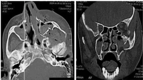
A female patient, suffered a zigomatic complex fracture on the left side, underwent surgical treatment under general anesthesia in order to achieve the reduction and fixation of fractures (Figure 1).
To access the zigomaticofrontal region, we opted for the creation of supra-tarsal approach. The supratarsal approach is performed in parallel with upper eyelid, over the tarsal plate to adequately expose zigomaticofrontal suture. When the goal is access to the anterior wall of the frontal sinus, the incision can be extended medially. The orbicularis muscle is dissected in the superior and lateral direction, exposing orbital periosteum, and the surgeon can palpate orbital rim to properly locate the fracture.
Figure 1. CT scan showing zygomatic-orbital fractures.
The zigomaticofrontal suture can be exposed by an incision in the periosteum from 2 to 3 mm along the
outer surface of the orbital margin (Figure 2). The incision is closed with interrupted sutures. A bandage compression is applied for 2 days to prevent the hematoma formation and help the repair process. In the postoperative period of 30 and 60 days the healing aspect of the surgical approach was satisfactory with no aesthetic sequelae (Figures 3 and 4).
Figure 2. Immediate postoperative.
Figure 3. Postoperative period of 30 days.
Arch Health Invest (2014) 3(6): 28-30 © 2014 - ISSN 2317-3009
Arch Health Invest 3(6) 2014 30
DISCUSSION
Various surgical techniques have been described for the repair of a zygomatic complex fracture. Extra and intraoral incisions have been proposed and intensively used for the development of simple access that promotes sufficient exposure to reduction and fixation of fractures with better results. [6-9]
The supratarsal incision is often used in plastic
surgery for blepharoplasty. For treatment of
zygomatic-orbital fractures it is known that Chuong and Kaban. [10] were the first to describe supratarsal approach to treat zygomatic fractures. The same, described the supratarsal access as a viable alternative for exposure of the suture zigomaticofrontal with exceptional aesthetic results with inconspicuous scars
[1,5]
as observed in present paper in Figures 3 and 4. In cases of zygomatic complex fractures and fractures of the anterior wall of the frontal sinus, we can use this approach to the direct visualization zigomaticofrontal suture. The disadvantages are similar of the eyebrow approach, because these areas are similar, with almost no vital structure.
For cosmetic improvement of incision, some precautions must be taken such as the gentle use of skin hooks in tissue handling, Ophthalmic ointments or temporary tarsorrhaphy to shelter eye, delicate tissue dissection during the procedure, and the use of flexible metal spatulas for protect the contents of the orbit. Careful positioning incision is vital to camouflage the scar completely, especially when the tissue is manipulated [9].
Complications of supratarsal access include the orbital fat exposure, lacrimal glands exposure due to the dissection of the orbital septum, but this can be avoided by careful dissection in subperiosteal plane.
CONCLUSION
The supratarsal fold incision provides excellent access to the supraorbital rim and zygomaticofrontal suture and uncomplicated design as well as its excellent cosmetic result. Complications may include exposure of orbital fat and lachrymal glands by dissection of the orbital septum, but this can be avoided by careful dissection in the subperiosteal.
REFERÊNCIAS
1. Hammer B. Fraturas orbitárias: diagnóstico, tratamento cirúrgico, correções secundárias. São Paulo:Santos; 2005, 2-5.
2. Khan AM, Varvares MA. Traditional Approaches to the Orbit. Otolaryngol Clin N Am. 2006; 39: 895–909.
3. Scolozzi P, Momjian A, Heuberger J, Andersen E, Broome M, Terzic A, Jaques B. Accuracy and predictability in use of AO three-dimensionally preformed titanium mesh plates for posttraumatic orbital reconstruction: a pilot study. J Craniofac Surg. 2009;20(4):1108-13.
4. Holtmann B, Wray RC, Little AG. A randomized comparison of four incisions for orbital fractures. Plast Reconstr Surg. 1981; 67:731-7.
5. Kung DS, Kaban LB. Supratarsal fold incision for approach to the superior lateral orbit. Oral Surg Oral Med Oral Pathol Oral Radiol Endod. 1996;81(5):522–5.
6. Keen WW. Surgery: its principles and practice. Philadelphia: Saunders; 1909.
7. Gillies HD, Kilner TP, Stone D. Fractures of the malar-zygomatic compound, with a description of a new X-ray position. Br J Surg.1927;14(56):651–6.
8. Snow RC, Parsons RW. Exposure through coronal
incision for initial treatment of facial fractures. Plast Recontr Surg. 1975;56 (3):254–9.
9. Gaia BF, Landgraf H, Pardo-Kaba SC, Shinohara EH. Access to the frontal sinus and zygomatico frontal suture through the supratarsal fold. Britsh J Oral Maxillofac Surg. 2008; 46(3): 226–8.
10.Chuong R, Kaban LB. Fractures of the zygomatic complex. J Oral Maxillofac Surg. 1986;44:283–8.
CONFLITO DE INTERESSES
Os autores declaram não haver conflitos de interesse.
AUTOR PARA CORRESPONDÊNCIA
Ellen Cristina Gaetti Jardim ellengaetti@gmail.com
Submetido em 12/09/2014