Factors that influence healing of chronic venous leg ulcers:
a retrospective cohort
*Marilia Formentini Scotton
1Hélio Amante Miot
1Luciana Patricia Fernandes Abbade
1DOI: http://dx.doi.org/10.1590/abd1806-4841.20142687
Abstract: BACKGROUND: Venous ulcers have a significant impact on patient quality of life, and constitute a worldwide public health problem. Treatment is complex, with high failure rates.
OBJECTIVES: To identify clinical and therapeutic factors that influence healing of venous ulcers.
METHODS: Retrospective cohort study of patients with venous ulcers. Ulcer area was measured at the first visit (T0) and after 6 months (T6) and 1 year (T12). A reduction in ulcer area of 50% or more at T6 and T12 was the outcome of interest, weighted by clinical, demographic and treatment aspects.
RESULTS: Ninety-four patients were included (137 ulcers). A reduction in ulcer area of 50% or more was seen in 40.1% of patients (95%CI 31.9 to 48.4%) at T6 and 49.6% (95%CI 41.2 to 58.1%) at T12. Complete healing occurred in 16.8% (95%CI 10.5 to 23.1%) at T6 and 27% (95%CI 19.5 to 39.5%) at T12. The lowest ulcer area reductions at T6 were associated with longstanding ulcer (RR=0.95; 95%CI 0.91 to 0.98), poor adherence to compression therapy (RR=4.04; 95%CI 1.31 to 12.41), and infection episodes (RR= 0.42; 95%CI 0.23 to 0.76). At T12, lower reductions were associated with longstanding ulcer (RR=0.95; 95%CI 0.92 to 0.98), longer topical antibiotic use (RR=0.93; 95%CI 0.87 to 0.99), and systemic antibiotic use (RR=0.63; 95%CI 0.40 to 0.99). CONCLUSIONS: Longstanding ulcer, infection, poor adherence to compression therapy, and longer topical and systemic antibiot-ic use were independently correlated with worse healing rates.
Keywords:Cohort studies; Leg ulcer; Lower extremity; Risk factors; Varicose ulcer; Wound healing
Received on 11.04.13.
Approved by the Advisory Board and accepted for publication on 17.06.2013.
* Work performed at the Department of Dermatology and Radiotherapy, Faculdade de Medicina de Botucatu – Universidade Estadual Paulista "Júlio de Mesquita Filho" (UNESP) – Botucatu (SP), Brazil.
Financial support: none. Conflict of interests: none.
1 Universidade Estadual Paulista “Júlio de Mesquita Filho” (UNESP) – Botucatu (SP), Brazil.
©2014 by Anais Brasileiros de Dermatologia INTRODUCTION
Chronic leg ulcers are defined as wounds locat-ed below the knee that do not heal within a 6-week period.1,2 There are three main types of leg ulcers: venous, arterial, and neuropathic. Venous ulcers (VUs) account for approximately 75% of all chronic leg ulcers, and are characterized by several factors that hinder and delay healing.
VUs affect patients of both sexes, and are most common in older adults. They constitute a public health issue in Brazil and worldwide, being associat-ed with absence from work, frequent physician visits, and high treatment-related expenses.1,3,4
The prevalence of VUs varies between studies, due to the heterogeneity in diagnostic methods and epidemiological characteristics of the sample and depending on whether foot ulcers are included as well.5,6 In Brazil, Maffei et al. (1986)7 found a 3.6% prevalence of active and/or healed VUs in a sample of 1,755 individuals aged >15 years.
VUs develop in the context of advanced chron-ic venous insuffchron-iciency (CVI) with dysfunction of the calf muscle pump.5,8,9The venous hypertension caused by venous valve incompetence, which is common in primary varicose veins and post-thrombotic syn-drome and may affect the superficial, communicating (perforator), or deep venous systems in isolation or concomitantly, is the key to ulcer development. Nevertheless, the exact mechanism whereby venous hypertension leads to VUs is unknown. Recent stud-ies suggest that the pathogenesis of the ulceration process is associated with the abnormal changes in microcirculation and consequent inflammatory response inherent to CVI.10,11
they are usually slow to develop, but onset may be sudden. Location is also variable, although the distal region of the legs – particularly over the medial malle-olus – is most commonly affected.6
Management of VUs is complex, and ranges from clinical treatment to surgical therapy of the underlying venous abnormality.11Compression thera-py is a key element.
Knowledge of the factors that contribute to fail-ure of VU healing is essential for prognostication and development of treatment strategies. Within this con-text, the present study seeks to identify clinical, demo-graphic, and treatment-related factors that influence VU healing.
MATERIALS AND METHODS
This was a retrospective cohort study of patients with chronic venous leg ulcers treated at the outpatient ulcer clinic of the Department of Dermatology, da Faculdade de Medicina de Botucatu – UNESP, from 2000 to 2010. The study was approved by the local research ethics committee, and was car-ried out in accordance with the principles set forth in the Declaration of Helsinki.
Clinical, demographic, and treatment-related variables were assessed in all selected patients over a 1-year period from their first visit to the outpatient ulcer clinic.
The inclusion criteria were: diagnosis of chron-ic leg ulcers of venous etiology; at least 12 months’ fol-low-up; and knowledge of ulcer area at the first visit (T0) and approximately 6 months (T6) and 12 months (T12) thereafter. The exclusion criteria were chronic leg ulcers of any other etiology and VUs associated with peripheral artery disease.
Chronic venous leg ulcers were defined as any VU located below the knee for over 6 weeks with one or more of the following manifestations: edema, hyperpigmentation (stasis dermatitis), eczema, lipo-dermatosclerosis, and varicose veins.
Peripheral arterial disease was defined by an ankle-brachial index (ABI), calculated using a 10-mHz handheld vascular Doppler device (DV 610B), of <0.9 and/or absence of distal pulses.
Ulcers were considered post-thrombotic when the patient had a history of deep venous thrombosis (DVT) of the affected limb or when patient records contained a history of DVT. Ulcers were considered due to primary varicose veins when varicose disease was present but there was no history of DVT.
Each ulcer was initially demarcated by tracing its borders onto clear plastic film. Each tracing was then transferred onto a blank sheet of paper, and a sticker of known size was placed next to the tracing for scale. The tracing and sticker were then
pho-tographed with a compact digital camera. Using the external scale reference (sticker), we established the pixel/cm relationship for each photo. Having estab-lished that relationship, we manually traced the perimeter of each ulcer in image editing software (Image J 1.4612) to calculate its area in pixels, which was then converted to cm2.
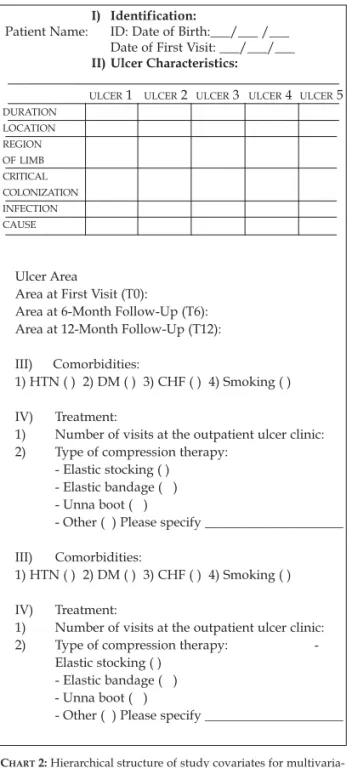
Information on the variables of interest was col-lected from patient records, into a form designed specifically for this purpose (Chart 1).
The sample size was defined on the basis of a pre-test of 60 ulcers using the Freeman formula (n=10*[k+1]), with attention to the covariates that indicated the composition of the final multivariate model and keeping a proportion of at least five events for each covariate with potential influence on the out-come of interest (p<0.25). As the pre-test yielded nine covariates and estimated that 50% of ulcers would heal (>50% reduction in area) during the period of interest, the final sample size was defined as 100 ulcers.13
Categorical variables were expressed as per-centages, and bivariate comparisons performed using the chi-square or Fisher’s exact tests. Quantitative variables were tested for normality with the Shapiro-Wilk test and expressed as means and standard devi-ations or medians and interquartile ranges (IQR = p25-p75) as appropriate. The Student t or Mann-Whitney U tests were used for bivariate comparisons as appropriate.
Longitudinal repeated measures of ulcer areas were assessed by a generalized linear mixed effects model (gamma probability distribution).
We then carried out a multivariate analysis with Poisson regression (log-linear model). Selection of variables for the final model was based on a hierar-chical structure; as long as a given covariate had a p-value <0.25 among all covariates within each hierar-chical block, it was kept for analysis of the next block (Chart 2). Covariates in which a suppression effect was identified were kept in the model.
The variables of the final model were tested for interaction effect in 2x2.
For multivariate analysis, missing data were filled in by means of the multiple imputation method, as long as they represented less than 10% of the data for the subject or variable of interest.
Effect size was estimated as relative risk (RR) with respective 95% confidence intervals (95%CI).
Data were tabulated and analyzed in IBM SPSS 20 software. The significance level was set at p<0.05.
RESULTS
74 (44.04%) were excluded for the following criteria: 17 ulcers for which 12-month follow-up was not avail-able (22.97%); eight ulcers of mixed etiology (10.81%); four cases of neuropathic plantar ulceration (mal per-forant) (5.40%); seven ulcers on which no data could be collected for the protocol (9.45%); seven ulcers of unknown etiology (9.45%); four ulcers secondary to bullous erysipelas (5.40%); and 27 ulcers due to other causes (36.48%), namely: photodynamic therapy, polycythemia vera, peripheral artery disease, hyper-tension, burns, rheumatoid arthritis, basal cell carci-noma, spindle cell carcicarci-noma, brown recluse spider bite, pyoderma gangrenosum, subungual verruca vul-garis, livedoid vasculopathy, and leprosy.
The overall profile of the 94 patients included is shown in table 1. Noteworthy findings include advanced age, a substantial proportion of ulcers larg-er than 20 cm2 (43%) at baseline (T0), and the female majority of the sample, for a total of 137 ulcers. The duration of active ulceration ranged from 12 months to 39 years (Table 1).
Graph 1 shows the distribution of ulcer areas at T0, T6, and T12. A ≥50% reduction in area was observed in 40.1% of ulcers (95%CI 31.9-48.4 %) at T6 and 49.6% of ulcers (95%CI 41.2-58.1%) at T12. Complete healing occurred in 16.8% of ulcers (95%CI 10.5-23.1%) at T6 and 27% (95%CI 19.5-39.5%) at T12. Statistically significant reductions in area occurred between T0 and T12 and between T6 and T12 (p<0.01), but not between T0 and T6 (p=0.74).
Table 2 shows bivariate and multivariate analy-ses of the variables of interest with regard to reduction in VU area to <50% of baseline at 6-month follow-up (T6). These analyses revealed that duration of ulcer progression, episodes of infection, and poor compli-ance with compression therapy had a significant neg-ative impact on healing.
Table 3 shows bivariate and multivariate analy-ses of the variables of interest with regard to reduction in VU area to <50% of baseline after 12 months of treat-ment (T12). Duration of ulcer progression and use of topical and systemic antibiotics were found to have significant negative impacts on long-term healing.
The 2x2 interactions of the final model variables for T6 and T12 did not yield significant products (p>0.05).
DISCUSSION
In this series of 94 patients with chronic venous leg ulcers, significant reductions in ulcer area were observed over a 12-month follow-up period, ratifying the need for persistence in the treatment of VUs.
Labropoulos et al.assessed the progression of 153 VUs in 127 patients in the New York region.14The factors associated with nonhealing were advanced age, increased body mass index, history of DVT,
non-First level: Patient-related / ulcer-related variables
Sex, age, active ulcer duration, ulcer location and area; episodes of critical colonization or infection; post-throm-botic etiology, high blood pressure, diabetes mellitus.
Second level: Treatment-related variables
Compliance with rest, occlusive dressings, and compres-sion therapy; duration of use of debriding dressings, topi-cal antibiotics, and systemic antibiotic use.
CHART2:Hierarchical structure of study covariates for multivaria-te analysis. First level, patient-relamultivaria-ted/ulcer-relamultivaria-ted variables;
second level, treatment-related variables
CHART1:Data collection protocol for patients with venous ulcers
I) Identification:
Patient Name: ID: Date of Birth:___/___ /___ Date of First Visit: ___/___/___
II) Ulcer Characteristics:
ULCER1 ULCER2 ULCER3 ULCER4 ULCER5
DURATION LOCATION REGION OF LIMB CRITICAL COLONIZATION INFECTION CAUSE
Ulcer Area
Area at First Visit (T0):
Area at 6-Month Follow-Up (T6): Area at 12-Month Follow-Up (T12):
III) Comorbidities:
1) HTN ( ) 2) DM ( ) 3) CHF ( ) 4) Smoking ( )
IV) Treatment:
1) Number of visits at the outpatient ulcer clinic: 2) Type of compression therapy:
- Elastic stocking ( ) - Elastic bandage ( ) - Unna boot ( )
- Other ( ) Please specify _____________________
III) Comorbidities:
1) HTN ( ) 2) DM ( ) 3) CHF ( ) 4) Smoking ( )
IV) Treatment:
1) Number of visits at the outpatient ulcer clinic:
2) Type of compression therapy: -
Elastic stocking ( ) - Elastic bandage ( ) - Unna boot ( )
GRAPH1:Representation of ulcer areas (Log2 of cm2) at T0, T6, and T12
compliance with compression therapy, and large ulcer area. In our study, duration of active ulcer, episodes of local infection, and use of antibiotics correlated with a poor prognosis for healing. These situations are very common and may reflect ineffective or inappropriate prior attempts at therapy.
In our sample, active ulcer duration was pro-longed, more so than in other studies.15This was prob-ably due to the fact that our center is a referral service for chronic ulcer care and receives patients from many other health facilities, including a large number of cases of treatment failure.
An influence of ulcer chronicity on the healing process was also found by Margolis et al., in a U.S. study conducted in Pennsylvania in the 1990s.15 The authors suggest that, the longer the active ulcer time, the harder it is to achieve healing, as a wide range of time-dependent changes may occur in the ulcer microenvironment, such as: excessive amounts of matrix metalloproteinases, collagenases, and elas-tases, which cause an early breakdown of collagen and growth factors; phenotypic alteration of wound cells, particularly fibroblasts, which would hinder their capacity for proliferation and movement; and a hypoxic microenvironment conducive to a high rate of fibroblast proliferation, leading to tissue fibrosis, as well as a greater tendency toward bacterial and fungal colonization.16
Some studies have shown that colonization and infection play a role in delaying the healing of chron-ic ulcers.16-18In our sample, these factors were present
in a substantial percentage of cases, demonstrating how often these conditions occur in patients with chronic leg ulcers. Episodes of critical colonization did not correlate with poor healing, but infection was a major factor associated with worse prognosis during follow-up.
The lack of association between critical colo-nization and poor prognosis in our patients may be justified by the fact that adequate therapy, such as sur-gical debridement and activated charcoal/silver or hydrofiber/silver dressings, was provided whenever this condition was diagnosed clinically. These meas-ures probably enabled early treatment of critical colo-nization, thus attenuating its influence on the healing process.
The chronic wound microenvironment is com-plex and usually contaminated by more than one species of bacteria. There may be formation of biofilms – communities of micro-organisms embedded within an extracellular polysaccharide matrix, which makes them more resistant to host defenses, antiseptics, and topical or systemic antibiotics.19In our sample, longer topical and systemic antibiotic therapy correlated with poor prognosis at 12-month follow-up, suggesting that antibiotics are ineffective in promoting long-term healing of VUs.
The clinical dimension of infection varies in direct relation to bacterial load and virulence and is inversely proportional to host defenses.20In our case series, infec-tion had a negative impact on the healing process at 6-month follow-up, with no influence from antibacterial treatment, which consisted of systemic antibiotic thera-py, often combined with surgical debridement of infect-ed tissue (particularly in cases of necrosis and a substan-tial amount of devitalized tissue).
In our study, compliance with compression therapy had a major favorable effect on healing at 6 months. Other risk factors reported in the literature as having a negative influence on healing of chronic venous leg ulcers, such as advanced age, ulcer area at baseline, and post-thrombotic etiology, were not iden-tified as such in our sample.
Labropoulos et al. found a history of DVT in 60% of patients with nonhealing ulcers.14In our study, 13% of ulcers were post-thrombotic; however, there was no significant difference between those with and those without a 50% reduction in area at 6 and 12 months.
External compression is regarded as essential to the VU healing process.21 Approximately 70% of patients in our sample used compressive elastic band-ages and 40% wore an Unna boot at some point dur-ing the follow-up period. Compliance with compres-sion therapy had a favorable effect on the healing process, but only at 6-month follow-up.
Observation times
Generalized linear mixed effects model (gamma regression) p<0.01 T0xT6 p=0.74 T0xT12 and T6xT12 p=0.01 (Bonferroni)
U
lc
er
ar
eas
(L
o
g
c
m
TABLE1:Overall profile of the sample
Variable Value
N % Sex
Female 67 71.3
Male 27 28.7
Age (years) - median (IQR) 60.5 52.1-70.2
Number of visits – mean (SD) 5.9 2.0
Duration of open ulcer (years) – median (IQR) 4.0 2.6-8.1
Ulcer area at baseline (cm2) – median (IQR) 11.4 4.0-38.4
Number of ulcers per patient
One 58 61.7
Two 29 30.9
More than two 7 7.4
Affected leg
Right 62 45.3
Left 75 54.8
Location on leg
Medial 48 35.6
Lateral 34 25.2
Anterior 24 17.8
Posterior 9 6.7
More than one location 20 14.8
Critical colonization during the first year of follow-up 48 35
Infection during the first year of follow-up 35 25.5
Etiology
Primary varicose veins 119 86.9
Post-thrombotic syndrome 18 13.1
Comorbidities
Hypertension 66 48.2
Diabetes mellitus 31 22.6
Heart failure 1 2.7
Smoking 14 14.3
Compression therapy
Elastic stocking 31 22.6
Elastic bandage 99 72.3
Unna boot 54 39.4
Type of dressing
Debriding 79 57.6
Topical antibiotic 39 28.4
Occlusive (hydrogel, activated charcoal with silver, hydrocolloid, hydrofiber) 82 59.8
Systemic antibiotic therapy
Therapeutic 36 26.3
Prophylactic 6 (4.4)
TABLE2:Bivariate and multivariate analysis of the outcome >50% ulcer area reduction at T6
Bivariate analysis Multivariate analysis
Variable >50% area No >50% area Relative risk p-value Relative risk p-value reduction reduction (95%CI) (95%CI)
at T6 (N=55) at T6 (N=82)
Female sexa 34 (62) 62 (76) 0.70 (0.47-1.06) 0.09
Patient age,b 58 (50-69) 61 (52-72) 0.99 (0.98-1.00) 0.14
in years
Duration of 3 (2-7) 5 (2-17) 0.97 (0.93-1.00) 0.09 0.95 (0.91-0.98) 0.01
active ulcer,b
in years
Ulcer locationa 0.55
Medial 25 (46) 23 (28) 1.76 (0.92-3.37)
Lateral 12 (22) 22 (27) 1.43 (0.69-2.98)
Anterior 8 (15) 16 (20) 1.42 (0.65-3.12)
Other / 10 (18) 21 (26) 1.00 (-)
More than one
Area at baseline,b 7 (4-25) 20 (4-87) 1.00 (0.99-1.00) 0.21 1.00 (0.99-1.00) 0.16
in cm2
Critical 15 (27) 33 (40) 0.69 (0.42-1.15) 0.15
colonizationa
Infectiona 8 (15) 27 (33) 0.47 (0.24-0.92) 0.03 0.42 (0.23-0.76) 0.01
Post-thrombotic 9 (16) 9 (11) 1.10 (0.61-1.97) 0.76
etiologya
Hypertensiona 24 (44) 42 (51) 0.80 (0.52-1.23) 0.32
Diabetes mellitusa 24 (44) 42 (51) 0.80 (0.52-1.23) 0.32
Compliance 39 (71) 49 (60) 1.21 (0.76-1.91) 0.42
with resta
Compliance 46 (84) 76 (93) 0.68 (0.42-1.09) 0.11 1.18 (0.42-3.30) 0.75
with occlusive dressingsa
Compliance 39 (71) 47 (57) 1.25 (0.79-1.98) 0.34 4.04 (1.31-12.41) 0.02
with compression therapya
Duration of 1 (0-4) 2 (0-9) 0.97 (0.92-1.01) 0.13
debriding dressing use,bin months
Duration of topical 0 (0-0) 0 (0-2) 1.02 (0.97-1.07) 0.49 antibiotic use,b
in months
Therapeutic use 9 (16) 28 (34) 0.33 (0.15-0.76) 0.01
of systemic antibioticsa
TABLE3:Bivariate and multivariate analysis of the outcome >50% ulcer area reduction at T12
Bivariate analysis Multivariate analysis
Variable >50% area No >50% area Relative risk p-value Relative risk p-value reduction reduction (95%CI) (95%CI)
at T12 (N=68) at T12 (N=69)
Female sexa 47 (69) 49 (71) 0.96 (0.67-1.38) 0.83
Patient age,b 60 (49-73) 60 (52-69) 1.00 (0.99-1.00) 0.91
in years
Duration of 3 (1-6) 6 (3-18) 0.95 (0.92-0.99) 0.01 0.95 (0.92-0.98) 0.02
active ulcer,b
in years
Ulcer locationa 0.74
Medial 25 (37) 27 (33) 1.28 (0.75-2.19)
Lateral 19 (28) 15 (22) 1.51 (0.87-2.61)
Anterior 11 (16) 13 (19) 1.42 (0.78-2.58)
Other / More 13 (19) 18 (26) 1.00 (-)
than one
Area at baseline,b 10 (4-38) 13 (5-82) 0.99 (0.99-1.00) 0.18
in cm2
Critical 19 (28) 29 (42) 0.76 (0.51-1.15) 0.19
colonizationa
Infectiona 14 (21) 21 (30) 0.63 (0.38-1.04) 0.07
Post-thrombotic 10 (15) 8 (12) 1.02 (0.61-0.69) 0.95
etiologya
Hypertensiona 29 (43) 37 (54) 0.82 (0.58-1.17) 0.28
Diabetes mellitusa 12 (18) 19 (28) 0.81 (0.51-1.28) 0.36
Compliance 45 (66) 43 (62) 1.08 (0.75-1.56) 0.68
with resta
Compliance 61 (90) 61 (88) 1.15 (0.65-2.03) 0.64
with occlusive dressingsa
Compliance 43 (63) 43 (62) 0.97 (0.68-1.39) 0.88
with compression therapya
Duration 1 (0-4) 2 (0-9) 0.97 (0.92-1.01) 0.13
of debriding dressing use,b
in months
Duration of 0 (0-0) 0 (0-3) 0.95 (0.89-1.02) 0.15 0.93 (0.87-0.99) 0.05
topical antibiotic use,bin months
Therapeutic 14 (21) 23 (33) 0.66 (0.40-1.09) 0.10 0.63 (0.40-0.99) 0.05
use of systemic antibioticsa
There is a consensus in the literature that the most effective intervention for treatment of VUs is strong com-pression, as it minimizes the effects of venous hyperten-sion on the affected leg.9,11,20,22-26Compression acts on the macrocirculation by increasing deep venous return, reducing pathological reflux during walking, and increasing the stroke volume during activation of the calf muscles. Limb compression increases tissue pres-sure, thus facilitating resorption of edema and improving lymphatic drainage. Furthermore, it acts on the microcirculation to decrease fluid and macro-molecule outflow from the capillaries and venules to the interstitial space, and can also stimulate fibrinolyt-ic activity.9,21,27,28
Compression therapy plays an essential role in promoting healing and prolonging the recurrence-free period after complete healing.29 It may consist of a multi-layer dressing, elastic stocking, elastic bandage, or Unna boot.26The current evidence is not sufficient to establish which provides greater benefit; instead, the adequate use of any of these methods is recom-mended.24Multi-layer compression wraps are current-ly considered the gold standard for treatment of leg VUs. However, this modality of compression therapy is still relatively unaffordable, particularly in the Brazilian Unified Health System, which was the set-ting of our study.25
Although diabetes mellitus may hinder healing of acute and chronic ulcerations, as poor glycemic control can have a negative impact on cytokine and
growth factor release and on collagen synthesis, dia-betes was not associated with poor prognosis in our sample; this finding is consistent with other stud-ies.15,30,31Like Margolis et al., we also failed to find any association between high blood pressure and treat-ment failure.15
While local ulcer care plays an important role in wound bed preparation to optimize healing, in our study, compliance with prescribed dressing care did not have an impact on the healing process. Overall, 88.6% of patients complied with prescribed dressings, but there were no statistically significant differences between those who did and those who did not. This finding is consistent with a systematic review of dressings for VUs, which showed that the type of dressing used in addition to compression therapy did not affect healing.25
CONCLUSIONS
MAILINGADDRESS:
Luciana Patricia Fernandes Abbade Distrito de Rubião Jr, S/N
18610-340 Botucatu, SP. E-mail: lfabbade@fmb.unesp.br
How to cite this article: Scotton MF, Miot HA, Abbade LPF. Factors that influence healing of chronic venous leg ulcers: a retrospective cohort. An Bras Dermatol. 2014;89(3):414-22
REFERENCES
Bergqvist D, Lindholm C, Nelzén O. Chronic leg ulcers: the impact of venous disea-1.
se. J Vasc Surg. 1999;29:752-5.
Lin P, Phillips T. Ulcers. In: Bolognia JL, Jorizzo JL, Rapini RP, editors. Dermatology. 2.
New York: Mosby; 2003. p.1631-49.
Evans CJ, Fowkes FG, Ruckley CV, Lee AJ. Prevalence of varicose veins and chronic 3.
venous insufficiency in men and women in the general population: Edinburgh Vein Study. J Epidemiol Community Health. 1999;53:149-53.
Castro e Silva M. Chronic venous insufficiency of the lower limbs and its socio-eco-4.
nomic significance. Int Angiol 1991; 10:152-7.
Fowkes FG, Evans CJ, Lee AJ. Prevalence and risk factors of chronic venous insuf-5.
ficiency. Angiology. 2001;52:S5-15.
Valencia IC, Falabella A, Kirsner RS, Eaglstein WH. Chronic venous insufficiency and 6.
venous leg ulceration. J Am Acad Dermatol. 2001;44:401-21.
Maffei FH, Magaldi C, Pinho SZ, Lastoria S, Pinho W, Yoshida WB, et al. Varicose 7.
veins and chronic venous insufficiency in Brazil: prevalence among 1755 inhabitants of a country town. Int J Epidemiol. 1986;15:210-7.
Gross EA, Wood CR, Lazarus GS, Margolis DJ. Venous leg ulcers: an analysis of 8.
underlying venous disease. Br J Dermatol. 1993;129:270-4.
Zimmet SE. Venous leg ulcers: modern evaluation and management. Dermatol Surg. 9.
1999;25:236-41.
Becker F. Mechanisms, epidemiology and clinical evaluation of venous insufficiency 10.
of the lower limbs. Rev Prat. 1994;44:726-31.
Abbade LPF, Lastória S. Afecções ulcerosas. In: Belda Junior W, Di Chiacchio N, 11.
Criado PR, eds. Tratado de Dermatologia. São Paulo: Atheneu; 2010. p. 2167 -97. Miot HA, Mendacolli TJ, Costa SV, Haddad GR, Abbade LPF. Úlceras Crônicas dos 12.
Membros Inferiores: Avaliação pela Fotografia Digital. Rev Ass Med Bras. 2009;55:145-8.
Miot HA. Tamanho da amostra em estudos clínicos e experimentais. J Vasc Bras. 13.
2011;10: 275-8.
Labropoulos N, Wang ED, Lanier ST, Khan SU. Factors associated with poor healing 14.
and recurrence of venous ulceration. Plast Reconstr Surg. 2012;129:179-86. Margolis DJ, Berlin JA, Strom BL. Risk factors associated with the failure of a venous 15.
leg ulcer to heal. Arch Dermatol. 1999;135:920-6.
Falanga V. The chronic wound: impaired healing and solutions in the context of 16.
wound bed preparation. Blood Cells Mol Dis. 2004;32:88-94.
Mertz PM, Eaglstein WH. The effect of a semiocclusive dressing on the microbial 17.
population in superficial wounds. Arch Surg. 1984;119:287-9.
Dagher FJ, Alongi SV, Smith A. Bacterial studies of leg ulcers. Angiology. 18.
1978;29:641-53.
O'Meara S, Al-Kurdi D, Ologun Y, Ovington LG. Antibiotics and antiseptics for venous 19.
leg ulcers. Cochrane Database Syst Rev. 2010;20;:CD003557.
Dow G, Browne A, Sibbald RG. Infection in chronic wounds: controversies in diag-20.
nosis and treatment. Ostomy Wound Manage. 1999;45:23-7,29-40.
Partsch H. Compression therapy of the legs. A review. J Dermatol Surg Oncol. 21.
1991;17:799-805.
Smith PC, Sarin S, Hasty J, Scurr JH. Sequential gradient pneumatic compression 22.
enhances venous ulcer healing: a randomized trial. Surgery. 1990;108:871-5. Bevis P, Earnshaw J. Venous ulcer review. Clin Cosmet Investig Dermatol. 2011;4:7-14. 23.
Fletcher A, Cullum N, Sheldon TA. A systematic review of compression treatment for 24.
venous leg ulcers. BMJ. 1997;315:576-80.
Palfreyman S, Nelson EA, Michaels JA. Dressings for venous leg ulcers: systematic 25.
review and meta-analysis. BMJ. 2007;335:244.
Kahle B, Hermanns HJ, Gallenkemper G. Evidence-based treatment of chronic leg 26.
ulcers. Dtsch Arztebl Int. 2011;108:231-7.
Partsch B, Partsch H. Compression stockings for treating venous leg ulcers: measu-27.
rement of interface pressure under a new ulcer kit. Phlebology. 2008;23:40-6. O'Meara S, Cullum NA, Nelson EA. Compression for venous leg ulcers. Cochrane 28.
Database Syst Rev. 2009:CD000265.
Abbade LPF, Lastoria S. Abordagem de pacientes com ulcera da perna de etiologia 29.
venosa. An Bras Dermatol 2006;81:509-22.
Abbade LP, Lastória S, Rollo Hde A. Venous ulcer: clinical characteristics and risk 30.
factors. Int J Dermatol. 2011;50:405-11
Brand FN, Dannenberg AL, Abbott RD, Kannel WB. The epidemiology of varicose 31.