Effects of the decongestive physiotherapy
in the healing of venous ulcers
EFEITOS DA TERAPIA FÍSICA DESCONGESTIVA NA CICATRIZAÇÃO DE ÚLCERAS VENOSAS
EFECTOS DE LA TERAPIA FÍSICA DESCONGESTIVA EN LA CICATRIZACIÓN DE ÚLCERAS VENOSAS
1 Physical Therapist. Professor, State University of Southeast of Bahia, Health department. Master Student, Federal University of Rio Grande do Norte, Health Sciences Program. Jequié, BA, Brazil. [email protected] 2 RN. PhD in Nursing. Professor, Federal University of Rio Grande do Norte, Health Sciences Program, Nursing Department, Graduate Program in Health Sciences. Natal, RN, Brazil. [email protected] 3 RN. MS in Nursing, Federal University of Rio de Janeiro State. PhD in Nursing, Federal University of Santa Catarina. Associate Professor, State University of Southeast of Bahia, Health Department, Master Program in Nursing. Leader of the Interdisciplinary Research Group in Health Sciences and Society. Member of the Brazilian Society of Geriatrics and Gerontology. Jequié, BA, Brazil. [email protected] 4 Physical Therapist. Specialist in Trauma Orthopedics. Professor, State University of Southeast of Bahia, Health Department. Coordinator of the University extension project Physical Therapy Care to Ulcerations in Lower Limbs. Jequié, BA, Brazil. [email protected] 5 Physical Therapist. Doctoral student, Federal University of Rio Grande do Norte, Program Health Sciences. Professor, Professor, State University of Southeast
O
RIGINAL
A
R
TICLE
RESUMO
Objetivou-se neste estudo verificar os efei-tos da terapia física descongestiva (TFD) na cicatrização de úlceras venosas. Trata-se de um estudo intervencionista, quase experi-mental, do qual participaram 20 clientes, divididos em 2 grupos: o grupo controle (n=10) e o grupo de intervenção (n=10). Os clientes do primeiro grupo foram tratados apenas com curativo convencional e os do segundo grupo, com curativo convencional e terapia física descongestiva (associação de técnicas: drenagem linfática manual, enfaixamento compressivo, elevação dos membros inferiores, exercícios miolinfoci-néticos e cuidados com a pele). Ambos os grupos foram tratados durante seis meses. Os clientes submetidos à TFD apresentaram significante redução de edema e da dor, além de melhora no processo cicatricial. Os resultados permitiram verificar que a tera-pia descongestiva estimula o processo de cicatrização de úlceras venosas, melhoran-do a qualidade de vida melhoran-dos indivíduos.
DESCRITORES Úlcera varicosa. Cicatrização de feridas. Modalidades de fisioterapia. Cuidados de enfermagem.
Roberta Azoubel1, Gilson de Vasconcelos Torres2, Luzia Wilma Santana da Silva3, Fabiano Veloso Gomes4, Luciana Araújo dos Reis5
ABSTRACT
The objective of this study was to verify the effects of the decongestive physiotherapy (DP) in the healing of venous ulcers. It is an interventionist, and almost experimental, study with the participation of 20 clients who were divided into 2 groups: the con-trol group (n=10) and the intervention group (n=10). Clients from the first group were only treated with conventional dress-ing and those in the second group were treated with conventional dressing and de-congestive physiotherapy (association of techniques: manual lymph drainage, com-pressive bandaging, elevation of the lower limbs, myolymphokinetic exercises and skin care). Both groups were treated during six months. The clients submitted to DP pre-sented significant reduction of the edema and the pain, besides an improvement in the healing process. Results allowed to verify that the decongestive therapy stimu-lated the healing process of venous ulcers, improving the quality of life of the subjects.
KEY WORDS Varicose ulcer. Wound healing.
Physical therapy modalities. Nursing care.
RESUMEN
En este estudio se objetivó verificar los efec-tos de la terapia física descongestiva (TFD) en la cicatrización de úlceras venosas. Se trató de un estudio intervencionista, casi experi-mental, del cual participaron veinte pacien-tes que constituyeron dos grupos: el grupo control (n=10) y el grupo de intervención (n=10). Los pacientes del primer grupo fue-ron tratados apenas con curaciones conven-cionales, mientras que los del segundo grupo recibieron curación convencional y terapia fí-sica descongestiva (asociación de técnicas: drenaje linfático manual, fajamiento compre-sivo, elevación de los miembros inferiores, ejercicios miolinfocinéticos y cuidados con la piel). Ambos grupos fueron tratados durante seis meses. Los pacientes sometidos a TFD presentaron significativa reducción de edema y dolor, y mejora en el proceso cicatricial. Los resultados permitieron verificar que la tera-pia descongestiva estimula el proceso de ci-catrización de úlceras venosas, mejorando la calidad de vida de los individuos.
INTRODUCTION
Studies addressing the healing process of venous ulcers
and quality of life have been viewed with importance(1-2). A
prevalence of 1% of venous ulcers in the adult population was identified in a European study and it has dramatically
increased in individuals older than 80 years of age(2). About
10% to 20% of the population in developed countries has varicose veins and the venous ulcer stands out as a preva-lent complication, which affects 0.5% to 2% of the world population. Studies of this nature are scarce in Brazil. One
study was carried out in Botucatu(3), SP, Brazil, which found
a prevalence of approximately 1.5% of cases of active or healed venous ulcers.
The precise physiopathological mechanisms that lead to ulceration have not yet been clarified and are discussed in the scientific community, though chronic venous hyper-tension, generally resulting from venous reflux, is the most
accepted factor in the majority of studies in the field(4-5).
In this context, considering that the underlying pathol-ogy of venous ulcers most accepted by the scientific com-munity is venous reflux, we considered in this
project the possibility of applying a specific technique to treat lymphedema, though di-rected to the treatment of ulcerations. This
technique is called Complex Physical Therapy(6)
(CFT), acknowledged and adopted by the Con-sensus Document of The International Society of Lymphology, The diagnosis and Treatment
of Peripheral Lymphedema(7) (1995), the
pro-cedure of which consists of combining manual lymphatic drainage, elastic compression,
myolymphoknetic exercise and skin care(8-9).
Manual Lymphatic Drainage (MLD) is intended to en-courage circulation of the lymph and interstitial fluid in order to reallocate it into the blood system, reabsorbing edema and treating different pathologies, through gentle circular motion of hands on the area to be treated in a
rhyth-mic and slow manner(10).
Elastic compression is applied after MLD to diminish the superficial venous system and diameter of the dilated vein, temporarily restoring vascular competence, impeding the venous reflux through inadequate perforating routes. Com-pression increases the contraction of the calf muscles, drain-ing deep veins as long as the blood flow remains unchanged. The effects of compression on microcirculation include ac-celeration of the blood flow into the capillaries, reduced capillary filtration and increased re-absorption through in-creased tissue pressure, improving local lymphatic draage and the effects of mediators involved in the local
in-flammatory response(11).
The systematic review carried out by Cullum and col-leagues shows that the range of high compression in
pa-tients with stasis ulcers is at least twice as effective as treat-ment with low compression in completely healing venous ulcers. Multi-layered compression bandages seem supe-rior to single-layered compression bandages, whereas the elastic multi-layered compression bandage is superior to the inelastic multi-layered compression bandage. Com-pression treatment, with bandaging or elastic stockings, is considered the first line of treatment when a venous ulcer occurs in the absence of a clinically important arte-rial disease(11).
Studies have shown that compressive therapy com-bined with rest and elevation of inferior limbs stimulates
the healing of venous ulcers(11), however exercise(12) and
manual lymphatic drainage(10) are not usually prescribed
to treat venous ulcers even though they are both tech-niques that stimulate venous and lymphatic return. We also stress that medical-scientific literature is scarce in terms of studies addressing the role of myolymphokinetic exercises in preventing the venous affection of lower
limbs(12). Therefore, this study proposes the unification of
these techniques, since they are isolated applied in clini-cal practice, so as to enable venous return and speed the
healing of venous ulcers.
OBJECTIVE
To verify the effectiveness of Deconges-tive Physical Therapy (DPT) in healing venous ulcers.
METHOD
This quasi-experimental intervention was carried out in the Physical Therapy Outpatient School at the State University of Southeast of Bahia (UESB), Jequié, BA, Brazil, which developed the University extension
project Physical Therapy Care to Ulcerations in Lower
Limbs. This study is an excerpt of the Master’s project Ef-fectiveness of decongestive physical therapy in healing venous ulcers developed in the Graduate Program in Health Sciences at the Federal University of Rio Grande do Norte (UFRN), Brazil.
The study’s population includes patients with venous ulcers cared for in the previously mentioned physical therapy outpatient clinic. A non-probabilistic sample com-posed of 20 patients who voluntarily visited the clinic be-tween June 2007 and May 2008 was used.
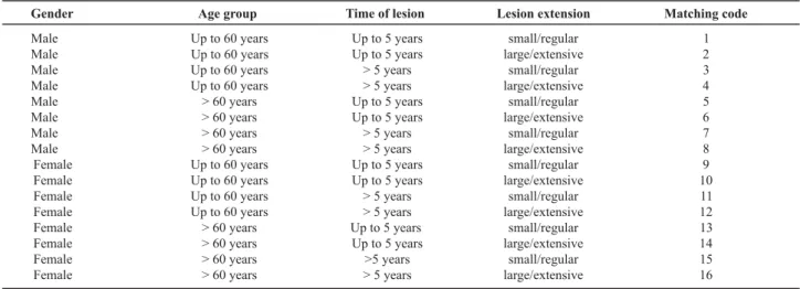
These patients formed two groups: the Control Group (CG) and the Intervention Group (IG), with 10 patients each. The groups were matched according to the follow-ing variables: gender, age range, time and extension of lesion. Matching codes were generated for these variables. (Table 1).
...exerciseand manual lymphatic drainage are
not usually prescribed to treat venous ulcers even though they are both techniques that stimulate venous and
Table 1 - Matching codes according to the variables: gender, age range, time and extension of lesion - Jequié, BA, Brazil - 2008
Gender
Male Male Male Male Male Male Male Male
Female Female Female Female Female Female Female Female
Up to 60 years
Age group
Up to 60 years Up to 60 years Up to 60 years Up to 60 years > 60 years > 60 years > 60 years > 60 years Up to 60 years
Up to 60 years Up to 60 years > 60 years > 60 years > 60 years > 60 years
Time of lesion
Up to 5 years Up to 5 years > 5 years > 5 years Up to 5 years Up to 5 years > 5 years > 5 years Up to 5 years Up to 5 years > 5 years > 5 years Up to 5 years Up to 5 years >5 years > 5 years
Lesion extension
small/regular large/extensive
small/regular large/extensive
small/regular large/extensive
small/regular large/extensive
small/regular large/extensive
small/regular large/extensive
small/regular large/extensive
small/regular large/extensive
Matching code
1 2 3 4 5 6 7 8 9 10 11 12 13 14 15 16
A balance between groups was attempted as the groups were being formed, that is, at each admission into one of the groups, another patient with the same code was allo-cated to the other group. The following inclusion criteria were used: patients with venous ulcer secondary to Chronic Venous Insufficiency (CVI) in one of the two lower limbs; being able to be undergo DPT in the healing process of venous ulcers according to the assessment of the angiologist member of the research team; being older than 18 years of age; attending the outpatient clinic to receive the DPT and dressings; being cognitively able to follow rec-ommendations during the study’s period; agreeing to vol-untarily participate in the study, and sign a free and in-formed consent form.
The exclusion criteria were: diabetic patients with neu-ropathic or arterial ulcer or any other type of ulcer in the lower limbs not related to CVI; local and/or systemic infec-tion; Deep Vein Thrombosis (DVT); those who missed the consultation three consecutive times or six alternated times and those who did not consent to participate.
All patients were referred to a nutritionist to control obesity and hypertension through hyposodic diet and to control other pathologies inherent to the condition of each patient.
This study was submitted to and approved by the Re-search Ethics Committee at the State University Southeast of Bahia according to Resolution 196/96 of the National Health Council that regulates research with human subjects (Protocol nº 59/2007).
The CG was submitted only to dressings performed by the nursing team (scholarship students and volunteers from the nursing undergraduate program at the UESB enrolled
in their 6th or later semesters) complying with the scientific
principles that guide nursing care provided for wounds. Dressings were composed of primary and secondary lay-ers, using gauzes wet with saline solution, and bandages. It is important to stress that the dressing technique was ap-plied daily and was identical for both groups.
In cases in which the wound developed fibrin and/or necrotic tissue in the ulcer bed, chemical debridement with papain cream at 10% was used in both groups whenever necessary up to the total removal of these undesirable tis-sues according to the percentage necessary for each patient. Papain was suspended after these tissues were removed. CG or IC patients underwent surgical debridement when needed. Chemical and surgical debridement was performed as many times as needed; chemical debridement was performed much more frequently (96%) than the surgical.
The IG had their dressings changed daily and the DPT was applied three times a week on alternating days by the same physical therapy team with a duration of 40 minutes for each session. During the weekends both the IC and CG were cared for at their homes. Therapy was applied in the following order: lower limbs were elevated to 30 degrees, manual lymphatic drainage, compression with elastic ban-dages up to the knee region, myolymphoknetic exercises, that is, flexo-extension of ankles, knees and hips three times with 30 repetitions performed with the limb under elastic compression. The elastic bandage was kept on a daily ba-sis, removed only to sleep and replaced in the morning upon waking.
The manual lymphatic drainage on the lower limbs started with the evacuation of popliteal and malleolar inguinal lymph nodes followed by rhythmic, slow and gentle pressure, around 30 to 40mmhg, directing the lymph to a closer group of lymph nodes in the caudal-cranial direction.
Before initiating the treatment, the patients were evalu-ated according to the Brazilian Society for Vascular Surgery. The following variables were considered: socio-demo-graphic variables (gender, age, schooling), alcoholism, smok-ing, associated diseases and leg ulcer extension, time of ulcer, site, and its degree of contraction. The level of pain was also assessed through a numerical scale from 0 to 10, and the edema of the affected limb.
determine: the largest horizontal and vertical lengths mea-sured with a ruler graduated in centimeters. Afterward, the
figure was input into the Autocad 2006(13), by the following
steps 1) the scale was compared in relation to the ruler
(cm2); 2) two lines (blue and red) were drawn between the
two measures (largest horizontal length and vertical length); these lines were measured and the average for the correct scale of the figure was computed; 3) the AutoCAD scale tool was used to put it in the Autocad scale; 4) the mea-sures were checked and confirmed; 5) the Polyline com-mand was used to draw the lesion area; 6) then the toolbar inquiry was used, and the option AREA was checked to measure the figures in cm².
Once the wound’s area was determined, the degree of wound contraction was computed and expressed as a
per-centage using the formula(14):
100 x (Wo – Wi) = % average of contraction Wo
Wo refers to the wound’s initial area and Wi •refers to the wound’s final area in the months 1, 2, 3, 4, 5 and 6.
To evaluate the intensity of pain, a numerical scale from 0 to 10 was applied: zero indicates absence of pain, one, two and three indicate mild pain (does not impede the per-formance of activities); four, five and six indicate moderate pain (hinders activities but does not impede them); seven, eight, and nine indicate strong or incapacitating pain (im-pedes any activity) and ten indicates extremely strong, un-bearable or excruciating pain (in addition to impeding
ac-tivity, it also leads to a lack of control)(15). The scale was
applied verbally and in writing and after the patients’ re-sponse, the observer noted the score.
The pitting test was used; pressure was applied to the pretibial region with the thumb for about 10 minutes in
order to observe whether an indentation was formed(16).
The edema was measured through a crosses scale, where one cross (+) = 0.25cm of indentation; two crosses (++) = 0.50cm of indentation; three crosses (+++) = 0.75cm of in-dentation, and four crosses (++++) = 1.0cm of indentation. For the purpose of comparison, one cross indicated mini-mum edema and four indicated maximini-mum edema.
The collected data were input into a spreadsheet in the SPSS, version 15.0, which performed descriptive analysis
and the Mann-Whitney and the t test to analyze the
aver-ages of wounds’ contraction.
RESULTS
Ten out of the 20 studied patients composed the con-trol group: seven were women (70%) and three were men (30%), with an average age of 61.9 (±11.66) years of age. The other ten patients composed the intervention group: three men (30%) and seven women (70%), with an average age of 65.5 (±10.28) years. The variables schooling, ulcer’s
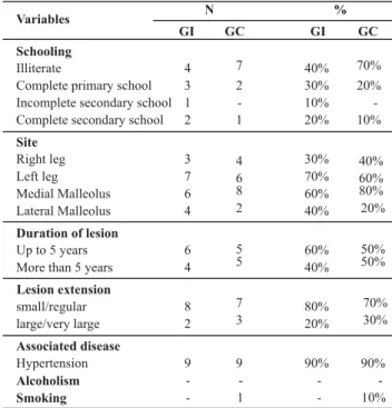
site and duration, associated diseases, alcoholism and smok-ing are presented in Table 2.
Table 2 - Distribution of patients with venous ulcers by groups (IG and CG) according to variables: schooling, ulcer’s site, duration and extension, associated diseases, alcoholism and smoking -Jequié, BA, Brazil - 2008
N %
Variables
Medial Malleolus Lateral Malleolus
Schooling
Illiterate
Complete primary school Incomplete secondary school Complete secondary school
Site
Right leg Left leg
Duration of lesion
Up to 5 years More than 5 years
Lesion extension
small/regular large/very large
Associated disease
Hypertension
Alcoholism Smoking
GI
4 3 1 2
3 7 6 4
6 4
8 2
9
-GC
7 2 -1
4 6 8 2
5 5
7 3
9 -1
GI GC
40% 70%
30% 20%
10% 20%
-10%
30% 70% 60% 40%
60% 40%
80% 20%
40% 60% 80% 20%
50% 50%
70% 30%
90% 90%
-
-- 10%
The lesion extension was classified according to the
COREN-MG-65/00 determination: small (less than 50cm2);
regular (more than 50cm2 and less than 150cm2); large
(more than 150cm2 and less than 250cm2), and extensive
(more than 250cm2).
In relation to the intensity of pain over time, the aver-age scores were significantly lower for the intervention group, as shown in Figure 1.
Intervention Control 0
1 2 3 4 5 6 7
1 Monthst
2nd
Month 3rd
Month 4th
Month 5th
Month 6th
Month
4.1(I)
3.6(I)
3.0(I)
2.0(I)
0.0(I) 0.0(I)
4.8(C)
4.6(C) 5.0
(C)
5.8(C)
3.6(C)
3.4(C)
P=0.026 P=0.001 P=0.000
Figure 1 - Distribution of average scores attributed to the intensity of pain through a numerical scale from 0 to 10 in relation to the time of healing of venous ulcers in patients in the intervention and
Analysis of the graph reveal that the results related to pain, when submitted to the Mann-Whitney test, did not show any significant difference between months 1, 2 and 3, though a significant difference was found between the groups in the months 4, 5 and 6.
Regarding edema, its averages in relation to time of the intervention and control groups are presented in Figure 2.
0 0.5
1 1.5
2 2.5
3 3.5
Month 1 Month 2 Month 3 Month 4 Month 5 Month 6
2.8(C)
2.4(C)
2.3(C)
1.1(I)
0.8(I)
0.8(I)
3.1(I)
2.3(I)
1.7(I)
2.3(C)
2.3(C)
2.3(C)
P= 0.035 P= 0.005 P= 0.005
Intervention Control
Figure 2 - Distribution of the average scores attributed to edema through the pitting test in relation to the time of healing of venous ulcers in patients in the intervention and control groups - Jequié, BA, Brazil - 2008
Test: U de Mann-Whitney (p<0.05)
In addition to the general and clinical characterization of patients, another important variable to be measured is the level of wound contraction. The stronger the
contrac-tion of a wound, the more advanced is its healing process(17).
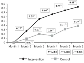
The averages of wound contraction of the intervention and control groups in relation to time are presented in Figure 3. It is important to note that the level of contraction in month 1 is zero because it is when patients were included in the study.
Test: U de Mann-Whitney (p<0.05) 0
0.1 0.2 0.3 0.4 0.5 0.6 0.7 0.8 0.9
0.27(I)
0.55(I)
0.66(I)
0.75(I)
0.83(I)
0.20(C)
0.16(C)
0.31(C)
0.39(C)
P=0.003 P=0.008 P=0.005
0.09(C)
Intervention Control
Month 1 Month 2 Month 3 Month 4 Month 5 Month 6
Figure 3 - Distribution of average scores attributed to the contraction of wounds in relation to time of healing of venous ulcers of patients from the intervention and control groups - Jequié,
Although the differences found in wound contraction are statistically significant only in months 4, 5 and 6, the intervention group consistently presented better results compared to the control group in all the remaining months.
It is noteworthy that at the end of the period (month 6), 100% of the intervention group sample presented con-traction greater than 50% while the level of concon-traction in the control group was 40%. Of the control group samples, 30% presented very unsatisfactory contraction with values below 10%. Additionally, 30% of the intervention group wounds were completely healed while only 10% of the con-trol group was in the same condition.
DISCUSSION
There was a prevalence of women among the 20 stud-ied patients. Statistical data indicate that venous ulcers are more frequent among women, though up to the age of 40, they are somewhat equally distributed among genders. It is possible that this difference is due to women’s longevity. The occurrence of venous ulcers has increased with the
growth of the elderly population. International studies
(18-19) have shown a prevalence between 0.06% to 3.6% in the
adult population and 3.6% in individuals older than 65 years of age.
The average age of the study’s participants (IG and CG) was 65.5 and 61.9 years old (+ 10.28; + 12.64) respectively; the 60 years old population predominated, which is in
ac-cordance with results found in the current literature(18).
However, there are others showing results below average(19),
where 85% of the sample was older than 76 years of age (women) and 78 years of age (men). These differences might be explained by the socio-economic, cultural and geo-graphic characteristics of each region. Most of the affected patients receive only one times the minimum wage, which confirms that low-income populations are more affected by the disease.
In relation to the ulcer’s site, the IG showed a higher predominance in the instep (40%), at the level of the me-dial malleolus (60%) and lateral malleolus (40%), while ul-cers were predominantly located on the calf (70%) at the level of the medial malleolus (80%) and lateral malleolus (20%) in the CG. The average duration of the lesions was
6.4 years (±3.5). In a study(3), carried out in Juiz de Fora,
MG, Brazil, which evaluated 169 cases of leg ulcers, 152 were located in the legs distal region: 85 (50.3%) on the
lateral side and 67 (39.6%) on the medial side. H y p e r
-tension is another disease that can interfere in the healing process because, in addition to its association with athero-sclerosis, experimental studies in mice show that this dis-ease also induces endothelial dysfunction, inhibition of collagen synthesis and decreased tissue oxygen due to
vaso-constriction, hindering the healing process(20).
in both groups). Hence, no specific group had an advantage or disadvantage in relation to the healing response associated with these pathologies.
There are, in addition to hypertension, other extrinsic factors such as smoking that can negatively interfere in the healing process by diverse mechanisms. Carbon monoxide produced during combustion of tobacco displays affinity for hemoglobin two hundred times greater than oxygen, reducing the release of this element on peripheral tissues. Nicotine, a component of cigarette smoke, causes vasocon-striction, increases blood pressure and mobilizes free fatty acids, and also decreases the proliferation of erythrocytes, fibroblasts and macrophages, which are key cells in the
healing process(21). In this study however smoking was not
prevalent in the studied patients; only one patient (10%) in the CG smoked.
Another complication possibly associated with wounds is pain, which in a chronic situation persists for too long a time for a lesion to heal or is associated with chronic patho-logical processes. It lasts more than three months and is either continuous or recurrent. In the case of ulcers, pain is caused by tissue injury, ischemia, hypoxia, inflammation, infection or adhesion of the covering to the wound bed. Pain causes adrenergic discharge causing vasoconstriction and, therefore, decreased tissue perfusion and alteration
of inflammatory mediators, resulting in delayed healing(22).
A statistically significant difference was found in rela-tion to pain between the groups in the last three months
of treatment (p=0.026; p=0.001; p=0.000, respectively),
which indicates the IG experienced lower levels of pain when compared to the control group. Another important finding was related to wounds contraction since a statisti-cally significant difference was also verified in the last three
months of treatment (p=0.003, p=0.008 and p=0.005,
re-spectively). In this context, it is believed that the interven-tion process to which patients were submitted contributed to reducing pain and consequently to speeding up the heal-ing process of venous ulcers.
Edema in the lower limbs is common(16-19) among
pa-tients with venous diseases and can present different de-grees. It is generally found in the perimalleolar region or extends to the leg’s lower third and is frequently associ-ated with CVI (20% of cases). In early phases, the venous component prevails, which then becomes a mixed edema – venous and lymphatic – as it extends to stasis.
It is important to highlight two mechanisms capable of reducing CVI edema, which are increased tissue oncotic pressure and blockage of local lymph. A statistical differ-ence was found in this study between the groups in the
last three months of treatment (p=0.035; p=0.005; p=0.005,
respectively).
These findings must be related to the association of the techniques used in the IG, especially MLD, specific manual technique, which mainly acts on the superficial lymphatic
system and its entire anatomic and physiologic structure, due to its ability to unblock the lymph nodes and drain the ex-cess fluid from the cells by stimulating protein re-absorption by the lymphatic system present in the interstitium and
main-taining a fluid balance between the interstitial spaces(10),
thereby creating an environment conducive to healing, es-pecially when acting in a compromised bloodstream return.
In addition to MLD helping venous return, the use of the compression technique is also essential because it in-creases the healing rate of venous ulcers when compared
to treatments without compression(11). It acts on micro and
macro circulation, diminishing pathological reflux during ambulation and increasing the volume of ejection during calf muscle activation, encouraging re-absorption of edema and improving lymphatic drainage.
More than 70% of the individuals with an active ulcer present impaired calf muscle function. Hence, muscle en-hancement may stimulate muscle hemodynamics, reduc-ing venous reflux and stimulatreduc-ing the healreduc-ing process. In patients with varicose veins, capability of ejection is im-paired to 60%, and in limbs with healed ulcer impairment achieves 76% while in limbs with an active ulcer this rate increases up to 90.5%(12).
Studies evaluating calf muscle(12) hemodynamics in
su-pervised exercise in limbs with active venous ulcers revealed results with significant improvement of venous volume ejec-tion, of the residual volume function and increased calf muscle resistance. This study used supervised exercise, ankle, knee and hip flexion-extension with a differential, that is, combined with exercise, the limb was under elastic compression and elevated to 30 degrees (myolymphokinetic exercises) to facilitate venous return.
Aiming to understand CVI clinical alterations the pre-dominant pathological characteristic of which is insufficient venous return, we stress the importance of unifying tech-niques capable of stimulating venous return. But it is also important to stress the need for interaction between pro-fessionals, patients and families in order to ensure higher patient adherence to treatment, behavioral changes, and the health team’s adapted and effective conduct.
CONCLUSION
Decongestive physical therapy accelerated the healing process, and reduced pain and edema in affected limbs. Hence, it is expected that the results of this study will con-tribute to the advancement of knowledge in the field, en-larging the use of decongestive physical therapy to reduce lymphedema and consequently heal venous ulcers.
REFERENCES
1. Ragnarson TG, Hjelmgren J. Anual cost of treatment for venous leg ulcers in Sweden and the United Kingdom. Wound Repair Regen. 2005;13(1):13-8.
2. Carpentier PH, Maricq HR, Biro C, Poncot-Makinen CO, Franco A. Prevalence, risk factors, and clinical patterns of chronic venous disorders of lower limbs: a population based study in France. J Vasc Surg. 2004;40(4):650-9.
3. Borges EL, Calin MHL, Hass VJ. Revisão sistemática do trata-mento tópico de úlcera venosa. Rev Lat Am Enferm. 2007;15 (6):350-7.
4. Valencia IC, Falabella A, Kirsner RS, Eaglstein WH. Chronic venous insufficiency and venous leg ulceration. J Am Acad Dermatol. 2001;44(3):401-21.
5. Figueiredo M. Ulcera venosa. Rev Virtual Med [periódico na Internet]. 2000 [citado 2008 mar. 25]:[cerca de 10 p.]. Disponí-vel em: http://www.medonline.com.br/med_ed/med9/ ulcera.htm
6. Andrade FMC. Curso de linfologia: diagnóstico e tratamento clínico do linfedema dos membros. Rev Ang Cirur Vasc. 2001;10(3):117-20.
7. Consensus Document of the International Society of Lymphology. The diagnosis and treatment of peripheral lymphedema. Lymphology. 2003;36(2):84-91.
8. Ciucci JL, Krapp JC, Soracco JE, Ayguavella J, Marcovecchio LD, Salvia C, et al. Clínica e evolução na abordagem terapêutica interdisciplinar em 640 pacientes com linfedema durante 20 anos. J Vasc Bras. 2004;3(1):72-6.
9. Soares MM, Sancho AG, Lucena RS, Silva DD. Abordagem fisioterapêutica no linfedema secundário pósvulvectomia com linfadenectomia inguinal. Rev Cient HCE [periódico na Internet]. 2008 [citado 2010 jan. 25];2(2):[cerca de 9 p.]. Disponível em: http://www.hce.eb.mil.br/rev/rev2008/abordagemfisio.pdf
10. Godoy MFG, Godoy JMP. Drenagem linfática manual: novo conceito. J Vasc Bras. 2004;3(1):77-80.
11. O’Meara S, Cullum NA, Nelson EA. Compression for venous leg ulcers. Cochrane Database Syst Rev. 2009;21(1):CD000265.
12. Alberti LR, Petroianu A, Corrêa D, Silva TF. Efeito da actividade física na insuficiência venosa crônica dos membros inferio-res. Acta Med Port. 2008;21(2):215-20.
13. Amorim DA, Ribeiro EM, Cordeiro GG, Silva MAS. O Progra-ma AUTOCAD 2000(r) como forProgra-ma de medida angular para articulações [monografia na Internet]. Itaúna: Faculdade de Fisioterapia; 2005 [citado 2008 out. 30].[cerca de 11 p.]. Dis-ponível em: http://www.wgate.com.br/conteudo/ medicinaesaude/fisioterapia/variedades/analise_autocad/ analise_autocad.htm
14. Ramsey DT, Pope ER, Wagner-Mann C, Berg JN, Swaim SF. Effects of three occlusive dressing materials on healing of full-thickness skin wounds in dogs. Am J Vet Res. 1995;56(7):941-9.
15. Carvalho DS, Kowacs PA. Avaliação da intensidade de dor. Rev Migrâneas Cefaléias. 2006;9(4):164-8.
16. Coelho EB. Mecanismos de formação de edemas. Medicina. 2004;37(1):189-98.
17. Kamamoto F. Contração de feridas: revisão bibliográfica e estudo da contração gerada por fibroblastos normais e de queloídes [dissertação]. São Paulo: Faculdade de Medicina, Universidade de São Paulo; 2007.
18. Scott TE, LaMorte WW, Gorin DR, Menzoian JO. Risk factors for chronic venous insufficiency: a dual case-control study. J Vasc Surg. 1995;22(5):622-8.
19. Nelzen O, Bergqvist D, Lindhagen A, Hallbook T. Chronic leg ulcers: an underestimated problem in primary health care among elderly patients. J Epidemiol Community Health. 1991;45(3):184-7.
20. Varo N, Iraburu MJ, Varela M, López B, Etayo JC, Diez J. Chronic AT1 blockadestimulates extra cellular collagen type I degra-dation and reverses myocardial fibrosis in spontaneously hy-pertensive rats. Hypertension. 2000;35(6):1197-202.
21. Broughton G, Janis JE, Attinger CE. A brief history of wound care. Plast Reconstr Surg. 2006;117(7 Suppl):6S-11S.