Structural and ultrastructural evidence for
telocytes in prostate stroma
Lara S. Corradi
a, Mariana M. Jesus
a, Ricardo A. Fochi
a, Patricia S. L. Vilamaior
a,
Luis A. Justulin-Jr
b, c, Rejane M. G
oes
a, S
ergio L. Felisbino
c, Sebasti~
ao R. Taboga
a,*
aDepartment of Biology, Institute of Biosciences, Humanities and Exact Sciences (IBILCE), Univ. Estadual Paulista
–UNESP, S~ao Jose do Rio Preto, S ~ao Paulo, Brazil
bDepartment of Morphology, Faculty of Pharmaceutical Sciences (FCF), Univ. Estadual Paulista
–UNESP, Araraquara, S~ao Paulo, Brazil
cDepartment of Morphology, Institute of Biology (IB), Univ. Estadual Paulista
–UNESP, Botucatu, S~ao Paulo, Brazil
Received: June 28, 2012; Accepted: December 28, 2012
Abstract
The prostate comprises a glandular epithelium embedded within a fibromuscular stroma. The stroma is a complex arrangement of cells and extracellular matrix (ECM) components in addition to growth factors, regulatory molecules, remodelling enzymes, blood vessels, nerves and immune cells. The principal sources of ECM components are fibroblasts and smooth muscle cells (SMC), which synthesize the structural and regulatory components of the ECM. Telocytes (TCs) were recently described as a novel stromal cell type that exhibited characteristic features. The aim of this study was to confirm the presence of TCs in prostate stromal tissue of gerbils, as the stromal compartment of this gland is a dynamic microenvironment. We used transmission electron microscopy (TEM), light microscopy and immunohistochemistry methods to pro-vide morphological epro-vidence for the presence of TCs. Cells that resembled TCs were observed in gerbil prostatic stroma. These cells had small cellular bodies with very thin and extremely long cellular processes. They were found primarily in the subepithelial area and also at the periphery of SMC layers. TCs also exhibited moniliform processes, caveolae and nuclei surrounded by small amounts of cytoplasm. Close contacts between TC podomers were evident, particularly in the adjacent epithelial compartment. This morphological evidence supported the presence of TCs in the gerbil prostatic stroma, which we report for the first time.
Keywords:Telocyteventral prostatestromal cellCD 34
Introduction
The prostate is an important accessory gland that is located in the mammalian genital tract. In conjunction with seminal vesicles, it produces the bulk of seminal fluid [1]. This gland comprises a glandular epithelium embedded within a fibromuscular stroma. The stromal compartment is a complex arrangement of stromal cells and extracellular matrix (ECM) components in addition to growth factors, regulatory molecules, remodelling enzymes, blood vessels, nerves and immune cells. These components act in a coordinated
manner to regulate cell function and maintain overall prostatic tis-sue homeostasis [2, 37].
The principal sources of ECM components are fibroblasts and smooth muscle cells (SMC), which synthesize the structural and reg-ulatory components of the ECM [2, 3]. The growth and development of the prostate depend on the levels of circulating androgens pro-duced by the testes. Its homeostatic state during adult life is main-tained by these steroid hormones, which act viastromal–epithelial interactions [4, 5]. Thus, in adulthood, a balance between cell prolifer-ation and cell death is established so that no further growth occurs in the prostate gland [6].
However, during ageing in humans and several other species, including dogs and some rodent strains, cellular hyperplasia may occur despite a decrease in the production of sexual hormones, such as testosterone, which results in age-dependent prostatic hyperpla-sias [7]. These alterations may evolve into prostate cancer, a disease that affects men worldwide. Prostate cancer results from a lesion *Correspondence to: Dr. Sebasti~ao Roberto TABOGA,
Departamento de Biologia - Ibilce/Unesp, Rua Cristov~ao Colombo, 2265 Jardim Nazareth, S~ao Jose do Rio Preto,
S~ao Paulo CEP 15054-000, Brazil. Tel: +55 17 3221-2386
Fax: +55 17 32212390 E-mail: [email protected]
doi: 10.1111/jcmm.12021
whose heterogeneous behaviour is still not completely understood. Thus, there is significant interest in the morphology, components and behaviours of the prostate during its normal development and in dif-ferent disorders.
Studies of the human prostate, as well as of breast and colon can-cer specimens, have identified stromal cells that are phenotypicaly altered cells, primarily fibroblasts and SMCs, in addition to modified ECM compositions, including new formation and deposition of abun-dant fibrillar collagens and increased capillary density [2, 8]. Corradi et al.[9, 10] noted general stromal rearrangements in the prostate glands of young, adult and old gerbils (Meriones unguiculatus) after blocking steroidogenic enzymes. These changes included the accu-mulation of fibrillar components and phenotypically altered SMCs and fibroblasts. However, detailed, specific information on these cells’ morphologies and immunophenotypes has not been acquired.
A novel stromal cell type was recently described. Popescuet al. [11, 12] noted that interstitial Cajal-like cells (ICLCs) were obviously not similar to the canonical interstitial cells of Cajal (ICC). Thus, they proposed the name Telocytes (TCs) to describe this novel cell type. In general, TCs have small cell bodies and exhibit variable numbers of long, thin cellular elongations designated telopodes (Tp). Since then, research on the presence and function of TCs has increased exponen-tially and provided relevant information to the study of this new cell type [11–13].
Variousin vitro(isolated cells in culture) andin vivo(fixed speci-mens) techniques and light, fluorescent, transmission and scanning electron microscopic have been used to characterize these cells mor-phologically [13, 14]. Also, immunohistochemistry is of fundamental importance to determine the phenotype(s) of TCs, in addition to eval-uating their pathological changes. Bucharest’s group tested numer-ous antibodies to find a single marker that could be considered specific for TCs [15–17]. However, TCs might have different immuno-histochemistry profiles among different organs and even within the same organ [13]. TCs have already been identified in the stroma of several organs, such as heart [11, 17–19], intestine [20], gallbladder [21], uterus and fallopian tube [22–24], mesentery [25], pulmonary veins [26], trachea and lungs [14, 27, 28], pleura [29], placenta [30], skeletal muscle [31], exocrine pancreas [32], skin [33], mammary gland [34] and parotid glands [35].
The identification of TCs, which have remarkably long, thin and moniliform processes, is based primarily on the recognition of Tp’s [11]. These Tp’s have a number of distinctive characteristics, includ-ing their length, thinness, moniliform processes and a branchinclud-ing net-work pattern, which forms a labyrinthine system by 3D convolution and overlapping and communications through junctions [12, 13]. Identifying these characteristics by ultrastructural analysis is essen-tial. Under higher magnification, the moniliform processes are char-acterized by alternating thin fibrillar-like segments (podomers) and dilated, cisternae-like regions (podoms) and the presence of caveolae, an endoplasmic reticulum, and mitochondria [35]. To date, it has been found that TCs are positive for c-kit (CD 117), CD 34 and vimen-tin [11] by immunohistochemistry.
The aim of this study was to confirm the presence of TCs in the prostatic stroma of gerbils, used as a rodent experimental model [7, 9, 10]. To provide morphological evidence for TCs, we used both
Transmission Electron Microscopy (TEM) and light microscopy. The morphological evidence and immunohistochemistry assay results presented in this study appear to confirm the presence of this novel cell type, TCs, in the gerbil prostatic stroma. To our knowledge, this is the first report of these cells in prostate stroma.
Materials and methods
Animal protocols
Thirty adult male gerbils (Meriones unguiculatus, Gerbilinae, Criscetidae) were housed under controlled conditions of temperature (25°C), relative humidity (40–70%) and light (12-hr light-dark cycle), and were allowed free access to standard chow and water. Gerbils were first weighed (80.0 gm), anaesthetized by CO2inhalation and decapitated. The
pros-tatic complex was dissected out, weighed and fixed according to the different protocols described below. The ventral prostate was carefully dissected out, weighed and fixed. Animal handling and experiments were in accordance with the guidelines of the Ethics Commission for Animal Experimentation (CEEA) of Campinas State University – UNI-CAMP, S~ao Paulo, Brazil (Process no.: 1236-1), which followed the Guide for Care and Use of Laboratory Animals.
Structural analysis
The entire prostate was dissected out and weighed. Only the ventral lobe was fixed by immersion in Karnovsky solution (4% parformaldehyde, 2.5% glutaraldehyde in 0.1 M phosphate buffer, pH 7.2) for 24 hrs or immediately frozen in liquid nitrogen for later cryosectioning. After fixa-tion, tissues were washed with running tap water, dehydrated in a graded ethanol series and embedded in glycol methacrylate resin (Leica Historesin Embedding Kit; Leica, Nussloch, Germany). Some prostatic fragments were fixed in 4% formaldehyde freshly prepared in phosphate buffer (pH 7.2) for 24 hrs. These were also dehydrated in a graded etha-nol series and embedded in Paraplast (Histosec-Merk, Darmstadt, Ger-many). Tissue sections (3–4lm) were obtained using a Leica automatic rotary microtome (Leica RM2155; Leica). For general studies, histologi-cal sections were stained with haematoxylin and eosin [36]. Microscopic analyses used a Zeiss-Jenaval (Zeiss-Jenaval, Jena, Germany) or an Olympus BX60 light photomicroscope (Olympus, Hamburg, Germany). Microscopic fields were digitized using Image-Pro Plus version 4.5 for WindowsTM
software (Media Cybernetics Inc., Bethesda, MD, USA).
Ultrastructural analysis
Prostate ventral lobe fragments were minced into small pieces and fixed by immersion in 3% glutaraldehyde plus 0.25% tannic acid solution in Millonig’s buffer, pH 7.3, containing 0.54% glucose for 24 hrs [9]. After washing with the same buffer, samples were post-fixed with 1% osmium tetroxide for 1 hr, washed in buffer, dehydrated in a graded acetone series and embedded in Araldite resin. Ultrathin sections (50– 75 nm) were prepared using a diamond knife and stained with 2% alco-holic uranyl acetate for 30 min. followed by 2% lead citrate in a 1 M sodium hydroxide solution for 10 min. Samples were evaluated by elec-tron microscopy using a LEO–Zeiss 906 TEM at 80 kV.
To provide evidence for TCs among several stromal structures of the gerbil prostate, we digitally coloured these cells on electron photomicro-graphs to make TCs more visible. All elements were carefully hand col-oured using Adobe Photoshop software (Adobe Systems, Adobe Acrobat 9 Pro Extended. Inc., New York, NY, USA).
Immunohistochemical analysis
We used 5-lm-thick cryosections for immunohistochemical analysis. Endogenous peroxidase activity was blocked using 0.3% H2O2in
metha-nol for 30 min. Then, sections were washed in PBS and incubated with 3% BSA for 1 hr. Incubation with a primary antibody was at 4°C over-night, followed by incubation with a peroxidase-labelled polymer (mouse on rat HRP polymer, BioCare Medical, ref. MRT621 H) for 60 min. and incubation with a substrate chromogen (3,3′diaminobenzidine tetrahy-dorchloride) for 15 min. Between each step, sections were thoroughly rinsed three times with phosphate buffered saline, pH 7.2. Counterstain-ing for nuclei was with haematoxylin for 10 sec. The primary antibody
Santa Cruz, CA, USA). Negative controls omitted the primary antibody. Images were acquired using an Olympus BX 60 microscope equipped with an Olympus digital camera (Olympus).
Results
Light microscopy and immunohistochemistry
results
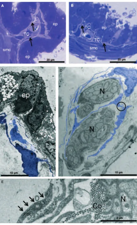
As shown in Figure 1A, for the acini of the gerbil ventral pros-tate, the glandular epithelial compartment was surrounded by vascularized connective tissue stroma in which smooth muscle bundles surrounded each acinus. The epithelium was simple or pseudostratified and comprised columnar cells. In the stroma, a complex arrangement of cells and ECM components, SMCs were well organized in a densely packed layer surrounding the acinar
A
B
C
D
E
epithelium. Moreover, collagen and reticular fibres were noted that were interspersed throughout the stroma, primarily along the basal aspect of epithelial structures, but also among SMCs. Immune cells, fibroblasts, blood vessels and nerves were also found in the stromal compartment.
In the area adjacent to the epithelium, TCs were readily identi-fied as small bodies with thin, long processes that were character-istic of the structures of Tp’s. Also, the same types of cells were noted at the periphery of the SMC layer. The small body of TCs could assume a piriform, spindle or triangular shape and Tp’s that extended from the body were thin (Figs 1B–E, 3A and B). Immuno-histochemical analysis showed that the prostatic stroma had cells that were strongly positive for staining with an anti-CD 34 antibody and had cytoplasmic elongations that corresponded to those described for TCs (Fig. 2).
Transmission electron microscopy results
Electron microscopy analysis confirmed the presence of cells having the ultrastructural features of TCs in the gerbil prostatic stroma (Figs 3C–E, 4A–D and 5A–K). These cells were mostly found adjacent to prostatic epithelium, although some were interspersed in the SMC layer and at the SMC layer periphery (Figs 3C, 4A and B). These results were in accordance with those obtained with light microscopy.
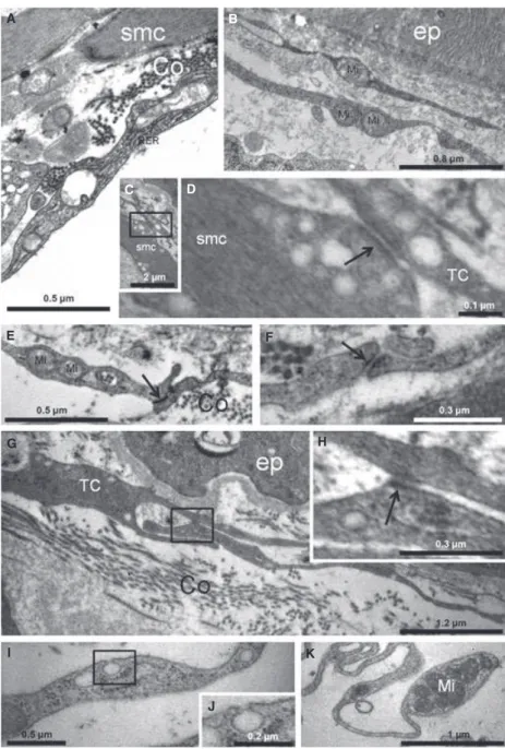
The major characteristics of the TC cell body are scarce cytoplasm around the nucleus and its spindle shape. The ultrastructural TC pattern in the stroma of the gerbil prostate exhibited a small, spindle-shaped body and two to three thin, very long Tp’s, with some that were in close contact similar to adherens junctions (Fig. 5C–H). In addition, several multiple contact connections and/or ‘adherens junc-tions’ between TCs were observed among these cells in the prostatic stroma (Figs 3D and 5D–H).
Contacts between Tp’s were found, especially among TCs in the subepithelial area. Regular moniliform processes, with alternating thin segments (podomeres; Fig. 4C) and dilations (podoms), which were rich in mitochondria (Fig. 5B, E and K), endoplasmic reticulum (Figs 3E and 5A) and caveolae (Fig. 5I and J) were also noted. Mito-chondria were typically noted in the dilation processes and the endo-plasmic reticulum was located primarily in the body region (Fig. 5B). Some TCs that were observed close to prostatic nerves had extremely thin, long processes that extended the cellular body with repeated curving (Fig. 3D and E).
Discussion
A number of animal models, particularly mice, have been used for research on prostate biology and prostate diseases [38]. These stud-ies have found both significant similaritstud-ies and differences in rat,
A B
C
D
E
F
G
Fig. 2Immunohistochemistry assay for CD34 in prostate cryosections. (A) General features of tissue immunoreactivity for CD34 in telocytes (TC) and its cellular elongations (arrows). (B–E) Details for isolated images of a TC and its elonga-tions (arrows). (F–G) Negative control. smc (smooth muscle cell), ep (epithe-lium), TC (telocytes).
mouse and human prostate glands and have analysed the potential of these models to recapitulate human prostatic disease [36, 39]. The rodent prostate complex comprises four distinct paired lobes, ventral, lateral, dorsal and anterior, each of which serves a particular function with regard to histology and secretory protein production [40]. The morphology of the human prostate is more compact, without distinct lobes, and is divided into three zones: central, transitional and periph-eral [41].
The Mongolian Gerbil is a small rodent of the Muridae family, sub-family Gerbillinae, speciesMeriones unguiculatus, which has become increasingly used as an experimental model for studies on male and female prostate histophysiology, as the female of this species has a functionally active prostate gland [42]. Unlike the prostate glands of mice and rats, the gerbil’s prostate morphology is similar to that of the human gland with respect to the fusion of its lobes in a compact structure [7]. The ventral gerbil prostate has been evaluated
morpho-C D
E
logically and quantitatively throughout postnatal development. Previ-ous results from our laboratory demonstrated that the histological, histochemical and ultrastructural features of the adult gerbil’s prostate were comparable with those of the human prostate [7, 9, 10, 42, 43].
Little research has been done on the prostatic stroma that forms a dynamic microenvironment, which is fundamentally important for prostate growth and development during foetal life and for homeosta-sis in adulthood and within which compartment TCs appear to be
present [44]. The suspected presence of this novel stromal cell type [11] was first noted in the gerbil prostate by light microscopy when a cell with a different morphology from those of typical fibroblasts was observed in the subepithelial region. The newly reported TC’s were described as having peculiar features, with very long, thin and monili-form processes called Tp’s. Identification of this cell was mostly based on the recognition of Tp’s [11].
These very long, thin processes could be clearly observed in the gerbil prostatic stroma, especially in the adjacent epithelial area, in
A
B
C
D Fig. 4Transmission electron microscopy
of prostate. (A) Telocytes (digitally col-oured in blue) are noted adjacent to the epithelial (ep) cells and in the periphery of the smooth muscle (smc) layer. In this region, the moniliform feature of a Telo-podes (digitally coloured in blue) can be observed, along with podomers (thin fibrillar-like segments) and podoms (dilated, cisternae-like regions). (B) In the stromal region between two acini, next to smc, numerous TCs were observed that exhibited very thin, long processes. (C) Phenotypic profile assessments of a TC showing a small, spindle-shaped body with a small amount of cytoplasm and thin, long processes in the prostate stroma. (D) General aspect of the isolated stromal TC surrounded by collagen fibrils (Co). This cell exhibits a thin podomer (arrows).
addition to the periphery of SMC layers. This suggested the hypothe-sis that these cells were not fibroblasts. However, to confirm the moniliform feature, in addition to a branching network pattern and communication through junctions between TCs, a higher magnifica-tion view with TEM is also required [32].
Ultrastructurally, we confirmed the presence of TCs in the prostate stroma of the adult gerbil. To our knowledge, this is the first report of TCs in prostatic stroma. This was based on finding moniliform processes with podomers (thin fibrillar-like segments) and podoms (dilated, cisternae-like regions). In addition, these regions were rich in caveolae, endoplasmic reticulum and
mitochondria, as observed for TCs in heart tissues [12, 16] and in parotid glands [32].
Immunohistochemistry has been performed for the gerbil prostate and TCs might show different immunohistochemical profiles among organs and even in the same organ. Bucharest’s group tested numer-ous antibodies in an effort to find a single marker that could be con-sidered specific for this cell type or, at least, specific for the TCs of a given organ. Determining the phenotype(s) of TCs is of fundamental importance, as this would provide for their unequivocal identification and would also aid in evaluating their size, shape, number and, ulti-mately, their movements, migration and pathological changes [13].
C D
E F
G H
I
J
K
C-kit-positive interstitial Cajal cells have been demonstrated in the human prostate [45], and a vimentin-positive population of interstitial cells has been demonstrated in the dog prostate [46]. Our results show for the first time that there are CD 34-reactive cells in the pros-tate stroma, which reinforces the presence of these TCs in prospros-tate stromal tissue.
Several roles have been suggested for TCs, most of which are plausible and not mutually exclusive. However, to date, none of these have been totally proven. As reported by Popescu and Faussone-Pel-legrini [11], TCs might provide mechanical support by guiding the migration of other cells to define the final organization of an organ or its repair or renewal. Intriguingly, a role in neurotransmission in the gut has also been suggested, which possibly contributes to spreading the slow waves generated by interstitial Cajal cells. TCs might also be involved in intercellular signalling.
Cardiac TCs have been suggested as having a nursing role, and those in the oviduct and myometrium are suggested to be sensors of steroid hormones [13]. For the prostate, TC functions and pathology have been suggested, including involvement in benign prostatic hyperplasia and cancer, although further studies are required. While it is too early to propose roles for gerbil prostate TCs, evidence of their involvement in maintaining prostate tissue architecture is justified based on their locations.
The TCs that appear in the subepithelial region along with those observed at the periphery of smooth muscle might ensure the homeo-stasis of the stromal compartment for generating the compartmentaliza-tion of prostatic tissue microenvironments. Studies of the lower urinary tract demonstrated that the guinea pig prostate contained a network of c-kit-positive interstitial cells that was between the glandular and stromal muscle layers of individual prostatic acini as well as between individual SMCs of the stroma [12, 47]. These authors’ discovery of slow waves and interstitial Cajal cells in the guinea pig prostate has prompted some interest in the prostate literature [45], as it suggests that slow-wave activity might be responsible for the spontaneous tonus of the SMC in the human prostate. Thus, this might be TCs function in the gerbil pros-tate, as their localization similar to that proposed by Lang and Klemm [47] is strategic for spontaneous contraction.
Telocytes might be novel target cells for the prevention and treatment of disease. This novel cell type within the prostatic microenvironment has become an important tool for our research group, as it provides insights into numerous unanswered ques-tions regarding the biology of the prostate and the effects of endo-crine disrupting chemicals on the prostatic stroma. The functions and pathology involving TCs in the prostate will require further studies, including studies on benign prostatic hyperplasia and can-cer. Investigating the biological functions of TCs within the pros-tate is important, as the stromal compartment plays key roles in both normal and abnormal tissues. The presence of TCs in the stroma of the gerbil prostate provides a new tool for understand-ing responses that have not yet been determined in prostate biology. The purpose of this study was primarily to confirm the presence of a novel cell type, TCs, in the prostate based on the specifics of their ultrastructural and immunohistochemical characteristics.
Acknowledgements
We thank Luiz Roberto Falleiros Junior of the Microscopy and Microanalyses Laboratory for his technical assistance with TEM and light microscopy. This work was supported by the following Grant sponsors: S~ao Paulo State Research Foundation (FAPESP); Brazilian National Research and Development Council (CNPq) and PROPE-UNESP, Univ. Estadual Paulista. This study is part of the Post-doctoral project developed by LSC for the Department of Biology– UNESP. The authors specific contributions were as follows: LSC and MMJ per-formed the research; SRT, SLF, PSLV, RMG and LAJ, Jr. designed the research study; SRT, PSLV and RMG contributed essential reagents or tools; LSC, MMJ, RAF and SRT analysed the data; LSC and SRT wrote the manuscript; all authors contributed to revising the final text.
Conflict of interest
The authors declare that there are no conflicts of interest associated with this work.
References
1. Hayward SW, Baskin LS, Haughney PC, et al. Stromal development in the ventral prostate, anterior prostate and seminal vesi-cle of the rat.Acta Anat. 1996; 155: 94–103. 2. Tuxhorn JA, Ayala GE, Rowley DR.Reactive stroma in prostate cancer progression. J Urol. 2001; 166: 2472–83.
3. Kalluri R, Zeisberg M.Fibroblast in cancer. Nat Rev Cancer. 2006; 6: 392–401. 4. Cunha GR, Donjacour AA, Sugimura Y.
Stromal-epitelial interactions and heteroge-neity of proliferative activity within the pros-tate.Biochem Cell Biol. 1986; 64: 608–14. 5. Marker PC, Donjacour AA, Dahiya R,et al.
Hormonal, cellular, and molecular control
prostatic development.Dev Biol. 2003; 253: 165–74.
6. Banerjee PP, Banerjee S, Brown TR. Increased androgen receptor expression correlates with development of age-depen-dent, lobo-specific spontaneous hyperplasia of the Brown Norway rat prostate. Endocri-nol. 2001; 142: 4066–75.
7. Pegorin de Campos SG, Zanetoni C, Goes RM,et al.Biological behavior of the Gerbil ventral prostate in three phases of postnatal development.Anat Rec A Discov Mol Cell Evol Biol. 2006; 288: 723–33.
8. Tuxhorn JA, Ayala GE, Smith MJ, et al. Reactive stroma in human prostate cancer:
induction of myofibroblast phenotype an extracellular matrix remodeling.Clin Cancer Res. 2002; 8: 2912–23.
9. Corradi LS, Campos SG, Santos FC,et al. Long-term inhibition of 5-alpha reductase and aromatase changes the cellular and extracellular compartments in Gerbil ventral prostate at different postnatal ages.Int J Exp Pathol. 2009; 90: 79–94.
10. Corradi LS, Goes RM, Vilamaior PS,et al. Increased androgen receptor and remodel-ing in the prostatic stroma after the inhibi-tion of 5-alpha reductase and aromatase in Gerbil ventral prostate.Microsc Res Tech. 2009; 72: 939–50.
ing way from Interstitial Cells of Cajal (ICC), viaInterstitial Cajal-Like Cells(ICLC) to telo-cytes.J Cell Mol Med. 2010; 4: 729–40. 12. Zheng Y, Bai C, Wang X.Telocyte
morpho-logies and potential roles in diseases.J Cell Physiol. 2012; 227: 2311–17.
13. Faussone-Pellegrini MS, Popescu LM. Telocytes.Biomol Concepts. 2011; 2: 481–9. 14. Popescu LM, Gherghiceanu M, Suciu LC, et al.Telocytes and putative stem cells in the lungs: electron microscopy, electron tomography and laser scanning microscopy. Cell Tissue Res. 2011; 345: 391–403. 15. Cretoiu D, Ciontea SM, Popescu LM,et al.
Interstitial Cajal-like cells (ICLC) as steroid hormone sensors in human myometrium: immunocytochemical approach. J Cell Mol Med. 2006; 10: 789–95.
16. Suciu L, Nicolescu MI, Popescu LM. Car-diac telocytes: serial dynamic images in cell culture.J Cell Mol Med. 2010; 14: 2687–92. 17. Gherghiceanu M, Popescu LM.Human
epi-cardium: ultrastructural ancestry of meso-thelium and mesenchymal cells.J Cell Mol Med. 2009; 13: 2949–51.
18. Gherghiceanu M, Manole CG, Popescu LM. Telocytes in endocardium: electron micro-scope evidence.J Cell Mol Med. 2010; 14: 2330–4.
19. Popescu LM, Manole CG, Gherghiceanu M, et al.Telocytes in human epicardium.J Cell Mol Med. 2010; 14: 2085–93.
20. Cantarero CI, Bartolome MJ, Escribano CJ. Identification of telocytes in the lamina propria of rat duodenum: transmission elec-tron microscopy.J Cell Mol Med. 2011; 15: 26–30.
21. Hinescu ME, Ardeleanu C, Gherghiceanu M, et al. Interstitial Cajal-like cells in human gallbladder.J Mol Histol. 2007; 38: 275–84.
22. Cretoiu SM, Cretoiu D, Suciu L,et al. Inter-stitial Cajal-like cells of human Fallopian tube express estrogen and progesterone recep-tors.J Mol Histol. 2009; 40: 387–94. 23. Popescu LM, Ciontea SM, Cretoiu D.
Inter-stitial Cajal-like cells in human uterus and fallopian tube. Ann NY Acad Sci. 2007; 1101: 139–65.
human fallopian tube.J Cell Mol. 2005; 9: 479–523.
25. Hinescu ME, Popescu LM, Gherghiceanu M, et al. Interstitial Cajal-like cells in rat mesentery: an ultrastructural and immuno-histochemical approach. J Cell Mol Med. 2008; 12: 260–70.
26. Gherghiceanu M, Hinescu ME, Andrei F, et al. Interstitial Cajal-like cells (ICLC) in myocardial sleeves of human pulmonary veins.J Cell Mol Med. 2008; 12: 1777–81. 27. Zheng Y, Manole CG, Sun A,et al.
Telo-cytes in trachea and lungs.J Cell Mol Med. 2011; 15: 2262–8.
28. Zheng Y, Bai C, Wang X.Potential signifi-cance of telocytes in the pathogenesis of lung diseases.Expert Rev Respir Med. 2012; 6: 45–9.
29. Hinescu ME, Gherghiceanu M, Suciu L, et al.Telocytes in pleura: two- and three-dimensional imaging by transmission elec-tron microscopy. Cell Tissue Res. 2011; 343: 389–97.
30. Suciu L, Popescu LM, Gherghiceanu M, et al. Telocytes in human term placenta: morphology and phenotype. Cells Tissues Organs. 2010; 192: 325–39.
31. Suciu LC, Popescu BO, Kostin S, et al. Platelet-derived growth factor receptor-b positive telocytes in skeletal muscle intersti-tium.J Cell Mol Med. 2012; 16: 701–7. 32. Nicolescu MI, Popescu LM.Telocytes in the
interstitium of human exocrine pancreas: ul-trastructural evidence.Pancreas. 2012; 41: 949–56.
33. Gherghiceanu M, Popescu LM. Interstitial Cajal-like cells (ICLC) in human resting mammary gland stroma. Transmission elec-tron microscope (TEM) identification.J Cell Mol Med. 2005; 9: 893–910.
34. Nicolescu MI, Bucur A, Dinca O,et al. Telo-cytes in parotid glands. Anat Rec. 2012; 295: 378–85.
35. Behmer AO, Tolosa EMC, Freitas-Neto AG. Manual de praticas para histologia normal e patologica . 1st edn. S~ao Paulo: Edart-Edusp; 1976.
36. Taboga SR, Vidal B. Collagen fibers in human prostatic lesions: histochemistry and
37. Vilamaior PSL, Felisbino SR, Taboga SR,et al.Collagen fiber reorganization in the rat ventral prostate following andro-gen deprivation: a possible role for the smooth muscle cells. Prostate. 2000; 45: 253–8.
38. Huss WJ, Maddison LA, Grenberger NM. Autochthonous mouse models for prostate cancer: past, present and future. Cancer Biol. 2001; 11: 245–59.
39. Sugimura Y, Cunha GR, Donjacour AA. Morphogenesis of ductal networks in the mouse prostate.Biol Reprod. 1986; 34: 961 –71.
40. McNeal JE.The prostate gland: morphology and pathobiology.Monogr Urol. 1983; 4: 3– 37.
41. Santos FC, Leite RP, Custodio AM,et al. Testosterone stimulates growth and secre-tory activity of the female prostate in the adult Gerbil (Meriones unguiculatus). Biol Reprod. 2006; 75: 370–9.
42. Rochel SS, Bruni-Cardoso A, Taboga SR, et al.Lobe identity in the mongolian Gerbil prostatic complex: a new rodent model for prostate study.Anat Rec. 2007; 290: 1233– 47.
43. Corradi LS, Goes RM, Carvalho HF,et al. Inhibition of 5a-reductase activity induces stromal remodeling and smooth muscle de-differentiation in adult Gerbil ventral prostate. Differentiation. 2004; 72: 198– 208.
44. Van der Aa F, Roskams T, Blyweert W, et al.Interstitial cells in the human prostate: a new therapeutic target? Prostate. 2003; 56: 250–5.
45. Aumuller G, Habenich UF, el Etreby MF. Pharmacologically induced ultrastructural and immunohistochemical changes in the prostate of the castrated dog. Prostate. 1987; 11: 211–8.
46. Lang RJ, Klemm MF.Interstitial cell of Ca-jal-like cells in the upper urinary tract.J Cell Mol Med. 2005; 9: 543–56.