Key words:
5-alpha Reductase Inhibitors; Receptors, Androgen; HOXB13 Protein; Prostate
Int Braz J Urol. 2013; 39: 875-83
__________________ Submitted for publication: February 01, 2013
__________________ Accepted after revision: August 01, 2013 Objectives: Five-alpha reductase inhibitors (5ARIs) are known as chemopreventive agents
in prostate cancer with a risk of high-grade disease. This study evaluated the effects of 5ARI on androgen receptor (AR) and proteins involved in prostate cell growth such as HOXB13 expression in human prostate tissue and LNCaP prostate cancer cells.
Materials and Methods: We retrospectively selected 21 patients who underwent TURP be-tween March 2007 and February 2010 for previously confirmed BPH by prostate biopsy. They were grouped into control (group 1, n = 9) and 5ARI treatment (group 2, n = 12) before TURP. AR and HOXB13 expression in prostate tissue was evaluated by immuno-histochemical staining. We tested the effect of 5ARI on the expression of AR, prostate specific antigen (PSA) and HOXB13 in LNCaP cells. Cells were assessed by Western blot analysis, MTT in vitro proliferation assay, and ELISA.
Results: Group 2 showed stronger reactivity for AR and HOXB13 than those of the group 1. MTT assay showed death of LNCaP cells at 25uM of 5ARI. At the same time, ELISA assay for PSA showed that 5ARI inhibited secretion of PSA in LNCaP cells. Western blot analysis showed that 5ARI did not greatly alter AR expression but it stimulated the ex-pression of HOXB13.
Conclusions: These results demonstrated that 5ARI influences AR and HOXB13 expres-sion in both LNCaP cells and human prostate tissue. In order to use 5ARI in chemo-prevention of prostate cancer, we still need to clarify the influence of 5ARI in ARs and oncogenic proteins and its regulation pathway.
Five-alpha Reductase Inhibitor Influences Expression of
Androgen Receptor and HOXB13 in Human Hyperplastic
Prostate Tissue
_______________________________________________
Chaeyong Jung, Youngwoong Park, Young-Rang Kim, Soo Bang Ryu, Taek Won Kang
Department of Urology and Department of Anatomy Research Institute of Medical Sciences, Chonnam National University Medical School, Gwangju, Korea
ABSTRACT
ARTICLE
INFO
_________________________________________________________ ___________________
INTRODUCTION
Five-alpha reductase inhibitor (5ARI) that is used to treat benign prostatic hyperplasia (BPH) blocks the conversion of testosterone (T) to dihydrotestosterone (DHT) and decreases intra-prostatic DHT to as low as 30% of normal value (1,2). Testosterone and the more potent DHT are both primary androgens in the prostate, and are
involved in the development of prostate cancer. During the past decades, 5ARIs have been investi-gated for their role in chemoprevention of prosta-te cancer (1-4).
cancer are strongly influenced by androgens. In a large-scale population-based prevention stu-dy, the Prostate Cancer Prevention Trial (PCPT), finasteride had a 24.8% reduction effect on the prevalence of prostate cancer (4). However, a hi-gher ratio of high-grade prostate cancer (6.4% vs. 5.1%) was found in men undergoing prostate cancer screening (4,5). There are several hypothe-ses for explaining this phenomenon. A reduction in gland volume may permit a greater degree of ascertainment in men receiving finasteride, resul-ting in a proportionally higher rate of detection of high-grade cancers. And the reduced contribution of prostate-specific antigen (PSA) to BPH as finas-teride makes changes in PSA more cancer specific (6-8). Li and Kim reported that molecular profiles on prostate carcinogenesis show that finasteride altered survival pathways and molecular adapta-tion associated with androgen receptor (AR) (9). Several studies have focused on the molecular effects of finasteride on prostate cancer develop-ment. A distinct biological involvement of chro-mosomal alterations and the AR gene were found in prostate cancer tissue that will change the ex-pression profile of AR and oncogenes. Therefore, this study evaluated the effects of 5ARI on expres-sion of AR and proteins involved in cell growth in human prostate tissue and cultured prostate can-cer cells.
MATERIALS AND METHODS
A total of 21 patients submitted to an ini-tial prostate biopsy and transurethral resection of the prostate (TURP) for BPH between March 2007 and February 2010 were reviewed, and 12 cases that had been treated with dutasteride 0.5 mg dai-ly for a minimum of 4 weeks before surgery were enrolled in the study in the 5ARI treatment group (group 2); nine patients who did not receive dutas-teride treatment were selected as controls (group 1). All patients in the 5ARI treatment and control groups had confirmed BPH by an initial prostate biopsy. Transrectal ultrasonography (TRUS) gui-ded prostate biopsy was performed in at least 8 cores or more of tissue targeting the peripheral zone at the apex, mid gland, and base on each side of the prostate. The tissue specimens obtained
from biopsy and TURP were stained for histolo-gical examination and for immunohistochemical (IHC) assay. The research attained ethical approval from the institutional review board of Chonnam National University Hospital (IRB No. 06-070). The recommendations of the Declaration of Helsinki for biomedical research involving human subjects were followed.
Tissues were fixed in 10% formalin for 24 h, transferred to 70% ethanol, cleared in xylene and then embedded in paraffin. Sections (5µm) were cut and mounted on slides. Slides were hydrated through xylene and graded alcohol and equilibrated in PBS. Antigen retrieval was per-formed with sodium citrate 10 mM pH 6, using a microwave for 10 min at 400W. Endogenous pe-roxidase activity was quenched with 3% H2O2 in methanol. Non-specific binding was blocked with normal serum (Pierce, Rockford, IL). Polyclonal antibody against AR and HOXB13 was used for immunohistochemical staining of prostate gland sections at dilution of 1:200. All the slides were then washed several times in PBS and incubated with biotinylated goat anti-rabbit IgG (Amersham Biosciences Europe GmbH, Milan, Italy) at dilution of 1:200, followed by peroxidase-labeled strep-tavidin (Amersham Biosciences Europe GmbH). The antigen-antibody complex was visualized by 10-min incubation with diaminobenzidine te-trahydrochloride (Sigma Chemicals, St Louis, MO). Negative controls, made by excluding polyclonal antibodies from the reaction, showed no specific staining. Counterstaining was performed with he-matoxylin (Sigma Chemicals, St Louis, MO) and cover slips were mounted on the slides with Eukitt (O. Kindler GmbH, Freiburg, Germany).
cell staining, l += 1-25% positively stained cells, 2 += 26-50% of cells, and 3 += >50% of cells. Results were analyzed by a pathologist blinded to the clinical data. Two independent observers sco-red the percentage of positively stained cells per high-power field in the tissue sections.
LNCaP prostate cancer cells were grown in RPMI media containing 5% charcoal/dextran-tre-ated fetal bovine serum (CDT-FBS) for two days and then plated on 96-well plates and grown to 30% confluence. After this, in order to decide the optimal concentration of finasteride, LNCaP cells were grown under testosterone-deprived condi-tion for 3 days. The next day, the cells were trea-ted with 1, 5, and 25µM of finasteride and grown for up to five days. Media and/or with finasteri-de concentrations were changed every two days. Then, the cells were stained with 5 mg/mL MTT (3-[4,5-dimethylthiazol-2-yl]-2,5-diphenyltetra-zolium bromide; thiazole blue, SIGMA, St. Louis, MO) solution and incubated for four hours at 37° C. The reaction, in which mitochondrial dehydro-genase activity reduces the yellow MTT dye to a purple formazan, was stopped by adding DMSO (Dimethyl sulfoxide). The absorbance was measu-red at 570 nm using a microplate reader with SOF-Tmax PRO software (Molecular Devices, Sunnyva-le, CA, USA).
LNCaP prostate cancer cells were plated in P100 culture dishes containing 5% FBS-RPMI media. To deprive the cells of androgens, the cells were grown in RPMI media containing 5% char-coal/dextran-treated fetal bovine serum (CDT--FBS) for 2 days. They were then treated with va-rious concentrations of finasteride for 24 and 48 h and lysed in protein extraction buffer (1x TBS, 1% NP-40, 0.5% sodium deoxycholate), 0.1% SDS and protease inhibitors. Twenty µg of total cell lysa-tes were loaded onto 10% Bis-Tris gel (Invitrogen) and separated using a Bio-rad electroporation sys-tem. After the proteins were transferred to a PVDF membrane, the primary antibodies were applied, followed by incubation with horse peroxidase--conjugated secondary antibodies. The following antibodies were used in this study. Polyclonal antibodies to HOXB13 have also been described previously (7). Antibodies to AR and β-actin were obtained from Santa Cruz Biotechnology Inc. The
blots were developed using the ECL detection sys-tem (Pierce).
LNCaP prostate cancer cells were plated in P100 culture dishes containing 5% FBS-RPMI media. To deprive the cells of androgens, the cells were grown in RPMI media containing 5% CDT--FBS for 2 days. They were then treated with va-rious concentrations of finasteride for 24 and 48 h
andlysedinlysisbuffer(25mMTris•HCl,150mM
NaCl, 1% NP-40, 1mM EDTA and 5% glycerol). This assay employs the quantitative sandwich enzyme immunoassay technique (R&D systems).
Before loading samples, each lysate was quanti-fied by Bradford assay and diluted with a buffered protein solution. The samples were added to each microplate well that had been precoated with a monoclonal antibody specific for kallikrein3/PSA and incubated at RT. After washing, an enzyme--linked polyclonal antibody specific for kalli-krein3/PSA was added to the wells. Following a wash, a substrate solution was added to the wells and color develops in proportion to the amount of kallikrein3/PSA bound in the initial step. The color development was stopped and the intensity of the color was measured at 570 nm using a mi-croplate reader with SOFTmax PRO software (Mo-lecular Devices).
Statistical analyses were performed with SPSS® software (version 13.0). Percentages were
calculated for categorical variables. Chi-square test or Fisher’s exact test was used to analyze ca-tegorical proportions. Multivariate analyses of AR immunoreactivity were conducted by fitted pro-portional odds polychotomous logistic regression model. P values less than 0.05 were considered significant.
RESULTS
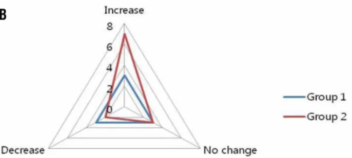
stai-ned for expressions of AR and HOXB13 and the expression scores were statistically evaluated. Ex-pressions of both AR and HOXB13 were confined to the nuclear compartments of prostatic luminal epithelial cells as shown in Figure-1. Expressions of both proteins were further evaluated by quan-titative analysis as mentioned and demonstrated in Figure-2. As shown in Figure-2, AR expres-sion was increased in group 2 (from 1.08 points to 1.57 points, p < 0.05) and on the contrary AR expression was decreased in group 1 (from 1.75
points to 1.5 points, p < 0.05). HOXB13 expres-sion was increased in both the groups (from 1.67 points to 2.75 points in group 1, from 0.71 points to 2.4 points in group 2, p < 0.05) (Figure-2). The-se results suggest that even the mid-term result of 5ARI treatment showed upregulation of transcrip-tion factors which are considered to be positive growth regulators, namely AR and HOXB13.
To further demonstrate the effect of 5ARI on cultured prostate cells, LNCaP cells were em-ployed. Currently, there are no hyperplastic
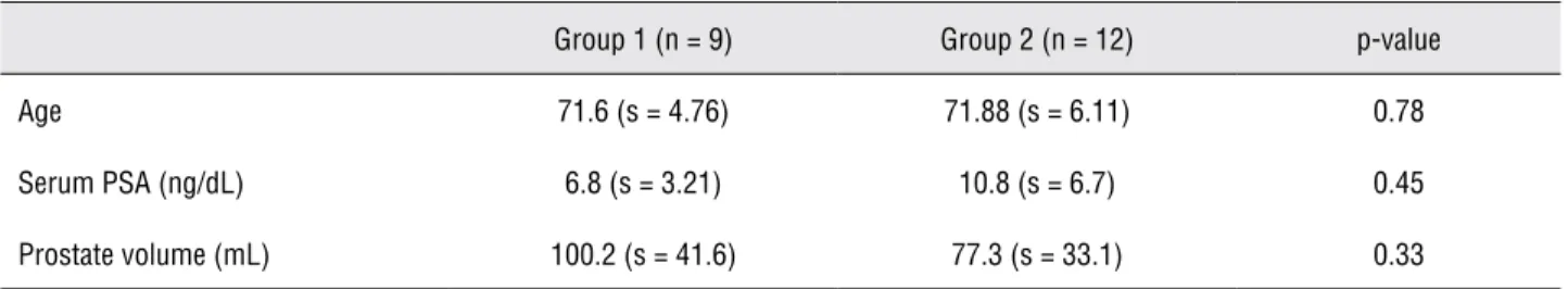
prosta-Table 1 - Patient characteristics.
Group 1 (n = 9) Group 2 (n = 12) p-value
Age 71.6 (s = 4.76) 71.88 (s = 6.11) 0.78
Serum PSA (ng/dL) 6.8 (s = 3.21) 10.8 (s = 6.7) 0.45
Prostate volume (mL) 100.2 (s = 41.6) 77.3 (s = 33.1) 0.33
PSA = Prostate specific antigen Group 1 = Control group Group 2 = Experimental group
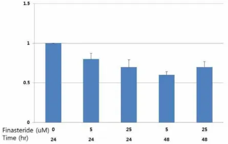
te cells or even normal prostate cells with intact AR expression. LNCaP cells are prostate cancer cells with a high androgen response. First, to de-cide the optimal concentration of finasteride, LN-CaP cells were grown under testosterone-deprived condition for three days. Various doses of finas-teride (final concentrations of 0, 1, 5, 25µM) were given to the cells every three days. Cell growth was measured by MTT in vitro proliferation assay every 2 days for up to 5 days. As shown in Fi-gure-3, androgen-independent growth of LNCaP cells started to slow down at a dose of 5µM of finasteride, more distinctly at a dose of 25µM of finasteride. Further studies were carried out at a
dose of 25µM of finasteride. At the same time, ELISA assay for PSA showed that 5ARI (at a dose of both 5 and 25µM) inhibited secretion of PSA in LNCaP cells (Figure-4). Next, LNCaP cells were grown under similar conditions as mentioned above and 25µM of finasteride was given for up to 48 hours. In Western blot analysis, finasteri-de slightly down-regulated AR expression but it stimulated the expression of HOXB13 in whole lysates of 5ARI-treated cells (Figure-5).
These results suggest that HOXB13 ex-pression was up-regulated by short-term finaste-ride treatment while AR expression was not very much affected by finasteride treatment.
Figure 2 - Diagram showing AR and HOXB13 immunoreactivity in a hyperplastic prostate tissue section. (A: changes in AR expression, B: changes in HOXB13 expression)
A B
DISCUSSION
Inhibition of the synthesis of DHT for the chemoprevention is based on the understanding that androgen and AR are essential for pathoge-nesis of both benign prostatic epithelial cells and prostate cancer cells and that DHT is the most potent androgen that acts on the prostate (10,11). Testosterone is converted to the more potent DHT by the enzymes 5AR type 1 and type 2 in the pros-tate. Both 5AR type 1 and type 2 are observed in
BPH. Currently, there are two types of 5ARIs avai-lable, finasteride inhibits 5AR type 2 and dutaste-ride inhibits both isoforms of 5AR. Dual inhibition by dutasteride has been shown to result in a gre-ater degree of DHT suppression; however, there is no significant clinical difference between the two types of isoforms.
The PCPT study showed that finasteride significantly decreased the 7-year risk of prostate cancer by 24.8% versus placebo (p < 0.001) in men with PSA 3.0ng/mL or less (4). However, high-gra-de tumors (Gleason score 7-10) were more com-mon in the finasteride group than in the placebo group (37% vs. 22.2%, p < 0.001). Some observers suggest that the likely cause of the bias could be that finasteride decreased PSA to a lesser extent in men with high-grade cancer, given that the sensi-tivity of PSA for detecting prostate cancer in the finasteride arm compared to the placebo arm was statistically significantly better (12). Another cau-se of the bias is likely due to the sampling density of a small gland volume and number of cores in the finasteride group, resulting in disproportio-nate sampling of the gland upon random needle biopsy as compared with the placebo group (13-15). Recently, in the Reduction by Dutasteride of Prostate Cancer Events (REDUCE) trial, there was
Figure 4 - Finasteride inhibits secretion of PSA in LNCaP cells: LNCaP cells were grown in RPMI media containing 5% CDT-FBS for 2 days. They were then treated with various concentrations of finasteride for 24 and 48 h and lysed. Quantikine human kallikrein3/PSA quantitative sandwich enzyme immunoassay technique (R&D systems) was used to measure PSA levels.
no significant increase in incidence of high-grade prostate cancer in the dutasteride arm, as com-pared with the placebo arm (1). There are some molecular biological hypotheses about this pheno-menon. One reasonable explanation is that 5ARI alters the histological appearance of prostate can-cers. It has been reported that androgen-deprived cancer has a significantly higher Gleason grade, lower nuclear grade, and smaller nucleolar diame-ter than untreated controls (16,17). Another hypo-thesis is that finasteride induces the development of high-grade prostate cancer by changing the in-traprostatic hormonal milieu (4). Finasteride can induce a low-DHT environment and this will pro-vide the growth advantage for DHT-independent, high-grade prostate cancer clone (18).
The expression of AR and 5AR are the main targets for the progression of prostate can-cer. Roger et al. concluded that type 2 5AR ex-pression decreased in prostate cancer, compared with benign prostate tissue and appears increased in high-grade than low-grade localized prostate cancer (19). The decrease in DHT with 5ARI was accompanied by a reciprocal increase in the serum and intraprostatic T levels. Due to the lower po-tency of T compared with DHT, 5ARI significantly decreased the total androgen effect (18). In this study, 5ARI significantly increased AR expression, matching the mechanism of hormone-refractory disease as AR overexpression or amplification. Also, HOXB13 expression was increased simulta-neously. This means that 5ARI could influence the progression of undiagnosed prostate cancer.
For interpreting PCPT data on the molecu-lar level, Bass et al. performed a study of patients receiving short-term, 30-day finasteride treatment before undergoing radical prostatectomy for lo-calized prostate cancer (20). The expression of nuclear AR was not significantly different by im-munohistochemistry quantification of benign and cancerous cells between the treatment and place-bo groups. Hsieh et al. reported that the result of a long-term study was that more than 30 days of finasteride treatment alters the expression of AR in prostate epithelial cells, but this phenomenon cannot be seen in fewer than 30 days or longer than 180 days of treatment (21). In this study, IHC study revealed significant upregulation of ARs by
5ARI treatment. These short-term results are con-sistent with the results of the study by Bass et al., however, different results were obtained with the long-term use of 5ARI (20). This data showed in-teresting results, but there is a limitation due to the inter-individual differences in AR expression. In this study, the authors minimized the bias by using the prostate tissue from the same person be-fore and after 5ARI use.
It is well established that Hox homeobox genes contain highly homologous homeodomains and are considered transcription factors that regu-late axial regional specification during embryonic development. Hox genes are expressed in a tis-sue-specific and frequently stage-related fashion (22). The HOX 13 paralog is important to the de-velopment of accessory sexual organs, including the prostate. HOXB13 is required for normal di-fferentiation and secretory function of the mouse prostate (23). In the previous studies, HOXB13 ex-pression was correlated with the AR in both cul-tured prostate cancer cells and prostate xenograft models (24). In this in vitro study, we investigated the pattern of expression of HOXB13 in LNCaP prostate cancer cells after treatment with 5ARI.
Although 5ARI has encouraging data sup-porting its role in chemoprevention, the long-term effects of 5ARI in high-grade prostate cancer are still controversial. Although several reports have ascribed the phenomenon to bias in tissue sam-pling, uncertainty in the reproducibility of histo-logical diagnosis, and increased PSA sensitivity, the main concern is the molecular profile of AR and the associated signaling pathway (25-27). In this study, 5ARI had been proven to be able to change the expression profile of AR and other on-cogenic factors such as HOXB13. In the immuno-histochemical study, we showed an increase in AR and HOXB13 expression in benign prostate tissue, and, we also showed an increase in the expression of HOXB13 in LNCaP prostate cancer cell model.
prostate cells adapt to the 5ARI effect or escape through a new growth pathway is yet to be determi-ned. Consequently, we need more study to realize the long-term effects of 5ARI in the molecular profile.
CONCLUSIONS
Five alpha reductase inhibitor treatment influenced AR and HOXB13 expression in both LNCaP cells and human prostate tissue. In order to use 5ARI in chemoprevention of prostate can-cer, we still need to clarify the influence of 5ARI in ARs and oncogenic proteins and its regulation pathway.
ACKNOWLEDGEMENTS
This study was financially supported by Chonnam National University and by a grant (CRI 120061-31) Chonnam National University Hospi-tal Research Institute of Clinical Medicine.
CONFLICT OF INTEREST
None declared.
REFERENCES
1. Andriole GL, Bostwick DG, Brawley OW, Gomella LG, Mar-berger M, Montorsi F, et al.: Effect of dutasteride on the risk of prostate cancer. N Engl J Med. 2010; 362: 1192-202. 2. Gormley GJ, Stoner E, Bruskewitz RC, Imperato-McGinley J,
Walsh PC, McConnell JD, Andriole GL, et al.: The effect of fin-asteride in men with benign prostatic hyperplasia. The Finas-teride Study Group. N Engl J Med. 1992; 327: 1185-91. 3. D’Amico AV, Barry MJ: Prostate cancer prevention and
fi-nasteride. J Urol. 2006; 176: 2010-2; discussion 2012-3. 4. Thompson IM, Klein EA, Lippman SM, Coltman CA, Djavan
B: Prevention of prostate cancer with finasteride: US/Euro-pean perspective.Eur Urol. 2003; 44: 650-5.
5. Thompson IM, Goodman PJ, Tangen CM, Lucia MS, Miller GJ, Ford LG, et al.: The influence of finasteride on the de-velopment of prostate cancer. N Engl J Med. 2003; 349: 215-24.
6. Cohen YC, Liu KS, Heyden NL, Carides AD, Anderson KM, Daifotis AG, et al.: Detection bias due to the effect of finas-teride on prostate volume: a modeling approach for analy-sis of theProstate Cancer Prevention Trial. J Natl Cancer Inst. 2007; 99: 1366-74.
7. Redman MW, Tangen CM, Goodman PJ, Lucia MS, Coltman CA Jr, Thompson IM: Finasteride does not increase the risk of high-grade prostate cancer: a bias-adjusted modeling approach. Cancer Prev Res (Phila). 2008; 1: 174-81. 8. Kaplan SA, Roehrborn CG, Meehan AG, Liu KS, Carides
AD, Binkowitz BS, et al.: PCPT: Evidence that finasteride reduces risk of most frequently detected intermediate- and high-grade(Gleason score 6 and 7) cancer.Urology. 2009; 73: 935-9.
9. Li J, Kim J: Molecular profiles of finasteride effects on prostate carcinogenesis. Cancer Prev Res (Phila). 2009; 2: 518-24.
10. Kim YR, Oh KJ, Park RY, Xuan NT, Kang TW, Kwon DD, et al.: HOXB13 promotes androgen independent growth of LNCaP prostate cancer cells by the activation of E2Fsignal-ing. Mol Cancer. 2010; 9: 124.
11. Koivisto PA, Schleutker J, Helin H, Ehren-van Eekelen C, Kallioniemi OP, Trapman J: Androgen receptor gene altera-tions and chromosomal gains and losses in prostate car-cinomas appearingduring finasteride treatment for benign prostatic hyperplasia. Clin Cancer Res. 1999; 5: 3578-82. 12. Thompson IM, Chi C, Ankerst DP, Goodman PJ, Tangen
CM, Lippman SM, Lucia MS, et al.: Effect of finasteride on the sensitivity of PSA for detecting prostate cancer. J Natl Cancer Inst. 2006; 98: 1128-33.
13. Kulkarni GS, Al-Azab R, Lockwood G, Toi A, Evans A, Trachtenberg J, et al.: Evidence for a biopsy derived grade artifact among larger prostate glands. J Urol. 2006; 175: 505-9.
14. Akduman B, Crawford ED: The PCPT: New findings, new insights, and clinical implications for the prevention of prostate cancer. Eur Urol Suppl. 2006; 5: 634-9.
15. Cohen YC, Liu KS, Heyden NL, Carides AD, Anderson KM, Daifotis AG, et al.: Detection bias due to the effect of finas-teride on prostate volume: a modeling approach for analy-sis of theProstate Cancer Prevention Trial. J Natl Cancer Inst. 2007; 99: 1366-74.
16. Bostwick DG, Qian J, Civantos F, Roehrborn CG, Montironi R: Does finasteride alter the pathology of the prostate and cancer grading? Clin Prostate Cancer. 2004; 2: 228-35. 17. Bostwick DG, Ramnani D, Cheng L: Treatment changes
in prostatic hyperplasia and cancer, including androgen deprivation therapy andradiotherapy. Urol Clin North Am. 1999; 26: 465-79.
18. Tindall DJ, Rittmaster RS: The rationale for inhibiting 5al-pha-reductase isoenzymes in the prevention and treatment of prostatecancer. J Urol. 2008; 179: 1235-42. Erratum in: J Urol. 2008; 179: 2490.
20. Bass R, Perry B, Langenstroer P, Thrasher JB, Dennis KL, Tawfik O, et al.: Effects of short-term finasteride on apop-totic factors and androgen receptors in prostate cancer cells. J Urol. 2009; 181: 615-9; discussion 619-20. 21. Hsieh JT, Chen SC, Yu HJ, Chang HC: Finasteride
upregu-lates expression of androgen receptor in hyperplastic prostate and LNCaP cells: implications for chemopreven-tion of prostate cancer. Prostate. 2011; 71: 1115-21. 22. Kondo T, Zákány J, Innis JW, Duboule D: Of fingers, toes
and penises. Nature. 1997; 390: 29.
23. Economides KD, Capecchi MR: Hoxb13 is required for normal differentiation and secretory function of the ven-tral prostate. Development. 2003; 130: 2061-9.
24. Jung C, Kim RS, Zhang HJ, Lee SJ, Jeng MH: HOXB13 in-duces growth suppression of prostate cancer cells as a re-pressor of hormone-activatedandrogen receptor signaling. Cancer Res. 2004; 64: 9185-92.
25. Klein EA, Thompson IM; Update on chemoprevention of prostate cancer. Curr Opin Urol. 2004; 14: 143-9.
26. Tindall DJ, Rittmaster RS: The rationale for inhibiting 5al-pha-reductase isoenzymes in the prevention and treatment of prostatecancer. J Urol. 2008; 179: 1235-42. Erratum in: J Urol. 2008; 179: 2490.
27. Thomas LN, Douglas RC, Lazier CB, Too CK, Rittmaster RS, Tindall DJ: Type 1 and type 2 5alpha-reductase expression in the development and progression of prostate cancer. Eur Urol. 2008; 53: 244-52.
______________________ Correspondence address: