RESEARCH ARTICLE
Development of an HIV-1 Subtype Panel in
China: Isolation and Characterization of 30
HIV-1 Primary Strains Circulating in China
Jingwan Han, Siyang Liu, Wei Guo, Zuoyi Bao, Xiaolin Wang, Lin Li, Yongjian Liu, Daomin Zhuang, Hanping Li, Lei Jia, Tao Gui, Hongshuai Sui, Tianyi Li, Jingyun Li*
Department of AIDS Research, State Key Laboratory of Pathogen and Biosecurity, Beijing Institute of Microbiology and Epidemiology, 0007, Beijing, P.R. China
Abstract
Background
The complex epidemic and significant diversity of HIV-1 strains in China pose serious chal-lenges for surveillance and diagnostic assays, vaccine development and clinical manage-ment. There is a lack of HIV-1 isolates in current canonical HIV-1 subtype panels that can represent HIV-1 diversity in China; an HIV-1 subtype panel for China is urgently needed.
Methods
Blood samples were collected from HIV-1 infected patients participating in the drug-resis-tance surveillance program in China. The samples were isolated, cultured and stored as neat culture supernatant. The HIV-1 isolates were fully characterized. The panel was used to compare 2 viral load assays and 2 p24 assays as the examples of how this panel could be used.
Results
An HIV-1 subtype panel for China composed of 30 HIV-1 primary strains of four subtypes (B [including Thai-B], CRF01_AE, CRF07_BC and G) was established. The samples were iso-lated and cultured to a high-titer (106-109copies/ml)/high-volume (40ml). The HIV-1 isolates
were fully characterized by the final viral load, p24 concentration,gag-polandenvC2V3 se-quencing, co-receptor prediction, determination of the four amino acids at the tip of theenv V3-loop, glycosylation sites in the V3 loop and the drug-resistance mutations. The compari-son of two p24 assays and two viral load assays on the isolates illustrated how this panel may be used for the evaluation of diagnostic assay performance. The Pearson value be-tween p24 assays were 0.938. The viral load results showed excellent concordance and agreement for samples of Thai-B, but lower correlations for samples of CRF01_AE.
PLOS ONE | DOI:10.1371/journal.pone.0127696 May 27, 2015 1 / 19
a11111
OPEN ACCESS
Citation:Han J, Liu S, Guo W, Bao Z, Wang X, Li L, et al. (2015) Development of an HIV-1 Subtype Panel in China: Isolation and Characterization of 30 HIV-1 Primary Strains Circulating in China. PLoS ONE 10(5): e0127696. doi:10.1371/journal.pone.0127696
Academic Editor:Rongge Yang, Chinese Academy of Sciences, Wuhan Institute of Virology, CHINA
Received:December 10, 2014
Accepted:April 17, 2015
Published:May 27, 2015
Copyright:© 2015 Han et al. This is an open access article distributed under the terms of theCreative Commons Attribution License, which permits unrestricted use, distribution, and reproduction in any medium, provided the original author and source are credited.
Data Availability Statement:All sequence files are available from the Los Alamos HIV Database or the GenBank (accession number(s) KP178420-KP178479). All other relevant data are within the paper.
Conclusion
The current panel of 30 HIV-1 isolates served as a basis for the development of a compre-hensive panel of fully characterized viral isolates, which could reflect the current dynamic and complex HIV-1 epidemic in China. This panel will be available to support HIV-1 re-search, assay evaluation, vaccine and drug development.
Introduction
It has been reported that the number of people living with HIV increased to 35 million (33.2 million–37.2 million) by the end of 2013 according to the United National Programme on HIV and AIDS (UNAIDS) [1]. Currently, the major source of the global pandemic is divided into four groups: M (Major), O (Outlier), N (Non-M and Non-O) and P. The M group includes nine major subtypes (i.e., A-D, F-H, J, and K), over 72 circulating recombinant forms (CRF), and numerous unique recombinant forms (URF). Their rapid evolution and comprehensive di-versity make them a serious challenge for the maintenance of a reliable serological and nucleic acid test for blood screening, epidemiological surveillance, diagnosis, and clinical management of the infected persons. Differences between subtypes in HIV-1 RNA quantitation and determi-nation, serological diagnostic and screening variations have been noted [2,3]. Many HIV-1 nu-cleic acid tests or serological assays designed and tested based on subtype B virus have
demonstrated an underestimation of some non-B subtypes [4–6]. Differences between subtypes in currently commercially available quantitative viral load tests have been reported [7–9]. Fur-thermore, viral diversity may also impact responses to antiretroviral therapies and vaccines [10–12]. Under these circumstances, a set of fully characterized viruses from HIV infections are needed to better represent current global HIV diversity and help control the worldwide HIV-1 epidemic through support of epidemiological testing, vaccines, and therapeutic efforts [13].
HIV-1 subtype B (Thailand’s variant of subtype B included), CRF01_AE, CRF07_BC and CRF08_BC are the major subtypes of HIV-1 strains circulating in China [14]. HIV-1 strains of subtype B from Thailand were transmitted to Yunnan province in the early 1990s. Strains of subtype C from India were transmitted to Yunnan soon after 1993 and these two subtypes caused the first wave of an HIV-1 epidemic in China among intravenous drug users (IDUs) in Yunnan province [15]. CRF07_BC and CRF08_BC were generated from subtypes B and C, and most likely originated in Yunnan [16–18]. CRF08_BC spread eastward to the southern coastal provinces around 1990 [16,18], while CRF07_BC spread to northwestern China along a drug traffic route around 1993 [15,18,19]. Meanwhile, unregulated and unsanitary commercial plasma collection between the years 1992 and 1995 in Henan caused an HIV-1 subtype Thai-B infection epidemic in central China [20–22]. On the other hand, since HIV-1 CRF01_AE was first found among heterosexuals and IDUs in southern China around 1996–1997, this subtype of the virus spread through sexual routes to other provinces quickly and broadly [23–25]. Sev-eral new circulating recombinant forms generated from the subtypes circulating in China were found recently [26–32], such as CRF55_01B, which was a recombinant of CRF01_AE and Thai-B and was discovered from men having sex with men (MSM) in the year 2013 [26]; CRF57_BC, which was a recombinant of Thai-B and India C and was discovered in western Yunnan in China in 2014 [27]; and CRF61_BC, which was comprised of CRF07_BC and CRF08_BC and was identified in the heterosexual population in two different regions in China [29]. The diversity of HIV-1 is extensive in China.
In the U.S., a pilot program called the NIAID External Quality Assurance Program Over-sight Laboratory (EQAPOL) established by the National Institutes of Health/National Institute of Allergy and Infectious Diseases (NIH/NIAID) in collaboration with other institutes
throughout the world developed an HIV subtype panel that encompassed the genetic and geo-graphic diversity currently present worldwide [13,33]. This program has collected over 100 viral samples, but only 7 of them were isolated from HIV-1 infections in China [13].
To establish an HIV-1 subtype panel that represents the diversity of HIV-1 strains circulat-ing in China, we collected 99 plasma specimens from HIV-1 infected individuals in 10 prov-inces of China through the HIV-1 drug-resistance surveillance program. Finally, we
successfully obtained 30 HIV-1 isolates of different subtypes. The HIV-1 p24 antigen concen-trations and viral loads of the isolates were evaluated. We also sequenced the three major struc-tural genes:gag,polandenvC2V3 for subtype analysis, prediction of co-receptor usage, the four amino acids at the tip of the V3-loop and the glycosylation sites onenvV3 sequence, as well as the drug-resistance mutations.
Materials and Methods
Patients and samples
Blood samples with the basic personal information of each patient were collected from HIV in-fected individuals who were participants in the HIV-1 drug-resistance surveillance program in 10 provinces of China (Guangxi, Guangdong, Sichuan, Henan, Xinjiang, Ningxia, Shanghai, Shandong, Beijing and Zhejiang). 10 ml of anticoagulated whole blood was obtained from each patient for virus isolation and expansion. The basic personal information of the patients showed that all the main routes of HIV-1 transmission were included among the cases (i.e., ho-mosexual sex, heterosexual sex, blood donation, blood transfusion, intravenous drug using and occupational exposure).
All the samples were identified by HIV-1 antibody enzyme linked immunosorbent assay (ELISA) screening, HIV-1 p24 antigen testing and the Western blot to confirm the infection of HIV-1.
Isolation and expansion of primary HIV-1 strains
Peripheral blood mononuclear cells (PBMCs) from the blood samples were separated immedi-ately after the collection, and they were co-cultured with PBMCs from healthy blood donors to separate and expand the virus in a three-step process: isolation, small-scale culture and large-scale culture.
For isolation, the healthy PBMCs were isolated from the blood of more than eight healthy blood donors by HISTOPAQUE-1077 (SIGMA). The isolated PBMCs were cultured in RPMI-1640 medium with 10% fetal bovine serum (FBS), 100 U/ml penicillin and 100μg /ml strepto-mycin. The healthy PBMCs were stimulated by 5μg/ml phytohemagglutinin (PHA) and 10 IU/ml interleukin-2 (IL-2) for three days. Then, the infected PBMCs were separated from the patients’blood in the same way and cultured in RPMI1640 medium with FBS, penicillin, strep-tomycin and IL-2 but without PHA. The patient's PBMCs and activated healthy donor PBMCs were centrifuged at 250×gfor 5 minutes, and then the PBMCs were re-suspended in RPMI-1640 medium to 2×10⁶cells/ml respectively. Infected and healthy PBMCs were mixed at a vol-ume ratio of 1:1 into 24-well plates and were then co-cultured in a cell incubator containing 5% CO2at 37°C. A total of 100μl of supernatant was sampled every seven days to measure the
con-centration of p24 antigen to observe the growth of every HIV-1 strain, and one half of the ini-tial volume of the cell supernatant was replaced by healthy donor cells to maintain a stable
Development of an HIV-1 Subtype Panel in China
environment for virus growth. The cultures were continued for up to 28 days or until p24 re-sults were greater than 2 pg/ml.
For small-scale culture, 4 ml of 2×10⁶cells/ml PBMCs from healthy donors were inoculated with 1 ml HIV-1 cell culture supernatant sampled at the end of the isolation, and the viruses were cultured for another 7 days until the 35thday or until the p24 results were greater than 10 pg/ml.
For large scale culture, 1 ml of cell supernatant from the small-scale culture were put into 39 ml of 1×10⁶cells/ml PBMCs, and cultured for another 7 days or until the p24 antigen results were greater than 25 pg/ml.
The cell suspensions were centrifuged at 800×gfor 5 minutes and the supernatant was ali-quoted into 1.5 ml centrifuge tubes at 1 ml/tube and stored in liquid nitrogen (-196°C).
HIV-1
gag,
pol,
envC2V3 and full-length genome amplification and
sequencing
The full-length ofgagandpolwere amplified and sequenced and then spliced together as a com-plete genetic region for subtyping. The C2V3 region ofenvwas also amplified and sequenced.
Viral RNA from the culture supernatant was extracted with a QIAamp Viral RNA Mini Kit and served as the template for amplification. The 1,502 bp full lengthgaggene (HXB2: 790– 2292), 3,011 bp full lengthpolgene (HXB2: 2,085–5,096) and 520 bpenvC2V3 (HXB2: 7021– 7541) fragments were amplified according to reference [34]. The full-lengthgagandpolgenes were connected together as a complete genetic region (HXB2: 790–5,096) for subtyping.
Phylogenetic analysis and subtype identification
In order to identify the subtypes of the isolated strains, we regarded thegag-polfragment as the target sequence. A neighbor-joining tree of these target sequences and reference strains (sub-types A–D, F–H, J, K and most kinds of circulating recombinant forms were included) was constructed by MEGA5.1 with bootstrap 1000. Virus samples that were unidentified in the
gag-polfragment were uploaded to REGA (http://jose.med.kuleuven.ac.be/genotypetool/html/
subtypinghiv.html) and NCBI HIV Subtyping. Recombinants and incompletegag-pol se-quences of the recombinants were analyzed using the jumping profile hidden Markov model (jpHMM) tool provided atwww.hiv.lanl.gov(http://jphmm.gobics.de/).
In order to identify the diversity of CRF01_AE isolates in our program, we performed phy-logenetic analysis for the CRF01_AE isolates in our panel in particular by using the full-length sequences of the genepolfrom the isolates, together with the HIV-1 CRF01_AE sequences used in Yi Feng and Xiang He’s study [35]. A neighbor-joining tree was constructed by MEGA5.1 with bootstrap 1000.
The prediction of co-receptor usage, the four amino acids at the tip of the
V3-loop and the glycosylation sites by
envV3
The genotype prediction of co-receptor usage of the HIV-1 isolates based on theenvV3 se-quence information was predicted with two online software programs: a Web-based genotypic algorithm—WebPSSM (http://intra.mullins.microbiol.washington.edu/webpssm/) and Geno2-Pheno (G2P) prediction, which is based on the bioinformatics prediction tool that uses the V3 sequence plus additional host-specific features to select false positive rates (FPR) at 1%, 2.5%, 5%, 15%, and 20%. The FPR represents the percentage of all samples that are X4-incapable will be predicted as X4-capable viruses, and FPR at 2.5% was used in this program;http://
Alamos HIV database (http://www.hiv.lanl.gov/content/sequence/GENE_CUTTER/cutter. html), and the top four amino acids at the tip of the V3-loop of each HIV-1 isolate. In addition, the glycosylation sites on the V3-loop were predicted by online prediction software N-Glyco-Site (http://www.hiv.lanl.gov/content/sequence/GLYCOSITE/glycosite.html).
The determination of viral drug resistance mutations
We submitted the full-lengthpolgene (2,085 bp–5,096 bp) to the Stanford University Network HIV-1 database (http://hivdb.stanfrod.edu/hiv/) after amplification and sequencing to deter-mine drug resistance mutations and tolerance of various antiviral drugs of the HIV-1 isolates.
Measurement of HIV-1 isolate p24 concentration and viral load after
isolation and expansion
We detected p24 antigen and measured the viral load to evaluate the correlation and agreement between the two indicators.
P24 antigen was quantified using a BioMerieux VIDAS HIV p24 II assay and one domestic p24 test assay. For the samples that were beyond the upper limits (400 pg/ml for the VIDAS HIV p24 II assay and 80 pg/ml for the domestic p24 test assay), the RPMI-1640 medium with 10% FBS was used for dilution in the performance of both of the assays.
The viral loads of all the 30 samples in the panel were measured using two commercial kits ac-cording to the manufacturers’instructions: the automated Roche Cobas AmpliPrep/Cobas Taq-Man HIV-1 test version 2.0 assay (CAP/CTM v2.0 assay) and Nuclisens HIV-1 EasyQ version 2.0 assay (EasyQ v2.0 assay). For the samples that were beyond the upper limits (5,757,126 IU/ml for EasyQ v2.0 assay and 17,000,000 IU/ml for the CAP/CTM v2.0 assay [36]), HIV-1-negative plas-ma was used to dilute the culture supernatant of the isolates in the perforplas-mance of the two assays. To compare the performance of the CAP/CTM v2.0 assay with that of the EasyQ v2.0 assay, all HIV-1 viral load values (expressed as copies/ml) were converted to IU/ml, and then into log IU/ml values for statistical analysis [36]. The correlation coefficient (R) of Pearson test was used to assess the strength of the linear association and the Bland-Altman method was used to assess the level of agreement between the paired measurements [37].
Statistical analysis
The mean differences and standard deviations (SD) were calculated using SPSS 22.0 software to compare the performance of the 2 viral load measurement assays. The Pearson correlation coefficient (R) was used to assess the strength of the linear association between the log-trans-formed levels in the positive samples measured by the two viral load assays and between the p24 concentration values tested by the two p24 assays. The Bland-Altman method was used to assess the level of agreement between the paired measurements using MedCalc 14.8.1.0 soft-ware [37]. The linear association between the p24 concentrations measured by the BioMerieux VIDAS HIV p24 II assay and the HIV-1 viral loads measured by a Roche Cobas AmpliPrep/ Cobas TaqMan HIV-1 test version 2.0 assay (CAP/CTM v2.0 assay) had also been evaluated.
Ethical statement
The study has been ethically approved by the Ethical Board of the Beijing Institute of Micro-biology and Epidemiology. Blood samples were collected from the patients participated in the drug-resistance surveillance program in China. The written informed consents were ob-tained from all the participants and the data were analyzed anonymously. The informed
Development of an HIV-1 Subtype Panel in China
consent process has also been approved by the Ethical Board of the Beijing Institute of Mi-crobiology and Epidemiology.
Results
Virus isolation and expansion
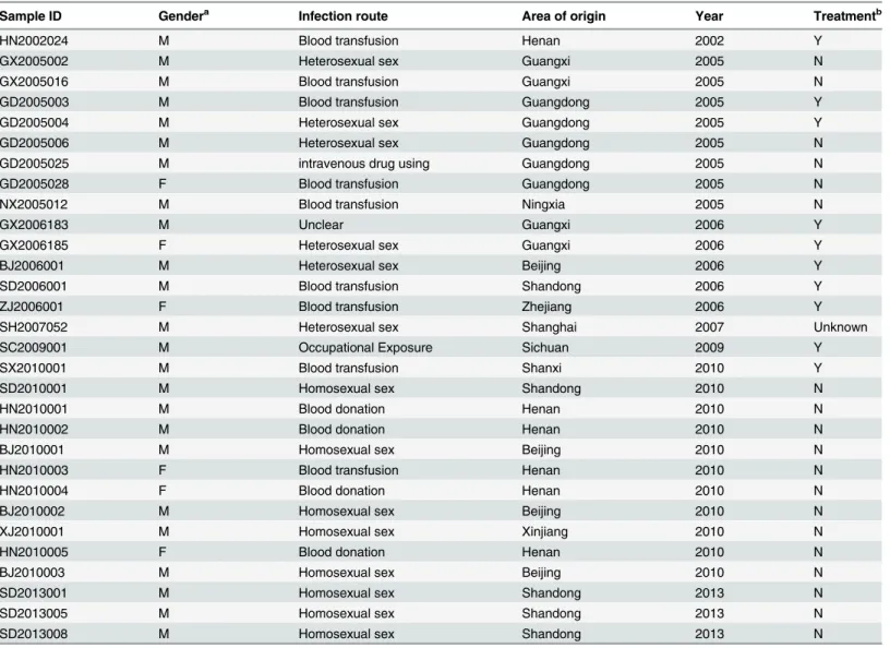
After seven times of isolation and expansion in different years, the p24 concentrations of 30 isolates were greater than 25 pg/ml. As for the 30 HIV-1 primary strains that were isolated and expanded successfully, strains from each of the 10 provinces in China still existed; i.e., Guangxi (n = 4), Guangdong (n = 5), Sichuan (n = 1), Henan (n = 11), Xinjiang (n = 1), Ningxia (n = 1), Shanghai (n = 1), Shandong (n = 4), Beijing (n = 1) and Zhejiang (n = 1). The basic personal in-formation of the cases is summarized inTable 1.
Table 1. Basic information on the 30 HIV-1 samples.
Sample ID Gendera Infection route Area of origin Year Treatmentb
HN2002024 M Blood transfusion Henan 2002 Y
GX2005002 M Heterosexual sex Guangxi 2005 N
GX2005016 M Blood transfusion Guangxi 2005 N
GD2005003 M Blood transfusion Guangdong 2005 Y
GD2005004 M Heterosexual sex Guangdong 2005 Y
GD2005006 M Heterosexual sex Guangdong 2005 N
GD2005025 M intravenous drug using Guangdong 2005 N
GD2005028 F Blood transfusion Guangdong 2005 N
NX2005012 M Blood transfusion Ningxia 2005 N
GX2006183 M Unclear Guangxi 2006 Y
GX2006185 F Heterosexual sex Guangxi 2006 Y
BJ2006001 M Heterosexual sex Beijing 2006 Y
SD2006001 M Blood transfusion Shandong 2006 Y
ZJ2006001 F Blood transfusion Zhejiang 2006 Y
SH2007052 M Heterosexual sex Shanghai 2007 Unknown
SC2009001 M Occupational Exposure Sichuan 2009 Y
SX2010001 M Blood transfusion Shanxi 2010 Y
SD2010001 M Homosexual sex Shandong 2010 N
HN2010001 M Blood donation Henan 2010 N
HN2010002 M Blood donation Henan 2010 N
BJ2010001 M Homosexual sex Beijing 2010 N
HN2010003 F Blood transfusion Henan 2010 N
HN2010004 F Blood donation Henan 2010 N
BJ2010002 M Homosexual sex Beijing 2010 N
XJ2010001 M Homosexual sex Xinjiang 2010 N
HN2010005 F Blood donation Henan 2010 N
BJ2010003 M Homosexual sex Beijing 2010 N
SD2013001 M Homosexual sex Shandong 2013 N
SD2013005 M Homosexual sex Shandong 2013 N
SD2013008 M Homosexual sex Shandong 2013 N
a:
“M”represents for male and“F”represents for female. b:
“Y”represents for treated and“N”represents for treated naive.
Phylogenetic analysis and subtype identification
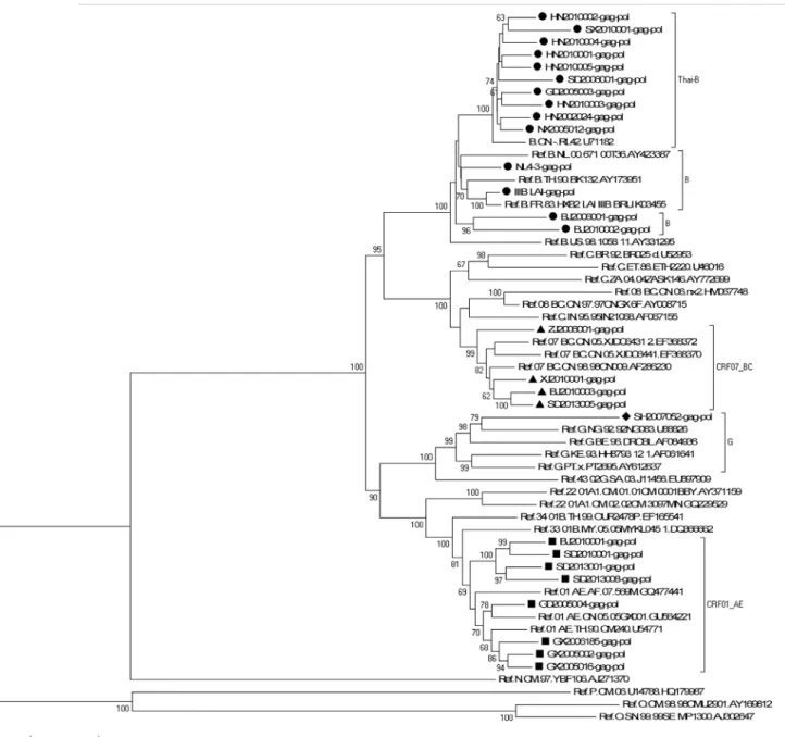
A neighbor-joining tree was constructed to determinate the subtype of each isolate based on 25 full-lengthgag-polgenetic region sequences of the isolates in our program (all thegag-pol se-quences are available from the Los Alamos Databases and their accession numbers of GenBank are KP178421-KP178445) (GD2005006, GD2005025 and GD2005028 failed ingag amplifica-tion; GX2006183, SC2009001 failed inpolamplification) and 30 standard subtype reference se-quences from the Los Alamos HIV sequence database (the reference sequence of subtype Thai-B: B.CN-RL42.U71182 was included) (Fig 1). Subtypes of five other strains (GD2005006, GD2005025, GD2005028, GX2006183 and SC2009001) were designated and identified by REGA Viral Subtyping and the NCBI HIV Subtyping tool based on their genegagsequences (GX2006183-gag: KP178446; SC2009001-gag: KP178447) or genepolsequences
(GD2005006-pol: KP178448; GD2005025-pol: KP178449; GD2005028-pol: KP178450). Sub-typing assessment results revealed that our panel included four subtypes currently (i.e., B, CRF01_AE, CRF07_BC and G). Thirteen of the 30 samples from this analysis were subtype B (11 strains of Thai-B were included), twelve were subtype CRF01_AE, four were subtype CRF07_BC, and one was subtype G, which was an exceptional subtype in China (Table 2). A neighbor-joining tree of 10 full-lengthpolgene sequences of all 11 CRF01_AE isolates
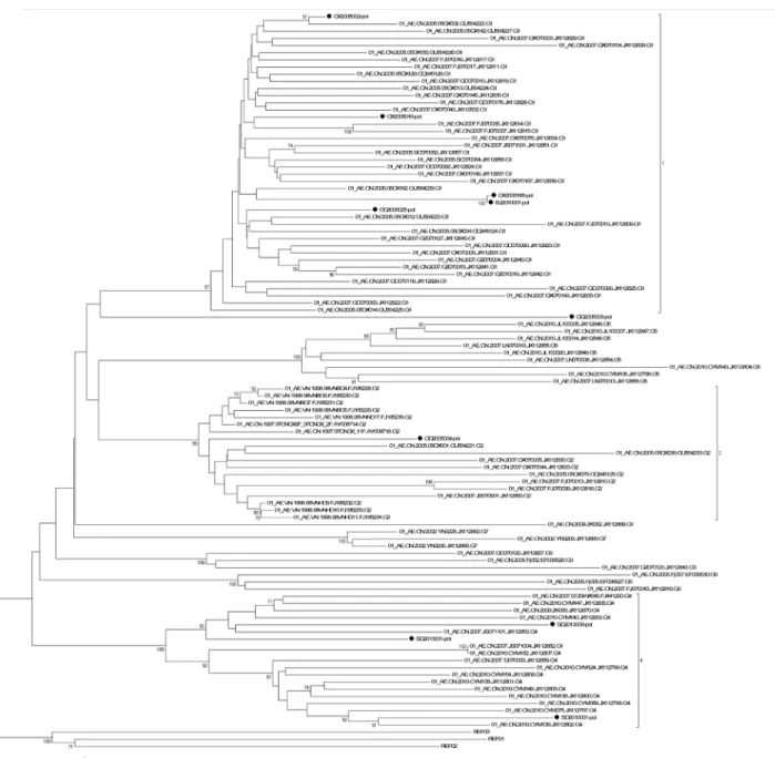
(GX2006183 failed inpolamplification) in our program and 93 CRF01_AE full-lengthpolgene sequences in Yi Feng and Xiang He’s study as the reference sequences was constructed to iden-tify the diversity of the CRF01_AE isolates in our panel (Fig 2). In Yi Feng and Xiang He’s study, seven distinct phylogenetic clusters of CRF01_AE were identified. The CRF01_AE iso-lates in our study were found in three of the seven clusters shown inFig 2.
The prediction of co-receptor usage, the four amino acids at the tip of the
V3-loop and the glycosylation sites by
envV3 sequences of each isolate
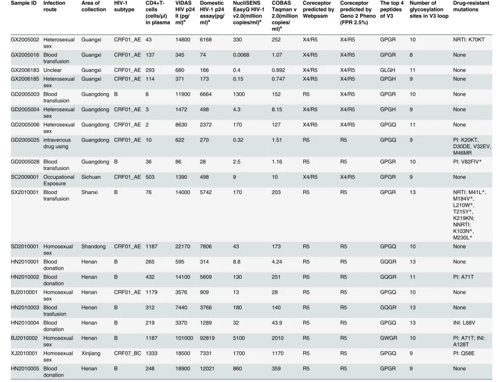
The predominant co-receptor usage of each of the virus isolates was determined based on ge-notype analysis by two online software programs: WebPSSM and Geno2Pheno (G2P) predic-tion (Table 2). Twenty-nineenvC2V3 sequences (KP178451-KP178479) of the thirty samples were amplified successfully. Fifteen of the thirty samples tested exhibited a CCR5 phenotype while fourteen of the thirty samples test exhibited a CXCR4/CCR5 phenotype in both assays (theenvC2V3 amplification of ZJ2006001 was negative). However, the predictions for GD2005003 and SD2013001 were opposite according to the two assays (Table 2). The four amino acids at the tip of the V3-loop and the number of glycosylation sites on the V3-loop for each isolate are shown inTable 2.
The determination of viral drug resistance mutations
Online tools at the Stanford University HIV-1 database (http://hivdb.stanfrod.edu/hiv/) were used to determine the drug-resistance mutations in the full-length sequences of the genepolof each isolate. Their tolerance for each kind of antiviral drug related to the mutations were also predicted to consummate the fully characterization of the isolates (Table 2).
Statistical analysis of the p24 concentrations and viral loads of the HIV-1
isolates
P24 antigen concentrations were quantified using a BioMerieux VIDAS HIV p24 II assay and a domestic p24 test assay. No isolates were below the lower limits of the assays (3.0 pg/ml for the VIDAS HIV p24 II assay and 5.0 pg/ml for the domestic p24 test assay). The results of the p24 concentration test by the BioMerieux VIDAS HIV p24 II assay ranged from 86.1 pg/ml to
Development of an HIV-1 Subtype Panel in China
1.01×10⁵pg/ml. The results of the p24 concentration test by the domestic p24 test assay ranged from 28.3 pg/ml to 1.29×10⁵pg/ml (Table 2). The viral loads were measured by the Cobas Ampli-Prep/Cobas TaqMan HIV-1 test version 2.0(CAP/CTM v2.0, Roche) and the NucliSens EasyQ HIV-1 version 2.0 (EasyQ v2.0, BioMerieux). No isolates were below the lower limits of the assays (5.747 IU/ml for the EasyQ v2.0 assay and 34 IU/ml for the CAP/CTM v2.0 assay). The viral load test by CAP/CTM v2.0 ranged from 7.47×10⁵copies/ml to 2.01×10⁹pg/ml. The viral load test with the EasyQ v2.0 ranged from 6.8×10³copies/ml to 5.1×10⁹copies/ml (Table 2).
Fig 1. Phylogenetic analysis of characterized HIV-1gag-polgene sequences.The tree was midpoint rooted. Horizontal branch lengths are drawn to scale (the scale bar represents 0.02 nucleotide substitution per site). Bracket separation is for clarity only. Numbers at the nodes indicate the bootstraps in which the cluster to the right was supported by 60% and higher. Markers in different shapes at the ends of the horizontal branch represent different subtypes to which the sequences belong. Triangle represents the subtype CRF07_BC, square represents the subtype CRF01_AE, rhombus represents the subtype G, and circle represents the subtype B (B represented by black round while Thai-B represented by dark red circle).
Table 2. Characterization of expanded viruses.
Sample ID Infection
route Area of collection HIV-1 subtype CD4+T-cells
(cells/μl)
in plasma VIDAS HIV p24 II (pg/ ml)a Domestic HIV-1 p24 assay(pg/ ml)a NucliSENS EasyQ HIV-1 v2.0(million copies/ml)a COBAS Taqman v 2.0(million copies/ ml)a Coreceptor predicted by Webpssm Coreceptor predicted by Geno 2 Pheno (FPR 2.5%)
The top 4 peptides of V3
Number of glycosylation sites in V3 loop
Drug-resistant mutations
GX2005002 Heterosexual sex
Guangxi CRF01_AE 43 14800 6168 330 252 X4/R5 X4/R5 GPGR 10 NRTI: K70KT
GX2005016 Blood transfusion
Guangxi CRF01_AE 137 345 74 0.0068 1.07 X4/R5 X4/R5 GPGR 8 None
GX2006183 Unclear Guangxi CRF01_AE 293 680 166 0.4 0.892 X4/R5 X4/R5 GLGH 11 None
GX2006185 Heterosexual sex
Guangxi CRF01_AE 114 371 173 0.15 0.747 X4/R5 X4/R5 GPGH 9 None
GD2005003 Blood transfusion
Guangdong B 8 11900 6664 1300 152 R5 X4/R5 GPGR 10 None
GD2005004 Heterosexual sex
Guangdong CRF01_AE 3 1472 498 4.3 8.15 X4/R5 X4/R5 GPGH 9 None
GD2005006 Heterosexual sex
Guangdong CRF01_AE 2 8630 2372 170 127 X4/R5 X4/R5 GPGQ 11 None
GD2005025 intravenous drug using
Guangdong CRF01_AE 10 622 270 0.32 1.51 R5 R5 GPGQ 9 PI: K20KT,
D30DE, V32EV, M46MR
GD2005028 Blood transfusion
Guangdong B 36 86 28 2.5 1.16 R5 R5 GPGR 10 PI: V82FIV*
SC2009001 Occupational Exposure
Sichuan CRF01_AE 503 1390 498 9 10 X4/R5 X4/R5 GPGR 9 None
SX2010001 Blood transfusion
Shanxi B 76 14000 5742 170 203 R5 R5 GPGR 13 NRTI: M41L*,
M184V*, L210W*, T215Y*, K219KN; NNRTI: K103N*, M230L* SD2010001 Homosexual sex
Shandong CRF01_AE 1187 22170 7806 43 173 R5 R5 GPGQ 10 None
HN2010001 Blood donation
Henan B 265 595 314 8.8 4.24 R5 R5 GQGR 13 None
HN2010002 Blood donation
Henan B 432 14100 5609 130 251 R5 R5 GQGR 11 PI: A71T
BJ2010001 Homosexual sex
Henan CRF01_AE 1179 3576 909 13 28 R5 R5 GPGQ 10 None
HN2010003 Blood trasfusion
Henan B 312 7440 3766 180 140 R5 R5 GQGR 13 None
HN2010004 Blood donation
Henan B 219 3370 1289 32 43.9 R5 R5 GPGQ 13 INI: L68V
BJ2010002 Homosexual sex
Henan B 1187 101000 92819 5100 2010 R5 R5 GWGR 10 PI: A71T; INI:
A128T
XJ2010001 Homosexual sex
Xinjiang CRF07_BC 1333 18500 7331 1700 1170 R5 R5 GPGQ 9 PI: Q58E
HN2010005 Blood donation
Henan B 248 18900 12021 860 359 R5 R5 GPGR 9 None
(Continued)
route collection subtype cells
(cells/μl)
in plasma HIV p24 II (pg/ ml)a HIV-1 p24 assay(pg/ ml)a EasyQ HIV-1 v2.0(million copies/ml)a Taqman v 2.0(million copies/ ml)a predicted by Webpssm predicted by Geno 2 Pheno (FPR 2.5%)
peptides of V3
glycosylation sites in V3 loop
mutations
BJ2010003 Homosexual sex
Henan CRF07_BC 1528 2630 554 250 101 R5 R5 GPGQ 12 INI: H51HQ
NX2005012 Blood transfusion
Ningxia B 21 237 65 5.8 2.12 R5 R5 GQGR 11 INI: L68V
SH2007052 Heterosexual sex
Shanghai G ND 363 50 0.66 0.823 R5 R5 APGQ 10 None
HN2002024 Blood trasfusion
Henan B ND 80000 128722 1700 618 X4/R5 X4/R5 GPGR 10 PI: I54M*,
L23IL, A71V
SD2013001 Homosexual sex
Shandong CRF01_AE 197 2575 1047 0.2 22.7 X4/R5 R5 GPGQ 11 INI: P145PS,
S153FS
SD2013005 Homosexual sex
Shandong CRF07_BC 715 107 46 14 3.11 R5 R5 GPGQ 10 None
SD2013008 Homosexual sex
Shandong CRF01_AE 486 21500 11583 40 254 X4/R5 X4/R5 GPGQ 8 None
BJ2006001 Heterosexual sex
Beijing B 40 1500 866 4.6 2.49 X4/R5 X4/R5 GRGR 12 NRTI: M41L*,
L210W*, T215F*; NNRTI: A98G, K103N*, V179E*, Y181C*, G190A*; PI: L63P, V77I
SD2006001 Blood trasfusion
Shandong B 74 1600 409 5.8 2.78 X4/R5 X4/R5 GPGR 13 NRTI: M184V*;
NNRTI: K103N*; PI: L63P, A71T, V77I, I93L
ZJ2006001 Blood transfusion
Zhejiang CRF07_BC 38 4060 1488 9.9 17 Unknown Unknown Unknown Unknown NRTI: K65R*,
T69D, K219R; NNRTI: V106M*, Y181C*; PI: L63P, I93L, L10X
IIIB_LAI (Ref)
Unknown France B Unknown 79000 43925 2600 708 X4/R5 X4/R5 GPGR 11 None
NL4-3(Ref) Recombinant France B Unknown 78000 44829 2200 686 X4/R5 X4/R5 GPGR 12 IN: V151I
ND: Not done,envC2V3 PCR was negative.
a: Samples for p24 antigen tests(VIDAS HIV p24 II and the domestic HIV-1 p24 assay) and viral load tests(NucliSENS EasyQ HIV-1 v2.0 and COBAS Taqman v 2.0) are culture
supernatant of the isolates after expansion.
*: NRTI/NNRTI/PI/INI Major drug-resistance mutations. Mutations without“*”are minor resistance mutations, accessory mutations or single nucleotide polymorphisms (SNP). Ref: Reference virus. IIIB_LAI and NL4-3 are reference virus with clear background information and high replication capacity but are not primary isolates.
The expanded virus was characterized in terms of infection route, area of origin, CD4+T cells concentration in blood sample, viral RNA concentration (Roche TaqMan v2.0 and BioMerieux NucliSENS EasyQ HIV-1 v2.0), p24 concentration (BioMerieux VIDAS HIV p24 II and One domestic HIV-1 p24 antigen assay), coreceptor usage (Webpssm and Geno
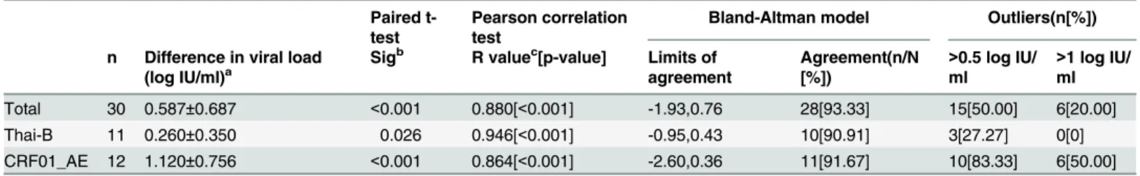
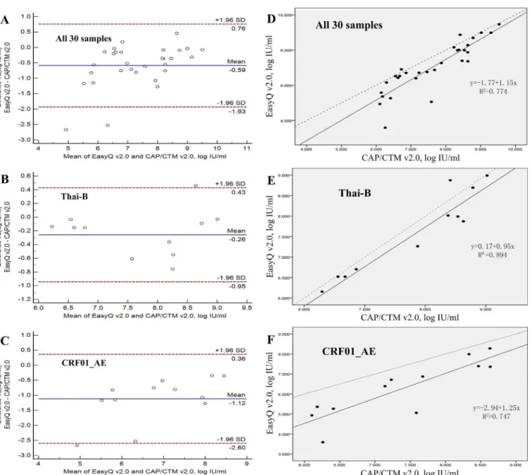
As for the two assays providing viral load tests, according to the results of the performance, EasyQ v2.0 showed a significant linear correlation (R = 0.880, p<0.001) and high agreement (93.33%, 28/30) with CAP/CTM v2.0 for all 30 samples (Table 3andFig 3A). The regression equation used for fitting was: EasyQ v2.0 = -1.77+1.15×CAP/CTM V2.0 (Fig 3D). The mean difference between the quantitative values measured by EasyQ v2.0 and CAP/CTM v2.0 was 0.587 log IU/ml (SD = 0.687;Table 3). For the different subtype isolates (subtype Thai-B and Fig 2. Neighbor-joining tree of HIV-1 CRF01_AEpolgene sequences.The phylogenetic tree was constructed with HIV-1 full-length genepolsequences. Horizontal branch lengths are drawn to scale (the scale bar represents 0.005 nucleotide substitution per site). The numbers“1”,“2”, and“4”marked on the bracket separation are clusters that correspond to the clusters in Yi Feng and Xiang He’s study. Numbers at the nodes indicate the bootstraps in which the cluster to the right was supported by 70% and higher. The red circles at the ends of the horizontal branch represent the HIV-1 CRF01_AE samples in our panel. The three reference sequences at the root of the neighbor-joining tree were HIV-1 CRF01_AE sequences from Central Africa. The accession numbers of the three references in the Los Alamos HIV Database are Ref.01_AE.CF.1990.90CF11697.AF197340, Ref.01_AE.CF.1990.90CF4071.AF197341 and Ref.01_AE.CF.1990.90CR402_CAR_E_4003.U51188.
doi:10.1371/journal.pone.0127696.g002
Development of an HIV-1 Subtype Panel in China
subtype CRF01_AE,Table 3), a significant linear correlation (R = 0.946 for subtype Thai-B, p <0.001; R = 0.864 for subtype CRF01_AE, p<0.001;Table 3) and high agreement (90.91% for
Thai-B and 91.67% for CRF01_AE;Fig 3) were observed. The mean difference between the val-ues measured by EasyQ v2.0 and CAP/CTM v2.0 was 0.260 log IU/ml (SD = 0.350) for subtype Thai-B (p = 0.260) and 1.120 log IU/ml (SD = 0.756) for subtype CRF01_AE (p<0.001). The number of samples with the quantitative differences between EasyQ v2.0 and CAP/CTM v2.0 exceeded 0.5 log IU/ml varied from each subtype: three samples (27.27%) for subtype Thai-B and 10 samples (83.33%) for subtype CRF01_AE. Furthermore, six subtype CRF01_AE sam-ples showed quantitative differences of>1 log IU/ml, while no samples of Thai-B showed quantitative differences of>1 log IU/ml (Table 3).
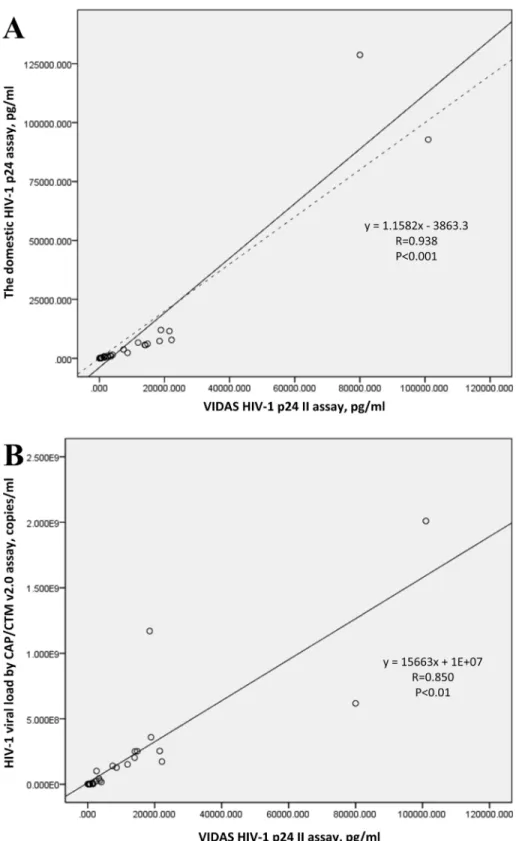
As for the two assays of p24 antigen tests, the Pearson correlation coefficient for the two HIV-1 P24 antigen assay based on all 30 samples in the panel was 0.938 (p<0.01). The correla-tion coefficient for the p24 concentracorrela-tions tested by the BioMerieux VIDAS HIV p24 II assay and the viral loads tested by the Roche COBAS Taqman version 2.0 assay based on all the 30 isolates was 0.850 (p<0.01;Fig 4).
Discussion
With the diversity of HIV-1 increasing, HIV-1 isolates of each subtype spread broader and faster around the world. The EQAPOL program was established by the NIH/NIAID and other cooperative institutions and their efforts were aimed at the development of an HIV subtype panel that encompassed the genetic and geographic diversity currently present worldwide. Ac-cordingly, we developed an HIV-1 isolate panel that can represent the diversity of HIV-1 strains circulating in China. This panel will help with the evaluation of HIV surveillance or di-agnostic assays and research on HIV vaccines or antiviral drugs in China [13]. In our study, blood samples from HIV infected people in China were collected from 2002 to 2013. 30 HIV-1 primary strains of high-titer and high-volume were obtained successfully. These isolates were all well preserved in liquid nitrogen and can be used as the standard substances or samples in evaluation of HIV-1 assays, antiviral drug screening or vaccine development conveniently. The subtypes of these isolates not only include China’s frequent subtype B, CRF01_AEand
CRF07_BC, but also a strain of subtype G which was infrequent in China was isolated from a plasma sample from Shanghai. Shown by Yi Feng and Xiang He’s research, the CRF01_AE epi-demic in China is remarkably complex [35]. In their study, seven distinct phylogenetic clusters of CRF01_AE were identified. The CRF01_AE isolates in our study were found in three of the seven clusters shown inFig 2, which means the CRF01_AE isolates in our panel can represent Table 3. Comparison of the EasyQ v2.0 and CAP/CTM v2.0 using samples belonging to different clades.
Paired t-test
Pearson correlation test
Bland-Altman model Outliers(n[%])
n Difference in viral load (log IU/ml)a
Sigb R valuec[p-value] Limits of
agreement
Agreement(n/N [%])
>0.5 log IU/ ml
>1 log IU/ ml
Total 30 0.587±0.687 <0.001 0.880[<0.001] -1.93,0.76 28[93.33] 15[50.00] 6[20.00] Thai-B 11 0.260±0.350 0.026 0.946[<0.001] -0.95,0.43 10[90.91] 3[27.27] 0[0] CRF01_AE 12 1.120±0.756 <0.001 0.864[<0.001] -2.60,0.36 11[91.67] 10[83.33] 6[50.00]
EasyQ v2.0: NucliSens EasyQ HIV-1 version 2.0; CAP/CTMv2.0: Cobas AmpliPrep/Cobas TaqMan HIV-1 test version 2.0. a: Values are expressed as mean±SD.
b
: Significance calculated using paired t-test (p-values). c
: Correlation coefficient (R value) calculated using Pearson correlation test.
the current complex diversity of this subtype to some extent. We amplified and sequenced the fundamental genes; i.e.,gag,pol,envC2V3 for each isolate. The co-recepter usage, glycosylation sites, four residues at the tip of the v3-loop for each isolate, also the drug-resistance mutations on genepolwere predicted to consummate the characterization of the isolates in this panel. This accurate and comprehensive characterization will make the panel convenient to use in the future.
Fig 3. Agreement and linear relationship between the Cobas AmpliPrep/Cobas TaqMan HIV-1 test version 2.0 (CAP/CTM v2.0) and the NucliSens EasyQ HIV-1 version 2.0 (EasyQ v2.0) calculated using the Bland-Altman model.(A) Agreement between EasyQ v2.0 and CAP/CTM v2.0 when used to measure all 30 samples in the panel. (B) Agreement between EasyQ v2.0 and CAP/CTM v2.0 when used to measure 11 clade Thai-B samples in the panel. (C) Agreement between EasyQ v2.0 and CAP/CTM v2.0 when used to measure 12 clade CRF01_AE samples in the panel. For A, B and C, solid horizontal lines indicate the mean values, and dashed horizontal lines indicate the +1.96SD and _1.96SD values. (D) The linear relationship between the CAP/CTM v2.0 and EasyQ v2.0 when used to measure all 30 samples. (E) The linear relationship between the CAP/CTM v2.0 and EasyQ v2.0 when used to measure 11 clade Thai-B samples. (F) The linear relationship between the CAP/CTM v2.0 and EasyQ v2.0 when used to measure 12 clade CRF01_AE samples. For D, E and F, solid line represents the fitted regression line and the dashed line represents the equality line.
doi:10.1371/journal.pone.0127696.g003
Development of an HIV-1 Subtype Panel in China
Fig 4. The linear relationship between the two assays of HIV-1 p24 measurement and the linear relationship between p24 antigen concentration and HIV-1 viral load based on the values of all 30 samples.(A) The linear relationship between the BioMerieux VIDAS HIV p24 II assay and the domestic HIV-1 p24 assay when used to measure all 30 samples. Solid line represents the fitted regression line and the dashed line represents the equality line. (B) The linear relationship between HIV-1 p24 antigen concentration and viral load based on all 30 isolates, tested by BioMerieux VIDAS HIV p24 II and Roche COBAS Taqman version 2.0, respectively. Solid line represents the fitted regression line.
The comparison of the p24 or viral load assays on the isolates illustrated how this panel may be used for evaluation of HIV-1 diagnostic assay performance. For example, according toFig 3, compared with CAP/CTM v2.0, the EasyQ v2.0 showed a greater variation in the values mea-sured for CRF01_AE samples than those for Thai-B samples. By Bland-Altman analysis, we could see that the distance between the mean values of the different results from the two assays in the viral loads of subtype CRF01_AE samples, which are shown as solid horizontal lines inFig 3C, and the origin, which is shown as the zero point inFig 3C(1.120), were much farther than that of Thai-B samples (0.260;Fig 3B). Furthermore, the proportion of subtype Thai-B samples showing quantitative differences between the two assays>0.5 log IU/ml or 1 log IU/ml was sig-nificantly lower than that for subtype CRF01_AE (Table 3). These data suggest higher levels of agreement between these two assays when measuring Thai-B samples than for CRF01_AE sam-ples. A similar conclusion was shown in Sihong Xu’s study [36]. Higher values measured by CAP/CTM v2.0 may because of the fact that CAP/CTM v2.0 reduces under-quantification of the HIV-1 viral load by using two dual-labeled hybridization probes targeting both thegagand LTR regions, while EasyQ v2.0 targeted the HIV-1gaggene only. The performance of the auto-mated extractor of CAP/CTM v2.0 may also contribute to the higher values and stable perfor-mance of CAP/CTM v2.0. The lower agreement between the two assays when measuring subtype CRF01_AE samples may also due to different hybridization probes, which indicates that the probe targets in the sequence may not be conservative enough in subtype Thai-B and CRF01_AE, and at least one of the assays under-quantified the viral loads of subtype CRF01_AE samples. To face this problem, it is strongly suggested that the same version of the same viral load assay be used consistently during clinical treatment. Much more importantly, the genetic diversity of HIV-1 (including the genetic variability within HIV-1 strains of the same subtype) must be taken into account when designing primers and probes for HIV-1 viral load assays. An-other example is the two different results for co-receptor usage prediction of GD2005003 and SD2013001 (Table 2) may be caused by the different calculation principles of the two software programs; i.e., Geno2pheno and PSSM, or the different FPR values chosen for Geno2pheno.
The entry of HIV-1 into host cells is mediated by interactions between the virus envelope (i.e.,env) glycoprotein (gp120/gp41) and host-cell receptor [38]. The third hypervariable re-gion 3 (V3) of the HIV-1 gp120 protein consists of 35 amino acids and plays an important role in viral infection by promoting the interaction between the virus and its co-receptor in the host cell membrane [39]. N-glycan represent approximately 50% of the molecular mass of gp120 and serves as a potential antigenic determinant and a shield against immune recognition [40]. It is convinced that HIV-1 escape from neutralizing antibodies through the extensive variability of the viral envelope glycoproteins, especially gp120 while the four amino acids at the tip of the V3 loop are subjected to strong purifying selection pressure due to their functional importance [41,42]. Therefore, using the consensus sequence onenvC2V3 to predict the co-receptor usage of each isolate, glycosylation sites on the v3-loop and identification of the four residues at the tip of the V3 loop is necessary for infection relevant experiments and studies.
In conclusion, by isolating, expanding, sequencing and analyzing the HIV-1 strains from blood samples of HIV-1 infected persons, our study produced a panel of HIV-1 primary strains of a variety of HIV-1 subtypes circulating in China with clear and complete fundamental se-quences and biological features. Our program will continue to collect HIV-1 strains for more subtypes to develop a more comprehensive panel that can represent the diversity, the current dynamic and complex epidemic of HIV-1 more completely and accurately. Especially for sam-ples of CRF08_BC, because it is one of the frequent subtypes circulating in China besides CRF07_BC, CRF01_AE and B. Also, more effort should be spent on the isolation of viruses from the acute/early stage of infection to analyze the transmitted or early founder viruses. This is an important step in achieving a molecular understanding of HIV-1 transmission and for
Development of an HIV-1 Subtype Panel in China
potentially developing an effective HIV/AIDS vaccine since direct analysis of HIV-1 at or near the moment of transmission is practically impossible [43]. In our study, we created a basic pro-tocol and standard for sample collection, inclusion and analysis by developing the panel. Under the circumstance of high variable HIV-1 strains of each subtype spread throughout the world, the creation of this panel will not only serves as the standard for assays, vaccines and drugs development andevaluation, but also as the samples for diversity related HIV-1 research to help improve the current situation when many researches on HIV-1 diversity still stay on the genome sequence level. By offering HIV-1 isolates isolated from blood samples directly, re-searches based on the panel will have high simulation and high reproducibility when compared with researches based on infectious clones of the viruses. In this way, our panel may also play a role in antiviral drug screening and vaccine development.
Acknowledgments
We would like to thank all the participants for their contributions and cooperation, as well as the local CDCs for sample provision.
Author Contributions
Conceived and designed the experiments: JYL JWH WG SYL. Performed the experiments: JWH WG SYL ZYB TYL. Analyzed the data: JWH XLW TG HSS LL YJL. Contributed re-agents/materials/analysis tools: SYL DMZ ZYB. Wrote the paper: JWH JYL WG LJ HPL.
References
1. UNAIDS, 2014. 2014 Gap report from UNAIDS. Available:http://www.unaids.org/en/resources /cam-paigns/2014/2014gapreport/gapreport
2. Sezgin E, Hendrickson SL, Jabs DA, Van Natta ML, Lewis RA, Troyer JL, et al. Effect of host genetics on incidence of HIV neuroretinal disorder in patients with AIDS. J Acquir Immune Defic Syndr. 2010; 54 (4):343–51. Epub 2010/06/10. doi:10.1097/QAI.0b013e3181deaf4dPMID:20531015; PubMed Cen-tral PMCID: PMC2908809.
3. Buonaguro L, Tornesello ML, Buonaguro FM. Human immunodeficiency virus type 1 subtype distribu-tion in the worldwide epidemic: pathogenetic and therapeutic implicadistribu-tions. Journal of virology. 2007; 81 (19):10209–19. Epub 2007/07/20. doi:10.1128/jvi.00872-07PMID:17634242; PubMed Central PMCID: PMC2045484.
4. Church D, Gregson D, Lloyd T, Klein M, Beckthold B, Laupland K, et al. Comparison of the RealTime HIV-1, COBAS TaqMan 48 v1.0, Easy Q v1.2, and Versant v3.0 assays for determination of HIV-1 viral loads in a cohort of Canadian patients with diverse HIV subtype infections. J Clin Microbiol. 2011; 49 (1):118–24. Epub 2010/11/19. doi:10.1128/jcm.00685-10PMID:21084515; PubMed Central PMCID: PMC3020439.
5. Kline NE, Schwarzwald H, Kline MW. False negative DNA polymerase chain reaction in an infant with subtype C human immunodeficiency virus 1 infection. Pediatr Infect Dis J. 2002; 21(9):885–6. Epub 2002/10/17. PMID:12380591.
6. Obaro SK, Losikoff P, Harwell J, Pugatch D. Failure of serial human immunodeficiency virus type 1 DNA polymerase chain reactions to identify human immunodeficiency virus type 1 clade A/G. Pediatr Infect Dis J. 2005; 24(2):183–4. Epub 2005/02/11. PMID:15702052.
7. Henquell C, Jacomet C, Antoniotti O, Chaib A, Regagnon C, Brunet S, et al. Difficulties in diagnosing group o human immunodeficiency virus type 1 acute primary infection. J Clin Microbiol. 2008; 46 (7):2453–6. Epub 2008/05/16. doi:10.1128/jcm.02217-07PMID:18480223; PubMed Central PMCID: PMC2446893.
8. Foglieni B, Candotti D, Guarnori I, Raffaele L, Berzuini A, Spreafico M, et al. A cluster of human immu-nodeficiency virus Type 1 recombinant form escaping detection by commercial genomic amplification assays. Transfusion. 2011; 51(4):719–30. Epub 2010/11/23. doi:10.1111/j.1537-2995.2010.02942.x
PMID:21087286.
breakthrough transmissions. Transfusion. 2009; 49(9):1850–8. Epub 2009/05/21. doi: 10.1111/j.1537-2995.2009.02212.xPMID:19453976.
10. Taylor BS, Hammer SM. The challenge of HIV-1 subtype diversity. N Engl J Med. 2008; 359(18):1965–
6. Epub 2008/10/31. doi:10.1056/NEJMc086373PMID:18971501.
11. Ntemgwa M, Gill MJ, Brenner BG, Moisi D, Wainberg MA. Discrepancies in assignment of subtype/re-combinant forms by genotyping programs for HIV type 1 drug resistance testing may falsely predict su-perinfection. AIDS Res Hum Retroviruses. 2008; 24(7):995–1002. Epub 2008/07/03. doi:10.1089/aid. 2008.0064PMID:18593348.
12. Snoeck J, Kantor R, Shafer RW, Van Laethem K, Deforche K, Carvalho AP, et al. Discordances be-tween interpretation algorithms for genotypic resistance to protease and reverse transcriptase inhibitors of human immunodeficiency virus are subtype dependent. Antimicrob Agents Chemother. 2006; 50 (2):694–701. Epub 2006/01/27. doi:10.1128/aac.50.2.694-701.2006PMID:16436728; PubMed Cen-tral PMCID: PMC1366873.
13. Sanchez AM, DeMarco CT, Hora B, Keinonen S, Chen Y, Brinkley C, et al. Development of a contem-porary globally diverse HIV viral panel by the EQAPOL program. J Immunol Methods. 2014; 409:117–
30. Epub 2014/01/23. doi:10.1016/j.jim.2014.01.004PMID:24447533; PubMed Central PMCID: PMC4104154.
14. He X, Xing H, Ruan Y, Hong K, Cheng C, Hu Y, et al. A comprehensive mapping of HIV-1 genotypes in various risk groups and regions across China based on a nationwide molecular epidemiologic survey. PLoS One. 2012; 7(10):e47289. Epub 2012/10/12. doi:10.1371/journal.pone.0047289PMID:
23056619; PubMed Central PMCID: PMC3466245.
15. Guan Y, Chen J, Shao Y, Zhao Q, Zeng Y, Zhang J, et al. [Subtype and sequence analysis of the C2-V3 region of gp120 genes among human immunodeficiency virus infected IDUs in Ruili epidemic area of Yunnan Province of China]. Zhonghua Shi Yan He Lin Chuang Bing Du Xue Za Zhi. 1997; 11(1):8–
12. Epub 1997/03/01. PMID:15619893.
16. Piyasirisilp S, McCutchan FE, Carr JK, Sanders-Buell E, Liu W, Chen J, et al. A recent outbreak of human immunodeficiency virus type 1 infection in southern China was initiated by two highly homoge-neous, geographically separated strains, circulating recombinant form AE and a novel BC recombinant. Journal of virology. 2000; 74(23):11286–95. Epub 2000/11/09. PMID:11070028; PubMed Central PMCID: PMC113233.
17. Su L, Graf M, Zhang Y, von Briesen H, Xing H, Kostler J, et al. Characterization of a virtually full-length human immunodeficiency virus type 1 genome of a prevalent intersubtype (C/B') recombinant strain in China. Journal of virology. 2000; 74(23):11367–76. Epub 2000/11/09. PMID:11070037; PubMed Cen-tral PMCID: PMC113242.
18. Tee KK, Pybus OG, Li XJ, Han X, Shang H, Kamarulzaman A, et al. Temporal and spatial dynamics of human immunodeficiency virus type 1 circulating recombinant forms 08_BC and 07_BC in Asia. Jour-nal of virology. 2008; 82(18):9206–15. Epub 2008/07/04. doi:10.1128/jvi.00399-08PMID:18596096; PubMed Central PMCID: PMC2546895.
19. Qin G, Shao Y, Liu G. [Subtype and sequence analysis of the C2-V3 region of gp120 genes among HIV-1 strains in Sichuan province]. Zhonghua Liu Xing Bing Xue Za Zhi. 1998; 19(1):39–42. Epub 1999/05/14. PMID:10322707.
20. Li YW, Shao YM, Luo XG. [Subtype and sequence analysis of the C2-V3 region of gp120 genes among HIV-1 strains in Hubei province]. Zhonghua Liu Xing Bing Xue Za Zhi. 1997; 18(4):217–9. Epub 1997/ 08/01. PMID:9812522.
21. Xu JQ, Wang JJ, Han LF, Xu C, Ruan YH, Xu ZH, et al. Epidemiology, clinical and laboratory character-istics of currently alive HIV-1 infected former blood donors naive to antiretroviral therapy in Anhui Prov-ince, China. Chin Med J (Engl). 2006; 119(23):1941–8. Epub 2007/01/04. PMID:17199937.
22. Wang FX, Zhou H, Ling H, Zhou HZ, Liu WH, Shao YM, et al. Subtype and sequence analysis of HIV-1 strains in Heilongjiang Province. Chin Med J (Engl). 2007; 120(22):2006–10. Epub 2007/12/11. PMID:
18067787.
23. Chen J, Young NL, Subbarao S, Warachit P, Saguanwongse S, Wongsheree S, et al. HIV type 1 sub-types in Guangxi Province, China, 1996. AIDS Res Hum Retroviruses. 1999; 15(1):81–4. Epub 1999/ 02/19. doi:10.1089/088922299311754PMID:10024057.
24. Yu XF, Chen J, Shao Y, Beyrer C, Liu B, Wang Z, et al. Emerging HIV infections with distinct subtypes of HIV-1 infection among injection drug users from geographically separate locations in Guangxi Prov-ince, China. J Acquir Immune Defic Syndr. 1999; 22(2):180–8. Epub 2000/06/08. PMID:10843533.
25. Xing H, Liang H, Wan ZY, Chen X, Wei M, Ma PF, et al. [Distribution of recombinant human immunode-ficiency virus type-1 CRF01_AE strains in China and its sequence variations in the env V3-C3 region]. Zhonghua Yu Fang Yi Xue Za Zhi. 2004; 38(5):300–4. Epub 2004/10/23. PMID:15498240.
Development of an HIV-1 Subtype Panel in China
26. Han X, An M, Zhang W, Cai W, Chen X, Takebe Y, et al. Genome Sequences of a Novel HIV-1 Circulat-ing Recombinant Form, CRF55_01B, Identified in China. Genome Announc. 2013; 1(1). Epub 2013/02/ 14. doi:10.1128/genomeA.00050-12PMID:23405298; PubMed Central PMCID: PMC3569284.
27. Wei H, Liu Y, Feng Y, Hsi J, Xing H, He X, et al. Genome sequence of a novel HIV-1 circulating recom-binant form (CRF57_BC) identified from Yunnan, China. AIDS Res Hum Retroviruses. 2014; 30 (4):384–8. Epub 2013/11/12. doi:10.1089/aid.2013.0228PMID:24205935; PubMed Central PMCID: PMC3976592.
28. Gao L, Hanson MN, Balakrishnan M, Boyer PL, Roques BP, Hughes SH, et al. Apparent defects in pro-cessive DNA synthesis, strand transfer, and primer elongation of Met-184 mutants of HIV-1 reverse transcriptase derive solely from a dNTP utilization defect. J Biol Chem. 2008; 283(14):9196–205. Epub 2008/01/26. M710148200 [pii] doi:10.1074/jbc.M710148200PMID:18218634; PubMed Central PMCID: PMC2431043.
29. Li X, Ning C, He X, Yang Y, Li F, Xing H, et al. Genome Sequences of a Novel HIV-1 Circulating Recom-binant Form (CRF61_BC) Identified among Heterosexuals in China. Genome Announc. 2013; 1(3). Epub 2013/07/03. doi:10.1128/genomeA.00326-13PMID:23814029; PubMed Central PMCID: PMC3695425.
30. Wei H, His J, Feng Y, Xing H, He X, Liao L, et al. Identification of a novel HIV-1 circulating recombinant form (CRF62_BC) in western Yunnan of China. AIDS Res Hum Retroviruses. 2014; 30(4):380–3. Epub 2013/10/30. doi:10.1089/aid.2013.0235PMID:24164474; PubMed Central PMCID: PMC3976572.
31. Hsi J, Wei H, Xing H, Feng Y, He X, Liao L, et al. Genome sequence of a Novel HIV-1 circulating recom-binant form (CRF64_BC) identified from Yunnan, China. AIDS Res Hum Retroviruses. 2014; 30 (4):389–93. Epub 2013/11/12. doi:10.1089/aid.2013.0234PMID:24205972; PubMed Central PMCID: PMC3976579.
32. Feng Y, Wei H, Hsi J, Xing H, He X, Liao L, et al. Identification of a novel HIV Type 1 circulating recombi-nant form (CRF65_cpx) composed of CRF01_AE and subtypes B and C in Western Yunnan, China. AIDS Res Hum Retroviruses. 2014; 30(6):598–602. Epub 2013/11/28. doi:10.1089/aid.2013.0233
PMID:24279591; PubMed Central PMCID: PMC4046203.
33. Manak M, Sina S, Anekella B, Hewlett I, Sanders-Buell E, Ragupathy V, et al. Pilot studies for develop-ment of an HIV subtype panel for surveillance of global diversity. AIDS Res Hum Retroviruses. 2012; 28(6):594–606. Epub 2011/12/14. doi:10.1089/aid.2011.0271PMID:22149143; PubMed Central PMCID: PMC3358106.
34. Liu W, Liang Sj Fau—Yang J-y, Yang Jy Fau—Li J-j, Li Jj Fau—Wang B, Wang B Fau—Chen L-l, Chen Ll Fau—Li L, et al. [Distribution of HIV-1 subtypes in Guangxi Zhuang Autonomous Region, 2008–
2009]. (0254–6450 (Print)). PMID:23648251
35. Feng Y, He X, Hsi JH, Li F, Li X, Wang Q, et al. The rapidly expanding CRF01_AE epidemic in China is driven by multiple lineages of HIV-1 viruses introduced in the 1990s. AIDS (London, England). 2013; 27 (11):1793–802. Epub 2013/06/29. doi:10.1097/QAD.0b013e328360db2dPMID:23807275; PubMed Central PMCID: PMC3819312.
36. Xu S, Song A, Nie J, Li X, Meng S, Zhang C, et al. Comparison between the automated Roche Cobas AmpliPrep/Cobas TaqMan HIV-1 test version 2.0 assay and its version 1 and Nuclisens HIV-1 EasyQ version 2.0 assays when measuring diverse HIV-1 genotypes in China. J Clin Virol. 2012; 53(1):33–7. Epub 2011/11/05. doi:10.1016/j.jcv.2011.10.001PMID:22051503.
37. Bland JM, Altman DG. Statistical methods for assessing agreement between two methods of clinical measurement. Lancet. 1986; 1(8476):307–10. Epub 1986/02/08. PMID:2868172.
38. Weissenhorn W, Dessen A, Harrison SC, Skehel JJ, Wiley DC. Atomic structure of the ectodomain from HIV-1 gp41. Nature. 1997; 387(6631):426–30. Epub 1997/05/22. doi:10.1038/387426a0PMID:
9163431.
39. Hwang SS, Boyle TJ, Lyerly HK, Cullen BR. Identification of the envelope V3 loop as the primary deter-minant of cell tropism in HIV-1. Science (New York, NY). 1991; 253(5015):71–4. Epub 1991/07/05. PMID:1905842.
40. Zhu X, Borchers C, Bienstock RJ, Tomer KB. Mass spectrometric characterization of the glycosylation pattern of HIV-gp120 expressed in CHO cells. Biochemistry. 2000; 39(37):11194–204. Epub 2000/09/ 14. PMID:10985765.
41. Kwong PD, Doyle ML, Casper DJ, Cicala C, Leavitt SA, Majeed S, et al. HIV-1 evades antibody-mediat-ed neutralization through conformational masking of receptor-binding sites. Nature. 2002; 420 (6916):678–82. Epub 2002/12/13. doi:10.1038/nature01188PMID:12478295.
43. Keele BF, Giorgi EE, Salazar-Gonzalez JF, Decker JM, Pham KT, Salazar MG, et al. Identification and characterization of transmitted and early founder virus envelopes in primary HIV-1 infection. Proceed-ings of the National Academy of Sciences of the United States of America. 2008; 105(21):7552–7. Epub 2008/05/21. doi:10.1073/pnas.0802203105PMID:18490657; PubMed Central PMCID: PMC2387184.
Development of an HIV-1 Subtype Panel in China