Connectivity in Healthy Subjects: Implications for
Antidepressant Drug Action
Milan Scheidegger1,2*., Martin Walter3,4,5
*, Mick Lehmann1, Coraline Metzger3,5, Simone Grimm2,6,7, Heinz Boeker2, Peter Boesiger1,9, Anke Henning1,8, Erich Seifritz2,8,9
1Institute for Biomedical Engineering, University and ETH Zurich, Zurich, Switzerland,2Clinic of Affective Disorders and General Psychiatry, Psychiatric University Hospital, Zurich, Switzerland, 3Department of Psychiatry, Otto-von-Guericke University, Magdeburg, Germany,4Department of Behavioral Neurology, Leibniz Institute for Neurobiology, Magdeburg, Germany,5Clinical Affective Neuroimaging Laboratory (CANLAB), Center for Behavioral and Brain Sciences, CBBS, Magdeburg, Germany,
6Cluster Languages of Emotion, Freie Universita¨t Berlin, Berlin, Germany,7Department of Psychiatry, Charite´, CBF, Berlin, Germany,8Zurich Center for Integrative Human Physiology (ZIHP), University of Zurich, Zurich, Switzerland,9Neuroscience Center Zurich, University of Zurich and ETH Zurich, Zurich, Switzerland
Abstract
Increasing preclinical and clinical evidence underscores the strong and rapid antidepressant properties of the glutamate-modulating NMDA receptor antagonist ketamine. Targeting the glutamatergic system might thus provide a novel molecular strategy for antidepressant treatment. Since glutamate is the most abundant and major excitatory neurotransmitter in the brain, pathophysiological changes in glutamatergic signaling are likely to affect neurobehavioral plasticity, information processing and large-scale changes in functional brain connectivity underlying certain symptoms of major depressive disorder. Using resting state functional magnetic resonance imaging (rsfMRI), the ’’dorsal nexus ‘‘(DN) was recently identified as a bilateral dorsal medial prefrontal cortex region showing dramatically increased depression-associated functional connectivity with large portions of a cognitive control network (CCN), the default mode network (DMN), and a rostral affective network (AN). Hence, Sheline and colleagues (2010) proposed that reducing increased connectivity of the DN might play a critical role in reducing depression symptomatology and thus represent a potential therapy target for affective disorders. Here, using a randomized, placebo-controlled, double-blind, crossover rsfMRI challenge in healthy subjects we demonstrate that ketamine decreases functional connectivity of the DMN to the DN and to the pregenual anterior cingulate (PACC) and medioprefrontal cortex (MPFC) via its representative hub, the posterior cingulate cortex (PCC). These findings in healthy subjects may serve as a model to elucidate potential biomechanisms that are addressed by successful treatment of major depression. This notion is further supported by the temporal overlap of our observation of subacute functional network modulation after 24 hours with the peak of efficacy following an intravenous ketamine administration in treatment-resistant depression.
Citation:Scheidegger M, Walter M, Lehmann M, Metzger C, Grimm S, et al. (2012) Ketamine Decreases Resting State Functional Network Connectivity in Healthy Subjects: Implications for Antidepressant Drug Action. PLoS ONE 7(9): e44799. doi:10.1371/journal.pone.0044799
Editor:Stefano L. Sensi, University G. D’Annunzio, Italy
ReceivedApril 18, 2012;AcceptedAugust 14, 2012;PublishedSeptember 24, 2012
Copyright:ß2012 Scheidegger et al. This is an open-access article distributed under the terms of the Creative Commons Attribution License, which permits unrestricted use, distribution, and reproduction in any medium, provided the original author and source are credited.
Funding:Imaging was carried out using an MR scanner financed by the ‘‘highly specilized medicine’’ grant of the canton of Zurich. The funders had no role in study design, data collection and analysis, decision to publish, or preparation of the manuscript.
Competing Interests:The authors have declared that no competing interests exist. * E-mail: [email protected] (MS); [email protected] (MW)
.These authors contributed equally to this work.
Introduction
Based on the increasing evidence of glutamate-modulating agents having strong and rapid antidepressant properties [1,2], the NMDA receptor antagonist ketamine has been firmly established as a research tool for the investigation of the neurobiology of the glutamatergic system in major depressive disorder (MDD) and novel molecular targets associated with rapid onset of antidepres-sant drug action [3–7]. Although its exact mechanism of action is still unknown, various neuronal and molecular pathways have been investigated in animal models and are proposed to critically mediate its antidepressant effects [8–11]. Here, we aim to investigate pharmacological changes in functional connectivity in the healthy human brain as a model for ketamine’s antidepressant action in order to elucidate its systems level biomechanisms. In the
following sections, we briefly review glutamatergic mechanisms that are relevant to ketamine’s drug action and that constitute a theoretical framework for the understanding of the neuronal adaptations that are accessible by pharmacological resting state functional magnetic resonance imaging (rsfMRI).
1. The glutamatergic system as a target for antidepressant intervention
depressed state can be characterized by the tendency to enter and to remain in an inappropriate mode of information processing in limbic-cortico-striato-pallido-thalamic circuits that subserve the regulation of mood [21–23]. Since glutamate is the most abundant and major excitatory neurotransmitter in the human brain, pathophysiological changes in glutamatergic signaling associated with chronic stress exposure and disease progression emerge as a powerful explanatory framework to integrate the observed findings into a comprehensive disease model and provide novel molecular targets for therapeutic interventions [1]. This notion is further supported by several findings at different levels of neuronal organization, demonstrating beneficial effects of ketamine on glutamatergic signaling [24–28], AMPA-to-NMDA-receptor throughput [11,29], intracellular signaling [8,10], and neuro-trophic factors [30,31]. In conclusion, those findings indicate that ketamine may have a stimulating effect on overall glutamate-glutamine-cycling, which is supposed to be reduced in MDD [12,17,32].
Multimodal imaging studies combining MR spectroscopy and fMRI raised the interest for the investigation of the neurochemical basis of blood oxygen level-dependent (BOLD) signal fluctuations during activity [17,33,34] and at rest [35,36]. Specifically in depressed patients, altered negative BOLD responses in the default mode network (DMN) [37] could be found during emotional processing tasks [38,39], with decreased negative BOLD signal amplitudes being positively correlated with lower glutamate concentrations within the pregenual anterior cingulate cortex (PACC) [17]. A current investigation by Salvadore et al. (2009) importantly revealed magnetoencephalographic activation in the PACC to be predictive of subsequent treatment response to ketamine in MDD, thus providing a link between ketamine efficacy and glutamatergic dysfunction in the ACC [40]. Interestingly, altered resting state functional connectivities can be traced back to specific glutamatergic abnormalities within distinct neuronal networks in depressed patients as well [35]. In conclusion, glutamatergic signaling and brain energy metabolism seem to be altered in MDD and might be reflected in changes of functional signals up to the systems level and explain some crucial aspects of depressive symptomatology. Targeting the glutamater-gic system by glutamate-modulating drugs such as ketamine might thus hold considerable promise for the development of new treatments for mood disorders.
2. Resting state functional connectivity as a biological marker for antidepressant intervention
Recent advances in resting state functional connectivity neuroimaging techniques suggest their utility for the investigation of (1) intrinsic brain connections in the healthy human brain, (2) pathophysiological alterations in disease states and (3) changes in neuronal network dynamics following therapeutic interventions [41,42]. The characterization of changes in functional connectivity between brain networks subserving distinct psychophysiological functions might explain how various psychiatric symptoms arise from disrupted connectivities between distinct functional networks. Several dysfunctions in cortico-limbic neurocircuits [43,44] as well as task-positive and task-negative systems [45] have been reported in previous studies of resting state functional connectivity in depressed patients. Recently, the ’’dorsal nexus’’ (DN) was defined as a bilateral dorsal medial prefrontal cortex (DMPFC) region showing dramatically increased depression-associated fMRI con-nectivity with large portions of the cognitive control network (CCN), the default mode network (DMN), and affective network (AN) [46]. Hence, reducing increased connectivity of the DN might play a critical role in reducing depressive symptomatology
and thus represent a potential therapeutic target for affective disorders. The hypothesis that decreasing the activity of the DN might be a potential marker of antidepressant drug intervention was tested in a recent study in healthy subjects, showing reduced connectivity between the left DN seed region and the left hippocampus after selective serotonin reuptake inhibitor (SSRI) administration (citalopram, 20 mg, given daily for seven days) in a double-blind placebo-controlled design [47]. Interestingly, in another study the SSRI citalopram and the selective norepineph-rine reuptake inhibitor (NARI) reboxetine reduced subcortical-cortical connectivities between the amygdala and the medial and orbitofrontal prefrontal cortices [48]. In conclusion, rsfMRI provides sufficient sensitivity and specificity for clinical applica-tions including research studies focussing on disease biomarkers and pharmacological interventions. Recent evidence points to altered functional connectivity within and between critical neurocircuits in MDD. Reducing abnormally increased connec-tivities in those functional networks might represent a general response pattern to antidepressant drug treatment.
3. Investigating large-scale neural network dynamics following ketamine administration
Little is known about how ketamine affects large-scale neural network dynamics in the healthy human brain and whether it has the potential to restore the aberrant functional connectivities seen in MDD. Based on previous findings with other antidepressants, we hypothesized that ketamine at subanaesthetic doses will decrease the cortico-limbic resting state connectivity in healthy subjects as a general response pattern. In addition, we aimed to evaluate the pharmacological effects of ketamine on resting state connectivities via the DN [46] as a model for antidepressant drug intervention. To test our hypothesis, we examined 19 healthy subjects in a randomized, placebo-controlled, double-blind, crossover study. Every subject underwent four MR scan sessions including two baseline and two post infusion measurements 24 hours following a subanaesthetic intravenous dose of S-ketamine, or saline, respectively (Fig. 1). The 24 h follow-up interval was based on the evidence that ketamine decreases depressive symptomatology most effectively one day after a single intravenous infusion [4]. We thus aimed at directly probing the effect of a glutamatergic antidepressant drug on resting state functional connectivity in healthy subjects.
Methods
Ethics Statement
The study was approved by the University of Zurich institutional review board, and all subjects gave written informed consent before screening.
Subjects
Study design
17 out of 19 subjects (one study dropout per ketamine or placebo run due to personal reasons) completed a total of four rsfMRI sessions in a double-blind, randomized, crossover study design (Fig. 1). The order of placebo and ketamine administration was assigned by an external third party. Participants were stratified for sex and age and randomly assigned to both groups (ketamine-placebo or (ketamine-placebo-ketamine) with a randomization ratio of 1:1 to assertain matched groups. The baseline rsfMRI scan was followed by an intravenous (i.v.) infusion (45 mins) of either S-ketamine (0.25 mg/kg, KetanestH S, Pfizer, Zurich, Switzerland) or saline (0.90% w/v of NaCl) outside the scanner. Previous clinical trials mostly used an i.v. dose of 0.5 mg/kg of racemic ketamine (R/S enantiomer ratio of 1:1). The S(+)-isomer of ketamine is characterized by a 3–4 times higher affinity or potency at specific receptors, so that a dose reduction of 50% is recommended [49]. Since the antidepressant effect of ketamine is most prominent after one day [4], the follow-up fMRI scans were scheduled 24 hours after the ketamine or placebo infusion in order to assess the related effects on neuronal network dynamics that might contribute to the understanding of its antidepressant efficacy. To avoid possible carry-over effects, the time lag between the two baseline measurements was set to at least ten days. The time of day for all the imaging sessions was kept constant for every participant.
Psychometric measures
Psychotomimetic side effects during ketamine infusion were assessed post hoc using the Altered States of Consciousness rating scale ‘5D-ASC’ [50]. The state-trait anxiety inventory (STAI X1) [51] and the Snaith–Hamilton Pleasure Scale (SHAPS) [52] were repeatedly used to assess subjective state and mood during the experiments (ratings before, 15 min, and 24 h after pharmacolog-ical intervention).
fMRI data acquisition and analysis
Measurements were performed on a Philips Achieva TX 3-T whole-body MR unit equipped with an 8-channel head array. The subjects were told to lie still in the scanner with their eyes closed during the acquisition of resting state data. The functional images were collected in 10 min runs (200 volumes) using a sensitivity-encoded single-shot echo-planar sequence (TE = 35 ms; field of view = 22 cm; acquisition matrix = 80680, interpolated to
1286128, 32 contiguous slices, voxelsize = 2.7562.7564 mm, and sensitivity-encoded acceleration factor R = 2.0) sensitive to BOLD contrast (T2* weighting). Using a midsagittal scout image, 32 contiguous axial slices were placed along the anterior-posterior commissure plane covering the entire brain and acquired with a repetition time of 3000 ms (h= 82u) in ascending slice order. A 3-dimensional T1-weighted anatomical scan was obtained for structural reference.
Data were analyzed using the SPM8 (Wellcome Trust Center for Neuroimaging, London, England) based data processing assistant for resting state fMRI (DPARSF, Yan Chao-Gan, State Key Laboratory of Cognitive Neuroscience and Learning, Beijing Normal University, China [53]) which includes a rsfMRI data analysis toolkit (REST, by Song Xiao-Wei et al. [54]). The preprocessing steps followed the standard protocol described by Yan and Zang [53]. Functional data was corrected for differences in slice acquisition time, motion-corrected using a least squares approach and a six-parameter (rigid body) linear transformation, spatially normalized (to 36363 mm isovoxels in standard space)
and smoothed using a 4-mm full-width-at-half-maximum Gauss-ian kernel. The data was linearly detrended and filtered by a band pass filter (0.01–0.08 Hz) to suppress cardiac and respiratory motion induced effects. An additional regression of nuisance covariates was applied during which the functional data was corrected for the six head movement parameters and for global mean signal as well as for white matter and cerebrospinal fluid signal (defined according to Yan and Zang, 2010) [53].
Seed region selection
We limited our analysis to a priori determined seed regions based on functional-anatomical network hypotheses and fMRI studies that yielded differential patterns of functional activation and connectivity in depressed patients and healthy subjects [21,44,46,48]. Primarily, we focused on seed regions of interest (ROI: x, y, z, in Montreal Neurological Institute (MNI) space) in the cognitive control network (CCN), the default mode network (DMN), and affective networks (AN) that have been shown to exhibit increased resting state connectivity via the DN in depressed patients (s. Fig. 2): the left and right DLPFC (sphere at636 27 29 with 10 mm radius), the left and right PCC (sphere at66–50 24 with 7 mm radius), and the sgACC (sphere at 2 28–5 with 5 mm radius). Since in a recent study, decreased connectivity of the amygdala to prefrontal areas has been shown following antide-pressant treatment [48], we further included anatomically defined ROIs for left and right amygdala, taken from the Automated Anatomical Labeling (AAL) atlas [55].
Statistical analysis
Using the DPARSF toolbox, whole brain functional connectiv-ity (FC) maps were obtained from the a priori determined seed regions of interest for each subject and every session separately. Statistical tests on regional functional connectivity maps were computed after application of Fisher’s r-to-z transform, which yields variates that are approximately normally distributed. Paired t-test significance maps were computed in SPM8, based on the individual FC maps of the sessions baseline (ketamine) vs.
follow-Figure 1. The randomized, double-blind, placebo controlled crossover design.Session 1 and 2 were completed by n = 19, session 3 and 4 by n = 17 (1 dropout per group). The blue and the red path indicate the randomly assigned order of administration.
up (ketamine) and baseline (placebo) vs. follow-up (placebo). For the three seed-based comparisons, the correction of the t contrasts was made with a voxelwise threshold of p,0.001 and 15 voxels, achieving a corrected cluster threshold of p,0.05, as determined by the Monte Carlo simulations via AlphaSim (http://afni.nimh. nih.gov/afni) across whole brain (Gaussian filter width (FWHM) computed from the estimation of spatial smoothness of the residuals using AFNI (http://afni.nimh.nih.gov/afni): sig-max = 2.70, FWHMx = 6.36; sigmay = 2.70, FWHMy = 6.35; sigmaz = 2.78, FWHMz = 6.54; cluster connection radius (rmm) = 5.2 mm, individual voxel threshold probability = 0.001, 1000 iterations). Using the DPARSF toolbox, functional time series were extracted within each of the seed regions and in the target ROI of the dorsal nexus and in the MPFC/PACC voxel cluster, which was determined from the paired t-test significance maps. To quantify changes in functional connectivity of the seed regions to the target voxel clusters, correlation coefficients between the extracted functional time series were computed in Matlab R2009b (The MathWorks, Inc., Natick, MA, USA) for each subject and every session separately. Paired t-tests were applied to compare differences in Fisher z-transformed correlation values between baseline and follow-up sessions. Graphs of the mean changes in z-transformed correlation values were created using SigmaPlot (Systat Software Inc.). Whole brain paired t significance
images were thresholded in SPM8 and visualized with the BrainNet Viewer (http://www.nitrc.org/projects/bnv/).
Results
Psychometric measures
Compared to placebo, subjects reported a significant increase in psychotomimetic symptoms following ketamine administration as assessed by the 5D-ASC questionnaire [50]. Ketamine treatment caused the most pronounced increase of scores in the scales of reduction of vigilance (n = 17, paired t-test: p,0.001), oceanic boundlesness (p = 0.005), anxious ego-dissolution (p,0.009), and visionary restructuralization (p,0.022). There was no significant correlation between psychotomimetic side effects and changes in functional connectivity of the AN und DMN seed regions to the DN. Ketamine or placebo treatment also did not affect subjective state and mood measured over the experimental period using the STAI X1 [51] and the Snaith-Hamilton Pleasure Scale (SHAPS) [52].
Default mode network
At the whole brain level, we observed a focal decrease in functional connectivity between the left and right PCC seed region and the bilateral dorsal medial prefrontal cortex (DMPFC), the pregenual anterior cingulate (PACC) and the medioprefrontal cortex (MPFC) following ketamine administration (n = 17, paired t-test: puncorr,0.001, extent threshold of k.15, resulting in a cluster-level pcorr,0.05; Fig. 3). There were no other brain regions showing significant reductions in functional connectivity to the PCC seed regions. The difference in mean Fisher z-transformed correlation values extracted from the corresponding seed (PCC) and projection region in the bilateral DMPFC and MPFC/PACC was significant for the ketamine condition (n = 17, paired t-test (baseline-follow-up): p,0.001), with no change after placebo administration (Fig. 3 and 5).
Affective network
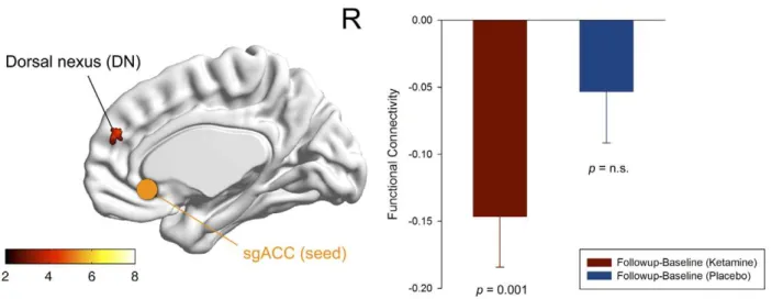
At the whole brain level, we observed a statistical trend for a reduction in functional connectivity between the sgACC seed region and the right dorsal medial prefrontal cortex (DMPFC) following ketamine administration compared to placebo (n = 17, paired t-test: puncorr,0.001, extent threshold of k.13, resulting in a cluster-level pcorr,0.1; Fig. 4). The difference in mean Fisher z-transformed correlation values extracted from the corresponding seed (sgACC) and projection region (DMPFC) was significant for the ketamine condition (n = 17, paired t-test (baseline-follow-up): p = 0.001), with no significant change after placebo administration (Fig. 4 and 5).
Other networks
No focal changes in functional connectivity with DLPFC were found in the whole brain paired t-test thresholded at puncorr,0.001 (extent threshold of k.15, resulting in a cluster-level pcorr,0.05) following ketamine administration. Likewise, no significant differ-ences in functional connectivity between amygdala and prefrontal cortical areas could be observed after drug administration. Additional reductions in functional connectivity were found between amygdala and posterior parietal and premotor areas but the pattern was similar for both the drug and placebo condition.
Whole brain dorsal nexus connectivity
In order to assess the specificity of our findings, we created an additional ’’dorsal nexus ‘‘seed (x: 6, y: 51, z: 24, volume:
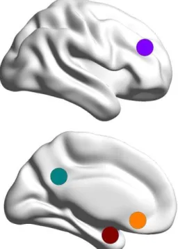
Figure 2. Seed region selection. Each of the four solid circles corresponds to a seed region in the Cognitive Control Network (CCN; purple): dorsolateral prefrontal cortex (DLPFC); in the Default Mode Network (DMN; green): posterior cingulate cortex (PCC); in the Affective Network (AN; orange): subgenual anterior cingulate cortex (sgACC); and in the amygdala (red).
513 mm3) based on the overlapping voxels that showed significant changes in functional connectivity to both the posterior and subgenual cingulate cortices after ketamine administration. At the whole brain level (using the same threshold as above), functional connectivity was reduced exclusively to the PCC but nowhere else in the brain.
Discussion
While pharmacological effects of ketamine on task-induced fMRI BOLD signals have been studied extensively [56–62], this is the first randomized, placebo-controlled, double-blind, crossover study demonstrating changes in resting state functional
connec-tivity in response to ketamine administration in healthy subjects. As our key finding we report a marked reduction of resting state functional connectivity between functional nodes of the default mode network (PCC) via the dorsal nexus (DN), pregenual anterior cingulate (PACC), and medioprefrontal cortex (MPFC) in healthy subjects 24 hours after ketamine administration compared to placebo. The term ’’dorsal nexus’’ was created recently by Sheline and colleagues (2010) to describe a functional node in the bilateral DMPFC with dramatically increased resting state connectivity to three important functional networks - the CCN, DMN, and AN - in patients suffering from major depression [46]. In our study, we aimed to model and identify ketamine-associated adaptations in healthy subjects within neural circuits that are
Figure 3. Functional connectivity of the default mode network (DMN).Significant voxels of the dorsal nexus (DMPFC) and pregenual anterior cingulate cortex (PACC) and medial prefrontal cortex (MPFC) showing reduced functional connectivity to the left posterior cingulate cortex (PCC) seed region (green) 24 hours after ketamine administration (n = 17, whole brain paired t-test: baseline(ketamine)-follow-up(ketamine); puncorr,0.001, extent threshold of k.15, corresponds to a cluster-level pcorr,0.05). The color bar indicates z values. The bar diagrams represent the change in functional connectivity (Fisher z-transformed correlation values) of the dorsal nexus (left) and the MPFC/PACC (right) to the left PCC from baseline to follow-up for the ketamine (red) and placebo condition (blue) (n = 17, paired t-test: p,0.001; error bars = s.e.m.).
relevant to the pathophysiology of MDD. Thus, the observed decrease in functional connectivity via the DN following ketamine administration in healthy subjects might have some implications for its therapeutic action in MDD patients. In light with the peak of ketamine’s antidepressant effect 24 hours after intravenous administration [4], our findings suggest that this effect may be mediated by reducing the hyperconnectivity of the DN as shown here. Importantly, this action differs from previously reported effects of acute administration.
Antagonism at NMDA receptors has been shown to induce behavioral and neuroplastic changes in animal models relevant to certain aspects of the pathophysiology of depressive disorders [8– 10]. The changes in resting state connectivity that we observed 24 hours post-infusion might thus result from adaptive changes in neuroglial glutamatergic throughput, neuroplasticity and informa-tion processing in specific neurocircuits. In strong support for such
a glutamatergic mechanism of action a recent study reported a direct relationship between aberrant resting state functional connectivities and glutamatergic imbalance in depressed patients across distinct functional networks [35]. This supports our hypothesis that glutamatergic modulation by specific drugs like ketamine exerts its antidepressant effects via reconfiguration of resting state functional connectivity.
The psychophysiological relevance of reducing functional hyperconnectivities within and between resting state networks like the DMN or the AN is given by their involvement in circumscribed aspects of the depressive psychopathology. Regions of the DMN commonly show the greatest activity at rest and decrease their level of activity during goal-directed tasks [37] and are thought to be involved in self-referential processes such as introspection, remembering, and planning [63]. In patients with major depression, a failure to normally down-regulate activity
Figure 4. Functional connectivity of the affective network (AN).Significant voxels of the dorsal nexus (DMPFC) showing reduced functional connectivity to the subgenual anterior cingulate cortex (sgACC) seed region (blue) 24 hours after ketamine administration (n = 17, whole brain paired t-test: baseline(ketamine)-follow-up(ketamine); puncorr,0.001, extent threshold of k.13, corresponds to a cluster-level pcorr,0.1 indicating trend-level significance). The color bar indicates z values. The bar diagram represents the change in functional connectivity (Fisher z-transformed correlation values) of the dorsal nexus to the sgACC from baseline to follow-up for the ketamine (red) and placebo condition (blue) (n = 17, paired t-test: p = 0.001; error bars = s.e.m.).
doi:10.1371/journal.pone.0044799.g004
Figure 5. Functional connectivity across the whole experiment.The bar diagrams represent the functional connectivity (Fisher z-transformed correlation values after global mean regression) for the following experimental conditions: baseline and follow-up (ketamine; red); baseline and follow-up (placebo; blue). From left to right: Dorsal nexus (DN) connectivity to the left posterior cingulate cortex (PCC); medioprefrontal cortex (MPFC) and pregenual anterior cingulate cortex (PACC) connectivity to the left PCC; subgenual anterior cingulate cortex (sgACC) connectivity to the DN (n = 17, paired t-tests; error bars = s.e.m.).
within the DMN during external stimulation was found [38,39], with increasing levels of DMN dominance being associated with higher levels of maladaptive, depressive rumination and lower levels of adaptive, reflective rumination [64]. Thus, the reduction in functional connectivity between anterior (PACC/MPFC) and posterior parts of the DMN (PCC) that we observed after ketamine administration in healthy subjects may have implications for antidepressant treatment in terms of a reduction of the increased level of DMN dominance (s. Fig. 6).
Moreover, the sgACC as a critical hub of the AN plays an important role in emotion processing and the pathogenesis of mood disorders and has become a promising target for deep brain stimulation in patients with severe, refractory depression [65]. A number of structural, metabolic and functional abnormalities has been identified in the sgACC of MDD patients [66]. Resting state sgACC functional connectivity with the DMN was significantly greater in depressed subjects and correlated positively with the length of the current depressive episode [44]. As proposed by Sheline et al. (2010) an attentional shift with increased self-focus might interfere with task performance in the CCN through increased resting state DMN connectivity with the DN [46]. The hot-wiring of the sgACC to those systems might further explain its maladaptive contribution to negative self-monitoring and reduced task-performance in MDD, given its role in the regulation of visceral functions and sad mood [66]. Compared to the reduction in DMN to DN connectivity after ketamine administration in healthy subjects, the reduction of AN to DN connectivity was less pronounced reaching statistical trend-level only and has therefore to be considered preliminary. The absence of a pre-existing hyperconnectivity of the sgACC to the DN in healthy subjects might explain the limited dynamic range in terms of a reduction in functional connectivity in our study, while this mechanism may become relevant in a clinical population (s. Fig. 6).
Our findings suggest that intravenous ketamine in healthy subjects affects primarily the DMN (PCC) connectivity via the DN and PACC/MPFC one day after infusion. We could not find any focal change in connectivity to the CCN following ketamine administration and contrary to resting state studies with seroto-nergic and noradreseroto-nergic antidepressants including citalopram and reboxetine [48], functional connectivity of the prefrontal cortex to the amygdala remained unaffected by ketamine. Hence, the circumscribed effect of ketamine on DMN connectivity to the DN supports the hypothesis that effective antidepressant treatment involves systematic alterations in connections among higher-order functional networks via nodes such as the DN. However, those putative implications for MDD have to be regarded as preliminary since the results reported here are based on healthy subjects. Apart from this limitation, our aim of addressing systems level mechanisms of ketamine’s antidepressant action is reflected in our elaborate study design including a 24 h post-infusion interval, appropriate dosage and duration of the ketamine infusion, and the selection of seed regions that are relevant to MDD. Therefore, our findings may serve as a model to elucidate potential biomechan-isms of drug action in the absence of any pre-existing homeostatic dysregulation as part of the disease process, medication status, or comorbidity. In a next step, the explanatory power of our observation has to be further confirmed in a randomized-controlled clinical trial in MDD patients receiving ketamine. Moreover, our results do not allow any conclusions to be drawn for the action of ketamine on the healthy human brain in general or in the context of ketamine as a model for schizophrenia.
In conclusion, we report a reduction of functional connectivity in networks that play a critical role in the pathophysiology of MDD in healthy subjects 24 hours after receiving an antidepres-sant dose of ketamine. Based on those findings we raise the hypothesis that reducing functional connectivity of the dorsal nexus reflects underlying molecular mechanisms relevant to the antidepressant efficacy of ketamine. Whether this circuit-level glutamatergic effect is likely to be associated with reversing aspects of emotional and behavioral dysregulation has to be further investigated in a clinical study involving MDD patients. This is in further support of the notion of using ketamine as a research tool into the neurobiology of mood disorders and to delineate potential biomarkers and action mechanisms of antidepressant treatment response.
Acknowledgments
We thank Rosilla Bachmann and Ulrich Heidecke for their support with ketamine administration, Philipp Staempfli and Jutta Ernst for their assistance with MR scanning, and Meng Li for his help with data analysis.
Author Contributions
Conceived and designed the experiments: MS MW SG HB PB AH ES. Performed the experiments: MS ML. Analyzed the data: MS MW CM. Wrote the paper: MS MW CM SG AH ES.
References
1. Sanacora G, Zarate CA, Krystal JH, Manji HK (2008) Targeting the glutamatergic system to develop novel, improved therapeutics for mood disorders. Nat Rev Drug Discov 7: 426–437. doi:10.1038/nrd2462
2. Vollenweider FX, Kometer M (2010) The neurobiology of psychedelic drugs: implications for the treatment of mood disorders. Nat Rev Neurosci 11: 642– 651. doi:10.1038/nrn2884
3. Berman RM, Cappiello A, Anand A, Oren DA, Heninger GR, et al. (2000) Antidepressant effects of ketamine in depressed patients. Biol Psychiatry 47: 351–354.
4. Zarate CA, Singh JB, Carlson PJ, Brutsche NE, Ameli R, et al. (2006) A randomized trial of an N-methyl-D-aspartate antagonist in treatment-resistant
Figure 6. Proposed hypothetical model of ketamine-associated changes in functional connectivity.In the healthy human brain, a single antidepressant dose of ketamine reduces functional connectivity of the dorsal nexus (DN) to the Default Mode Network (DMN; green). The reduction in functional connectivity of the Affective Network (AN; orange) to the DN reached trend-level significance only (s. Fig. 4), possibly due to the absence of any pre-existing hyperconnectivity in healthy subjects. This action may serve as a model for the discovery of novel antidepressant biomechanisms in major depression where functional connectivity of the DMN and AN via the DN is increased. The solid circles correspond to seed regions in the posterior cingulate cortex (PCC; green) and subgenual anterior cingulate cortex (sgACC; orange). The correspondingly colored open circles and dotted lines represent regions with decreased connectivity with the respective seed regions after ketamine administration. This hypothetical model is based on results from previous rsfMRI studies in MDD patients and on data obtained in healthy subjects after ketamine administration and needs to be further verified in MDD patients receiving ketamine.
major depression. Arch Gen Psychiatry 63: 856–864. doi:10.1001/arch-psyc.63.8.856
5. Phelps LE, Brutsche N, Moral JR, Luckenbaugh DA, Manji HK, et al. (2009) Family history of alcohol dependence and initial antidepressant response to an N-methyl-D-aspartate antagonist. Biol Psychiatry 65: 181–184. doi:10.1016/ j.biopsych.2008.09.029
6. Aan Het Rot M, Collins KA, Murrough JW, Perez AM, Reich DL, et al. (2010) Safety and efficacy of repeated-dose intravenous ketamine for treatment-resistant depression. Biol Psychiatry 67: 139–145. doi:10.1016/j.biopsych.2009.08.038 7. Machado-Vieira R, Salvadore G, Diazgranados N, Zarate CA (2009) Ketamine
and the next generation of antidepressants with a rapid onset of action. Pharmacol Ther 123: 143–150. doi:10.1016/j.pharmthera.2009.02.010 8. Li N, Lee B, Liu RJ, Banasr M, Dwyer JM, et al. (2010) mTOR-Dependent
Synapse Formation Underlies the Rapid Antidepressant Effects of NMDA Antagonists. Science 329: 959–964. doi:10.1126/science.1190287
9. Autry AE, Adachi M, Nosyreva E, Na ES, Los MF, et al. (2011) NMDA receptor blockade at rest triggers rapid behavioural antidepressant responses. Nature 475: 91–95. doi:10.1038/nature10130
10. Li N, Liu R-J, Dwyer JM, Banasr M, Lee B, et al. (2011) Glutamate N-methyl-D-aspartate receptor antagonists rapidly reverse behavioral and synaptic deficits caused by chronic stress exposure. Biol Psychiatry 69: 754–761. doi:10.1016/ j.biopsych.2010.12.015
11. Maeng S, Zarate CA, Du J, Schloesser RJ, McCammon J, et al. (2008) Cellular mechanisms underlying the antidepressant effects of ketamine: role of alpha-amino-3-hydroxy-5-methylisoxazole-4-propionic acid receptors. Biol Psychiatry 63: 349–352. doi:10.1016/j.biopsych.2007.05.028
12. Yu¨ksel C, Ongu¨r D (2010) Magnetic resonance spectroscopy studies of glutamate-related abnormalities in mood disorders. Biol Psychiatry 68: 785– 794. doi:10.1016/j.biopsych.2010.06.016
13. Cotter D, Mackay D, Landau S, Kerwin R, Everall I (2001) Reduced glial cell density and neuronal size in the anterior cingulate cortex in major depressive disorder. Arch Gen Psychiatry 58: 545–553.
14. Chana G, Landau S, Beasley C, Everall IP, Cotter D (2003) Two-dimensional assessment of cytoarchitecture in the anterior cingulate cortex in major depressive disorder, bipolar disorder, and schizophrenia: evidence for decreased neuronal somal size and increased neuronal density. Biol Psychiatry 53: 1086– 1098.
15. Choudary PV, Molnar M, Evans SJ, Tomita H, Li JZ, et al. (2005) Altered cortical glutamatergic and GABAergic signal transmission with glial involvement in depression. Proc Natl Acad Sci USA 102: 15653–15658. doi:10.1073/ pnas.0507901102
16. Bechtholt-Gompf AJ, Walther HV, Adams MA, Carlezon WA, Ongu¨r D, et al. (2010) Blockade of Astrocytic Glutamate Uptake in Rats Induces Signs of Anhedonia and Impaired Spatial Memory. Neuropsychopharmacology 35: 2049–2059. doi:10.1038/npp.2010.74
17. Walter M, Henning A, Grimm S, Schulte RF, Beck J, et al. (2009) The relationship between aberrant neuronal activation in the pregenual anterior cingulate, altered glutamatergic metabolism, and anhedonia in major depres-sion. Arch Gen Psychiatry 66: 478–486. doi:10.1001/archgenpsychia-try.2009.39
18. Keedwell PA, Andrew C, Williams SCR, Brammer MJ, Phillips ML (2005) The neural correlates of anhedonia in major depressive disorder. Biol Psychiatry 58: 843–853. doi:10.1016/j.biopsych.2005.05.019
19. Portella MJ, de Diego-Adelin˜o J, Go´mez-Anso´n B, Morgan-Ferrando R, Vives Y, et al. (2011) Ventromedial prefrontal spectroscopic abnormalities over the course of depression: a comparison among first episode, remitted recurrent and chronic patients. J Psychiatr Res 45: 427–434. doi:10.1016/j.jpsychires.2010. 08.010
20. Drevets WC, Price JL, Furey ML (2008) Brain structural and functional abnormalities in mood disorders: implications for neurocircuitry models of depression. Brain Struct Funct 213: 93–118. doi:10.1007/s00429-008-0189-x 21. Price JL, Drevets WC (2010) Neurocircuitry of mood disorders.
Neuropsycho-pharmacology 35: 192–216. doi:10.1038/npp.2009.104
22. Mayberg HS (1997) Limbic-cortical dysregulation: a proposed model of depression. J Neuropsychiatry Clin Neurosci 9: 471–481.
23. Holtzheimer PE, Mayberg HS (2011) Stuck in a rut: rethinking depression and its treatment. Trends Neurosci 34: 1–9. doi:10.1016/j.tins.2010.10.004 24. Stone JM, Dietrich C, Edden R, Mehta MA, De Simoni S, et al. (2012)
Ketamine effects on brain GABA and glutamate levels with 1H-MRS: relationship to ketamine-induced psychopathology. Mol Psychiatry. doi:10.1038/mp.2011.171
25. Kim S-Y, Lee H, Kim H-J, Bang E, Lee S-H, et al. (2011) In vivo and ex vivo evidence for ketamine-induced hyperglutamatergic activity in the cerebral cortex of the rat: Potential relevance to schizophrenia. NMR Biomed: n/a–n/a. doi:10.1002/nbm.1681
26. Rowland LM, Bustillo JR, Mullins PG, Jung RE, Lenroot R, et al. (2005) Effects of ketamine on anterior cingulate glutamate metabolism in healthy humans: a 4-T proton MRS study. Am J Psychiatry 162: 394–396. doi:10.1176/ appi.ajp.162.2.394
27. Lorrain DS, Baccei CS, Bristow LJ, Anderson JJ, Varney MA (2003) Effects of ketamine and N-methyl-D-aspartate on glutamate and dopamine release in the rat prefrontal cortex: modulation by a group II selective metabotropic glutamate receptor agonist LY379268. NSC 117: 697–706.
28. Moghaddam B, Adams B, Verma A, Daly D (1997) Activation of glutamatergic neurotransmission by ketamine: a novel step in the pathway from NMDA receptor blockade to dopaminergic and cognitive disruptions associated with the prefrontal cortex. J Neurosci 17: 2921–2927.
29. Alt A, Nisenbaum ES, Bleakman D, Witkin JM (2006) A role for AMPA receptors in mood disorders. Biochem Pharmacol 71: 1273–1288. doi:10.1016/ j.bcp.2005.12.022
30. Martinowich K, Manji H, Lu B (2007) New insights into BDNF function in depression and anxiety. Nat Neurosci 10: 1089–1093. doi:10.1038/nn1971 31. Garcia LSB, Comim CM, Valvassori SS, Re´us GZ, Barbosa LM, et al. (2008)
Acute administration of ketamine induces antidepressant-like effects in the forced swimming test and increases BDNF levels in the rat hippocampus. Prog Neuropsychopharmacol Biol Psychiatry 32: 140–144. doi:10.1016/ j.pnpbp.2007.07.027
32. Valentine GW, Sanacora G (2009) Targeting glial physiology and glutamate cycling in the treatment of depression. Biochem Pharmacol 78: 431–439. doi:10.1016/j.bcp.2009.04.008
33. Northoff G, Walter M, Schulte RF, Beck J, Dydak U, et al. (2007) GABA concentrations in the human anterior cingulate cortex predict negative BOLD responses in fMRI. Nature Publishing Group 10: 1515–1517. doi:10.1038/ nn2001
34. Muthukumaraswamy SD, Edden RAE, Jones DK, Swettenham JB, Singh KD (2009) Resting GABA concentration predicts peak gamma frequency and fMRI amplitude in response to visual stimulation in humans. Proceedings of the National Academy of Sciences 106: 8356–8361. doi:10.1073/pnas.0900728106 35. Horn DI, Yu C, Steiner J, Buchmann J, Kaufmann J, et al. (2010) Glutamatergic and resting-state functional connectivity correlates of severity in major depression - the role of pregenual anterior cingulate cortex and anterior insula. Front Syst Neurosci 4. doi:10.3389/fnsys.2010.00033
36. Duncan NW, Enzi B, Wiebking C, Northoff G (2011) Involvement of glutamate in rest-stimulus interaction between perigenual and supragenual anterior cingulate cortex: A combined fMRI-MRS study. Hum Brain Mapp: n/a–n/a. doi:10.1002/hbm.21179
37. Raichle ME, MacLeod AM, Snyder AZ, Powers WJ, Gusnard DA, et al. (2001) A default mode of brain function. Proc Natl Acad Sci USA 98: 676–682. doi:10.1073/pnas.98.2.676
38. Grimm S, Boesiger P, Beck J, Schuepbach D, Bermpohl F, et al. (2009) Altered negative BOLD responses in the default-mode network during emotion processing in depressed subjects. Neuropsychopharmacology 34: 932–843. doi:10.1038/npp.2008.81
39. Sheline YI, Barch DM, Price JL, Rundle MM, Vaishnavi SN, et al. (2009) The default mode network and self-referential processes in depression. Proceedings of the National Academy of Sciences 106: 1942–1947. doi:10.1073/ pnas.0812686106
40. Salvadore G, Cornwell BR, Colon-Rosario V, Coppola R, Grillon C, et al. (2009) Increased anterior cingulate cortical activity in response to fearful faces: a neurophysiological biomarker that predicts rapid antidepressant response to ketamine. Biol Psychiatry 65: 289–295. doi:10.1016/j.biopsych.2008.08.014 41. Fox MD, Greicius M (2010) Clinical applications of resting state functional
connectivity. Front Syst Neurosci 4: 19. doi:10.3389/fnsys.2010.00019 42. Friston K (2011) Functional and Effective Connectivity: A Review. Brain
Connect 1: 1–24. doi:10.1089/brain.2011.0008
43. Anand A, Li Y, Wang Y, Wu J, Gao S, et al. (2005) Activity and connectivity of brain mood regulating circuit in depression: a functional magnetic resonance study. BPS 57: 1079–1088. doi:10.1016/j.biopsych.2005.02.021
44. Greicius MD, Flores BH, Menon V, Glover GH, Solvason HB, et al. (2007) Resting-state functional connectivity in major depression: abnormally increased contributions from subgenual cingulate cortex and thalamus. Biol Psychiatry 62: 429–437. doi:10.1016/j.biopsych.2006.09.020
45. Zhou Y, Yu C, Zheng H, Liu Y, Song M, et al. (2010) Increased neural resources recruitment in the intrinsic organization in major depression. J Affect Disord 121: 220–230. doi:10.1016/j.jad.2009.05.029
46. Sheline YI, Price JL, Yan Z, Mintun MA (2010) Resting-state functional MRI in depression unmasks increased connectivity between networks via the dorsal nexus. Proceedings of the National Academy of Sciences 107: 11020–11025. doi:10.1073/pnas.1000446107
47. Mccabe C, Mishor Z, Filippini N, Cowen PJ, Taylor MJ, et al. (2011) SSRI administration reduces resting state functional connectivity in dorso-medial prefrontal cortex. Mol Psychiatry 16: 592–594. doi:10.1038/mp.2010.138 48. McCabe C, Mishor Z (2011) Antidepressant medications reduce
subcortical-cortical resting-state functional connectivity in healthy volunteers. NeuroImage 57: 1317–1323. doi:10.1016/j.neuroimage.2011.05.051
49. Sinner B, Graf BM (2008) Ketamine. Handb Exp Pharmacol: 313–333. doi:10.1007/978-3-540-74806-9_15
50. Dittrich A (1998) The standardized psychometric assessment of altered states of consciousness (ASCs) in humans. Pharmacopsychiatry 31 Suppl 2: 80–84. doi:10.1055/s-2007-979351
51. Spielberger CD, Gorsuch RL, Edward LR (1970) STAI manual for the state-trait anxiety inventory (‘‘Self-evaluation questionnaire’’).
53. Chao-Gan Y, Yu-Feng Z (2010) DPARSF: A MATLAB Toolbox for ‘‘Pipeline’’ Data Analysis of Resting-State fMRI. Front Syst Neurosci 4: 13. doi:10.3389/ fnsys.2010.00013
54. Song X-W, Dong Z-Y, Long X-Y, Li S-F, Zuo X-N, et al. (2011) REST: a toolkit for resting-state functional magnetic resonance imaging data processing. PLoS ONE 6: e25031. doi:10.1371/journal.pone.0025031
55. Tzourio-Mazoyer N, Landeau B, Papathanassiou D, Crivello F, Etard O, et al. (2002) Automated anatomical labeling of activations in SPM using a macroscopic anatomical parcellation of the MNI MRI single-subject brain. NeuroImage 15: 273–289. doi:10.1006/nimg.2001.0978
56. Musso F, Brinkmeyer J, Ecker D, London MK, Thieme G, et al. (2011) Ketamine effects on brain function - Simultaneous fMRI/EEG during a visual oddball task. NeuroImage 58: 508–525. doi:10.1016/j.neuroimage.2011.06.045 57. Nagels A, Kirner-Veselinovic A, Krach S, Kircher T (2011) Neural correlates of S-ketamine induced psychosis during overt continuous verbal fluency. Neuro-Image 54: 1307–1314. doi:10.1016/j.neuroimage.2010.08.021
58. Abel KM, Allin MPG, Kucharska-Pietura K, Andrew C, Williams S, et al. (2003) Ketamine and fMRI BOLD signal: distinguishing between effects mediated by change in blood flow versus change in cognitive state. Hum Brain Mapp 18: 135–145. doi:10.1002/hbm.10064
59. Abel KM, Allin MPG, Kucharska-Pietura K, David A, Andrew C, et al. (2003) Ketamine alters neural processing of facial emotion recognition in healthy men: an fMRI study. Neuroreport 14: 387–391. doi:10.1097/01.wnr.0000058031. 29600.31
60. Honey RAE, Honey GD, O’Loughlin C, Sharar SR, Kumaran D, et al. (2004) Acute ketamine administration alters the brain responses to executive demands in a verbal working memory task: an FMRI study. Neuropsychopharmacology 29: 1203–1214. doi:10.1038/sj.npp.1300438
61. Northoff G, Richter A, Bermpohl F, Grimm S, Martin E, et al. (2005) NMDA hypofunction in the posterior cingulate as a model for schizophrenia: an exploratory ketamine administration study in fMRI. Schizophr Res 72: 235– 248. doi:10.1016/j.schres.2004.04.009
62. Fu CHY, Abel KM, Allin MPG, Gasston D, Costafreda SG, et al. (2005) Effects of ketamine on prefrontal and striatal regions in an overt verbal fluency task: a functional magnetic resonance imaging study. Psychopharmacology 183: 92– 102. doi:10.1007/s00213-005-0154-9
63. Buckner RL, Andrews-Hanna JR, Schacter DL (2008) The brain’s default network: anatomy, function, and relevance to disease. Ann N Y Acad Sci 1124: 1–38. doi:10.1196/annals.1440.011
64. Hamilton JP, Furman DJ, Chang C, Thomason ME, Dennis E, et al. (2011) Default-Mode and Task-Positive Network Activity in Major Depressive Disorder: Implications for Adaptive and Maladaptive Rumination. Biol Psychiatry 70: 327–333. doi:10.1016/j.biopsych.2011.02.003
65. Mayberg HS, Lozano AM, Voon V, McNeely HE, Seminowicz D, et al. (2005) Deep brain stimulation for treatment-resistant depression. Neuron 45: 651–660. doi:10.1016/j.neuron.2005.02.014