ASSOCIAÇÃO ENTRE OS POLIMORFISMOS DOS
GENES GSTM1 E GSTT1 E A LEUCOPLASIA BUCAL
ELIZA CARLA BARROSO DUARTE
ASSOCIAÇÃO ENTRE OS POLIMORFISMOS DOS GENES
GSTM1 E GSTT1 E A LEUCOPLASIA BUCAL
Dissertação apresentada ao Curso de Mestrado em Odontologia, área de Concentração Patologia Bucal, da Faculdade de Odontologia da Universidade Federal de Minas Gerais, como requisito parcial para a obtenção do grau de Mestre em Odontologia, área de Patologia Bucal
Orientador: Prof. Dr. Ricardo Santiago Gomez
Pai,
AGRADECIMENTOS
A DEUS razão do meu viver.
À minha mãe, por me amar e me apoiar nessa conquista.
À minha querida irmã Aninha, pela amizade e por estar sempre ao meu lado.
À minha cunhada Michele por ser minha grande amiga e por me ajudar sempre. Ao Dé e ao João por serem importantes em minha vida.
À Déia e Deza, minhas queridas primas-irmãs, pela amizade e por serem pessoas fundamentais em minha vida.
À minha querida madrinha Marina pela amizade.
À minha avó Pequena, meus tios Fátima, André, Graça, José Geraldo, Afonso, Naia e aos primos Bianca, Paola, Renato e Rui por serem especiais em minha vida.
Ao Leandro, pelo amor, carinho e confiança.
Ao professor Ricardo Santiago Gomez, pela orientação, disponibilidade e por abrir as portas do laboratório para que eu buscasse esta conquista.
Às professoras Cássia e Dorinha pela amizade, carinho, apoio.
Ao professor Ricardo Mesquita pelo apoio, disponibilidade e ensinamentos.
Ao professor Vagner Santos pela atenção no início da minha experiência didática.
Ao grande amigo Júlio Lacerda pela convivência, confiança e por ajudar em meu crescimento profissional.
As amigas Daniela Pacheco, Christiane Salgado e Larissa Santoro pela amizade verdadeira.
Ao amigo Luiz Henrique Lage pelo carinho, oportunidade e confiança profissional.
Aos colegas da Clinidonto pela excelente convivência e por torcerem por mim.
Aos colegas do laboratório de patologia bucal Jeane, Júnia, Alessandra, Carolina, Luciano, Flávio, André, Wagner Castro, e ao colega de mestrado Bruno Jhan.
Aos queridos alunos de iniciação científica Marcus e Marina por me ajudarem nos trabalhos laboratoriais.
Às funcionárias Inês, Heloísa e Cristiane e aos funcionários do Hospital Municipal Odilon Behrens pela ajuda profissional.
Aos meus alunos de graduação pelo carinho e confiança.
Aos pacientes por contribuírem para que este trabalho fosse realizado.
LISTA DE ABREVIAÇÕES
A Adenina
C Citosina
°C Graus Celsius
C6H12O6 Glicose
CYP450 Citocromo p-450
CYP1A1 Gene citocromo P-450, subfamília 1A,
polipeptídeo 1
CYP2E1 Gene citocromo P-450, subfamília 2E,
polipeptídeo 1
DNA Ácido desoxirribonucléico
DNTPs Desóxi-nucleotídeos trifosfato
G Guanina
GST Glutationa S-transferase
GSTM1 Gene glutationa S-transferase classe μ GSTT1 Gene glutationa S-transferase classe θ
KCl Cloreto de potássio
mM Mili molar
MgSO4 Sulfato de magnésio
MgCl2 Cloreto de magnésio
NaCl Cloreto de sódio
OR Odds ratio
pb Pares de base
PCR Reação em cadeia da polimerase
RNA Ácido ribonucléico
T Timina
Taq Thermus aquaticus DNA polimerase
Tris Tris-hidroximetilaminometano
LISTA DE TABELAS
Página
Tabela 1- Freqüência dos genótipos do gene GSTM1 nos grupos 29
casos e controles. Distribuição em relação à graduação
histológica e localização. Artigo GSTM1 e leucoplasia
bucal.
Tabela 1- Freqüência dos genótipos do gene GSTM1 nos grupos 40
casos e controles. Distribuição em relação à graduação
histológica e localização. Artigo GSTT1 e a leucoplasia
RESUMO
A leucoplasia bucal é a lesão cancerizável mais comum da cavidade
bucal e apresenta como principal fator de risco o consumo de tabaco em suas
várias formas. As substâncias carcinogênicas do tabaco são metabolizadas por
enzimas específicas em duas etapas denominadas fases I e II. Estas enzimas
apresentam polimorfismos genéticos que influenciam na susceptibilidade individual
a lesões cancerizáveis e ao câncer. A associação entre estes polimorfismos
genéticos e o câncer é conflitante. O objetivo deste estudo foi investigar a
associação entre a deleção dos genes GSTM1 e GSTT1 e a leucoplasia bucal em
pacientes fumantes em uma população brasileira. Neste estudo do tipo
caso-controle foi extraído DNA de 52 pacientes fumantes com o diagnóstico clínico de
leucoplasia bucal e histopatológico de hiperqueratose com ou sem displasia epitelial
(grupo caso) e 52 indivíduos fumantes sem nenhuma patologia bucal (grupo
controle) a partir de raspados da mucosa clinicamente normal. A análise dos
genótipos dos genes foi realizada através da reação em cadeia da polimerase e os
produtos foram visualizados através da eletroforese em gel de poliacrilamida a 6,5%
e coloração pela prata. Os resultados demonstraram associação entre a deleção
SUMÁRIO
PÁGINA
1. INTRODUÇÃO 11
1.1 LEUCOPLASIA BUCAL 12
1.2 O METABOLISMO DE SUBSTÂNCIAS CARCINOGÊNICAS 14
1.3 POLIMORFISMOS GENÉTICOS 15
1.4 FAMÍLIA DAS GLUTATIONAS S-TRANSFERASES 15
2. ARTIGO-GSTM1 E LEUCOPLASIA BUCAL 18
3.ARTIGO-GSTT1 E LEUCOPLASIA BUCAL 31
4. CONSIDERAÇÕES FINAIS 43
5. CONCLUSÕES 48
6. REFERÊNCIAS BIBLIOGRÁFICAS 50
7. ANEXOS 56
ANEXO A- ELETROFORESE EM GEL DE POLIACRILAMIDA A 6,5%-GENE GSTM1 57
1 INTRODUÇÃO
1.1 LEUCOPLASIA BUCAL
A leucoplasia bucal é definida como “uma placa predominantemente
branca na mucosa bucal que não pode ser caracterizada clinicamente ou
patologicamente como outra doença” (KRAMER et al., 1978; VAN DER WAAL &
AXÉLL, 2002). O diagnóstico final desta doença somente pode ser feito após
avaliação histológica e, quando necessário, outros exames laboratoriais
complementares (GOMEZ, 2005).1 A leucoplasia bucal é a lesão cancerizável mais
comum da cavidade bucal, no entanto o epitélio displásico ou carcinoma invasivo é
encontrado apenas em 5% a 25% dos casos. A probabilidade de transformação
maligna varia nas diferentes formas de leucoplasia e algumas características como
presença de displasia epitelial, aparência clínica e localização da lesão aumentam
esse risco (VAN DER WAAL & AXÉLL, 2002). O risco anual de transformação
maligna das leucoplasias não excede 1% (SCHEIFELE & REICHART, 2003).
A leucoplasia bucal apresenta uma predileção por pacientes do sexo
masculino, (70%) e acima dos 40 anos de idade (MEHTA et al.,1981). Esta doença
pode ocorrer em qualquer mucosa de revestimento da cavidade bucal. As lesões
localizadas no soalho bucal, palato mole e língua são caracterizadas como de alto
risco, enquanto lesões em outros sítios são consideradas como de baixo risco
(ZHANG et al., 2001). Clinicamente podem ter aspecto homogêneo,
apresentando-se como uma lesão branca, uniforme, superfície plana, aparência fina podendo
1
exibir fendas rasas, ou possuir um padrão não-homogêneo, caracterizado por uma
lesão branca ou branca-vermelha, podendo assumir a forma nodular ou verrucosa
(AXÉLL et al.,1996). As leucoplasias podem ser únicas ou múltiplas na mucosa
bucal (VAN DER WALL & AXÉLL, 2002).
Os aspectos histológicos da leucoplasia bucal são caracterizados pela
presença de uma espessa camada de queratina na superfície do epitélio
(hiperqueratose) e/ou espessamento da camada espinhosa (acantose).
Frequentemente células inflamatórias crônicas estão presentes no tecido conjuntivo
subjacente. Displasia epitelial pode ser ou não observada. As alterações
histopatológicas das células displásicas são caracterizadas por pleomorfismo
nuclear e celular, hipercromatismo nuclear, nucléolos proeminentes e irregulares,
alteração na relação núcleo-citoplasma, aumento do número de figuras de mitose,
figuras de mitoses atípicas, alteração na polarização da camada basal, queratose
intraepitelial, perda de estratificação e projeções epiteliais em forma de gota. A
partir da presença dessas alterações morfo-citológicas, do nível de acometimento
das camadas epiteliais e da presença ou não de projeções epiteliais em forma de
gota, as lesões podem ser graduadas em: hiperqueratose com ou sem displasia
epitelial. A intensidade da displasia epitelial pode ser dividida em três tipos, discreta,
moderada ou acentuada. As lesões de hiperqueratose não apresentam alterações
morfo-citológicas. Nas lesões com displasia epitelial discreta nota-se a presença de
uma ou mais alterações morfo-citológicas, mas sem projeções epiteliais em gota.
Em lesões com displasia epitelial moderada observa-se a presença de uma ou mais
alterações morfo-citológicas acometendo até a metade da espessura do epitélio e
possuem uma ou mais alterações morfo-citológicas acometendo mais da metade da
espessura do epitélio e projeções epiteliais em gota (PAULA & GOMEZ, 2001).
O maior fator de risco para leucoplasia bucal é o consumo de tabaco em
suas várias formas (cigarro, tabaco mascado) e o uso de pasta de betel (JHONSON
et al., 1996; NEVILLE & DAY, 2002). Numerosos estudos têm demonstrado uma forte associação entre leucoplasia bucal e o tabaco, confirmada pela relação
dose-resposta (GUPTA, 1984; EVSTIFEEVA & ZARIDZE, 1992; LEE et al., 2003). Além
disso, a susceptibilidade ao carcinoma de células escamosas de cabeça e pescoço
tem sido associada a polimorfismos genéticos em genes que codificam enzimas
humanas relacionadas ao metabolismo de substâncias tóxicas e que influenciam na
susceptibilidade individual aos efeitos deletérios dos carcinógenos químicos (HAHN
et al., 2002).
1.2 O METABOLISMO DE SUBSTÂNCIAS CARCINOGÊNICAS
O tabaco contém mais de 50 compostos incluindo hidrocarbonetos
policíclicos aromáticos (PAHs), nitrosaminas específicas do tabaco, aminas
aromáticas e radicais livres que apresentam um importante papel na
carcinogênese, sendo os principais responsáveis pela transformação neoplásica do
epitélio bucal (SAPP et al., 1997; SCULLY et al., 2000).
O metabolismo de substâncias químicas exógenas é realizado em duas fases, denominadas I e II. A principais reações da fase I são hidrólise, redução e
oxidação realizadas pelo complexo enzimático citocromo P450 (CYP450) em que
eliminados diretamente ou sofrer ação das enzimas de fase II. As principais
reações de fase II são glicuronidação, sulfatação, metilação e conjugação
realizadas pela família das glutationas S-transferases, gerando compostos
hidrossolúveis e facilmente excretados (CONTRAN et al.,1999).
1.3 POLIMORFISMOS GENÉTICOS
Os polimorfismos genéticos são variações na sequência do DNA
caracterizados por substituição, deleção ou inserção de nucleotídeos e deleção ou
duplicação de genes. Estes polimorfismos ocorrem em uma freqüência de pelo
menos 1% da população normal. Estas variações podem ou não causar alterações
na função do produto protéico (WORMHOUDT et al., 1999; MILLER et al., 2001). A
eficácia na eliminação dos carcinógenos do tabaco pode ser influenciada por
polimorfismos nas enzimas metabolizadoras dessas substâncias. O grau de
expressão e a atividade dessas enzimas podem estar alterados e relacionar-se à
susceptibilidade individual a lesões cancerizáveis e ao câncer (NAIR et al., 1999;
1.4 FAMÍLIA DAS GLUTATIONAS-S-TRANSFERASES (GSTs)
As glutationas S-transferases são codificadas por uma superfamília de
genes que compreendem cinco grupos enzimáticos - ∝, π, θ, μ, Z (LAZARIUS et al.,
2000). Os compostos GST-conjugados são geralmente hidrofílicos e atóxicos sendo
facilmente excretados (WIENCKE et al., 1990).
O gene GSTM1, classe μ, responsável pela codificação da enzima
GSTM1 localiza-se no cromossomo 1q13.3. O gene GSTM1 apresenta um
polimorfismo genético caracterizado pela deleção total do gene (genótipo GSTM1
nulo ou 0/0), sendo que o indivíduo com genótipo nulo não apresenta atividade
enzimática (SREELEKHA et al., 2001).
Existe uma variação étnica e geográfica em relação à prevalência do
genótipo GSTM1 nulo. Na população brasileira a freqüência da deleção do gene
GSTM1 foi de 40,2% no estudo de AMORIN et al. (2000).
A associação entre a deleção do gene GSTM1 e carcinoma de células
escamosas de laringe, orofaringe e boca é controversa. Alguns estudos sugerem
que o polimorfismo do gene GSTM1 está relacionado à maior susceptibilidade a
estes carcinomas (SATO et al., 1999; NOMURA et al., 2000; SREELEKHA et al.,
2001; DRUMMOND et al., 2004; JHAVAR et al.2004), entretanto outros trabalhos
contradizem tal associação (PARK et al., 1997; MATTHIAS et al., 1998; AMADOR
et al., 2002). No estudo de Nair et al. (1999) uma correlação positiva foi encontrada
entre o genótipo GSTM1 nulo e leucoplasia bucal.
O gene GSTT1, classe Theta ou θ, se localiza no cromossomo 22q11.2 e
é responsável pela codificação da enzima GSTT1. O gene GSTT1 apresenta um
nulo ou 0/0), sendo que o indivíduo com genótipo nulo não apresenta atividade
enzimática (PEMBLE et al., 1994; WEBB et al., 1996; SREELEKHA et al., 2001).
As freqüências do genótipo GSTT1 nulo variam de acordo com a etnia e a
localização geográfica. Na população brasileira a freqüência da deleção do gene
GSTT1 foi de 25,0% no estudo de AMORIN et al. (2000).
A associação entre o polimorfismo do gene GSTT1 e carcinoma de
células escamosas de laringe, orofaringe e boca é controversa. Alguns estudos
sugerem que a deleção gene GSTT1 está relacionada à maior susceptibilidade a
carcinomas do trato aéreo digestivo superior (TRIZNA et al., 1995; SREELEKHA et
al., 2001; AMADOR et al., 2002; JHAVAR et al., 2003; DRUMMOND et al., 2005).
Outros trabalhos contradizem tal associação (GRONAU et al., 2003;
NAZAR-STEWART et al., 2003). A associação entre o genótipo GSTT1 nulo e o risco de
desenvolvimento de leucoplasia bucal em usuários de pasta de betel foi descrito por
Nair et al. (1999).
Considerando que o tabaco é o principal fator etiológico para o
desenvolvimento de câncer de boca e que a leucoplasia é a lesão cancerizável mais
prevalente, associado ao fato de que os polimorfismos dos genes GSTM1 e GSTT1
aumentam o risco para o carcinoma de células escamosas de boca, o objetivo
desse trabalho foi investigar a associação entre a deleção dos genes GSTM1 e
GSTT1 e a leucoplasia bucal em pacientes fumantes em uma população brasileira.
A seguir apresentamos os dois artigos enviados para publicação que abordam os
Title: GSTM1 polymorphism and oral leukoplakia
Authors:
Eliza Carla Barroso Duarte, DDS, Department of Oral Pathology and Surgery, School of
Dentistry, Universidade Federal de Minas Gerais.
Marina Sena Lopes da Silva, Undergraduate Student, Department of Oral Pathology
and Surgery, School of Dentistry, Universidade Federal de Minas Gerais,
Marcus Vinícius Gomez, PhD, Department of Pharmacology, Universidade Federal de
of Minas Gerais.
Ricardo Santiago Gomez. DDS, PhD, Department of Oral Surgery and Pathology,
School of Dentistry, Universidade Federal de Minas Gerais.
Correspondence to:
Prof. Ricardo Santiago Gomez Departmento de Patologia e Cirurgia Faculdade de Odontologia
Universidade Federal de Minas Gerais Av. Antonio Carlos, 6627
SUMMARY
We investigated the frequency of the GSTM1 genotypes in 104 tobacco smoking
subjects in a Brazilian population. The GSTM1 genotypes were studied by PCR-based
methods. The frequency of the GSTM1 null genotype in the group with oral leukoplakia
(57.7%) was statistically different from the controls (34.6%) (Odds ratio, O.R.=2.57,
95% C =1.16-5.69, P<0.05). The stratification of the samples according to the level of
dysplasia showed increased prevalence of GSTM1 null genotype on lesions with
moderate/severe histological dysplasia (68.2%) compared to the control group (31.9%).
This difference was statistically significant (O.R.=4.59, 95% CI=1.29-16.33, P<0.05). In
conclusion, the GSTM1 null genotype may increase the risk for oral leukoplakia
development.
INTRODUCTION
Oral leukoplakia is defined as “a chronic white mucosal patch which cannot
be characterized clinically or pathologically as any other disease”. 1-2 The development
of oral leukoplakia is strongly associated with exogenous exposure to carcinogens,
mainly smoking, chewing tobacco, and betel nut. The use of tobacco is the most
important, and has been present in 80% of the cases. 3-4 Oral leukoplakia shows
predilection for man (70%), generally up to 40 years old 5 and may occur as a single or multiple lesion. 6 Histologically, the leukoplakia may show mild, moderate or severe
dysplasia. 6-7 Although the presence of nodular, verrucous or erythroplastic, or severe
dysplasia may relate to increased risk for malignant transformation, these markers are
not useful in individual cases. The annual transformation rate of oral leukoplakia seems
not to exceed 1%. 8
Molecular epidemiological studies have now provided evidence that an individual
susceptibility to cancer is mediated by genetic and environmental factors. 9 Numerous
carcinogenic components present in the tobacco smoking require metabolic activation
by phase I enzymes (e.g. cytochrome P450 oxidases like CYP1A1 and CYP2E1) or
deactivation by phase II enzymes (e.g. glutathione transferase). Glutathione
S-transferase (GSTs) are a supergene family coding for five multigene enzyme groups
referred to as μ or M, θ or T, π, α and κ, which conjugate glutathione to genotoxic
eletrophiles such as polycyclic aromatic hydrocarbons, epoxybutanes, ethylene oxide,
and halomethanes. GST-conjugated compounds are generally rendered hydrophilic and
non-toxic, and are easily excreted. 10-11
Genetic polymorphisms have been described for enzymes involved in the
expression and/or activity of enzymes involved in carcinogen activation or
deactivation.11 The GSTM1 null polymorphism results in a loss of expression, resulting
in decreased ability to detoxify GST-μ-conjugated carcinogens.12 Considering that
tobacco is the main etiological factor in oral cancer and that oral leukoplakia is the most
prevalent potentially malignant lesion, together with the fact that GSTM1 polymorphism
increases the risk for oral squamous cell carcinoma13, the purpose of this study was to
investigate the GSTM1 null polymorphism and the risk for oral leukoplakia in individuals
with tobacco smoking habit in a Brazilian population.
MATERIALS AND METHODS
Subjects and sample collection
Fifth-two smokers patients (mean age = 47,9 years; range 25-87 years) with oral
leukoplakia and 52 healthy smokers volunteers (mean age = 48,6 years; range 29-81
years) were included in the study. The criteria for the diagnosis of oral leukoplakia were
previously reported. 2 All subjects were selected at the Dental Clinics of the School of
Dentistry and were of the same geographical region as well as socio-economic status.
The sex and age of both groups were matched. All subjects had smoked at least 10
cigarettes/day over a 20 years period. There were 31 males (59,6%) and 21 (40,4%)
females in the patient’s group. Ethnicity was not established as the hazards of judging
Brazilians by color, race and geographical origin. 14 The lesions located at the floor of
the mouth, tongue and soft palate were grouped together as high risk site group, while
all the oral sites were included in the group of low risk site. The grade of epithelial
dysplasia was established as described elsewhere. 15 The histopathological features of
than one layer of cells having a basaloid appearance, increased nuclear-cytoplasmic
ration, drop-shaped rete process, irregular epithelial stratification, increased number of
mitotic figures, presence of mitotic figures in the superficial half of the epithelium,
cellular pleomorphism, nuclear hyperchromatism, enlarged nucleoli, reduction of cellular
cohesion, and diskeratosis. 6 Local ethical committee approved the study protocol, and
informed consent was obtained from all patients.
Oral swabs were taken from each subject. The procedure was performed on the
contra-lateral intact mucosa of the patients with oral leukoplakia. The swabs from the
control subjects were collected from their labial mucosa. The swabs were performed
with sterile plastic tips and placed immediately in Eppendorf microtubes contain 500 μL
of Krebs buffer (NaCl 20%, KCl 2%, CaCl22H2O 2%, MgSO4, KH2PO4, C6H12O6). The
pellet obtained after 10 min of centrifugation at 17,900 x g was stored at –20°C until
processing. The DNA extraction was carried out as described by Boom et al.16
Polymerase chain reaction
The GSTM1 genotypes were studied by polymerase chain reaction (PCR) as previously
described. 17 The amplification was performed in a final volume of 25 μL containing 5 μL
of genomic DNA, 0,75 mM MgCl2, 40 mM KCl, 10 mM Tris HCl (ph=8,4) 0,1 % Triton
X-100, 11,4 μL H2O, 2,5 μL dNTPs and 2,5 U Taq DNA polymerase. Samples were
subjected to 5 min at 94°, followed by 35 cycles of amplification at 95° for 30 s, 64° for 1
min and 72° for 1 min. The run was terminated by a 7 min elongation step at 72°. A
negative control reaction without DNA and samples with known GSTM1 genotype were
always used. The reaction produced a 220bp product. Amplification of the β-globin gene
(PTC – Programmable Thermal Controller). The product was analyzed in a 6.5%
polyacrylamide gel electrophoresis followed by silver stain.
Statistical analysis
Statistical analysis was performed by Chi-square and Fisher’s tests; and significance
was set at p-value of < 0.05.
RESULTS
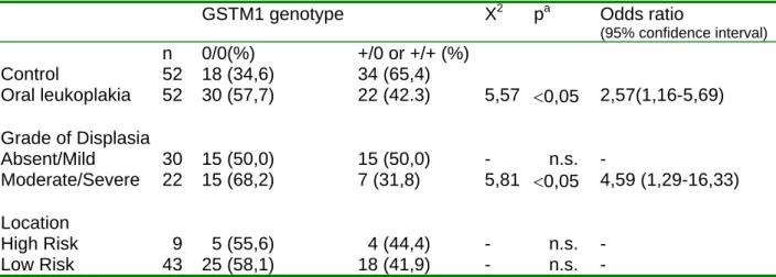
The frequencies of the GSTM1 genotypes between cases and controls are show in
Table 1. Individuals homozygous for the wild type GSTM1 (+/+) and heterozygous (+/0)
were grouped together. The frequency of the GSTM1 null genotype in the group with
oral leukoplakia (57.7%) was statistically different from the controls (34.6%) (Odds ratio,
O.R.=2.57, 95% C =1.16-5.69, P< 0.05). When the groups were stratified according to
the gender and site of occurrence, no statistical difference was observed. However, the
stratification of the samples according to the level of dysplasia showed increased
prevalence of GSTM1 null genotype on lesions with moderate/severe histological
dysplasia (68.2%) compared to the control group (31.9%). This difference was
DISCUSSION
Epidemiological studies have demonstrated a significant influence of tobacco and
alcohol consumption on the risk for oral cancer. Oral leukoplakia is the most common
potentially malignant lesion of the oral mucosa. Although tobacco use is related to oral
leukoplakia, alcohol consumption has not a significant association with it. 18 Since
exposure to chemical carcinogens acts as an important mechanism of oral cancer
development, the investigation of genetic polymorphisms of drug-metabolizing enzymes
and cancer susceptibility is of significant interest.
GSTM enzymes may offer protection against DNA damage induced by free
radicals and metabolites of polycyclic aromatic hydrocarbons.19 The lack of the GSTM1
activity is a result of a homozygous deletion (null genotype) of the GSTM1 gene. The
null genotype results in a loss of expression, resulting in decreased ability to detoxify
GST-μ-conjugated carcinogens. 11 The GSTM1 null genotypes (0/0) polymorphism has
been linked to increased susceptibility to oral squamous cell carcinoma development. 13, 17, 20, 21
. In addition, this polymorphism seems to be a risk factor for developing multiple
primary neoplasms in the upper aero-digestive tract. 22 In the present study, a positive
association between the GSTM1 null genotype and oral leukoplakia in Brazilian subjects
was observed. This polymorphism was also demonstrated to be a risk factor for
developing oral leukoplakia in ethnic Indian betel quid/tobacco chewers. 23
In our study, the frequencies of female or male patients in the case group who
were null for the GSTM1 genotype was not statistically different from the female or male
control group, respectively. No statistical difference was also observed when the
samples were stratified by the location of the lesion. These results may be due to the
increased the risk factor to lesions showing histological moderate/severe dysplasia. We
may speculate that the lack of GSTM1 activity would make the oral tissues more
susceptible to the action of tobacco carcinogens and to the development of a high grade
level of dysplasia. Longitudinal studies evaluating the impact of this polymorphism on
malignant transformation of oral leukoplakia would be of interest and could be an
interesting tool for targeting patients with oral leukoplakia at particular risk for future
cancerous lesion.
In conclusion, the present study shows a positive association between the
presence of GSTM1 null genotype and the development of oral leukoplakia. Indeed, this
association varies in accordance to the histological grade of dysplasia.
ACKNOWLEDGEMENT
This study was supported in part by grants from Fundação de Amparo à Pesquisa do
Estado de Minas Gerais (FAPEMIG), Programa de Excelência (PRONEX) and
Conselho Nacional de Desenvolvimento Científico e tecnológico (CNPq), Brazil. Dr.
REFERENCES
1. Kramer IRH, Lucas RB, Pindborg JJ, Sobin LH, WHO Collaborating Centre for
Oral Precancerous Lesions. Definition of leukoplakia and related lesions: an aid
to studies on oral precancer. Oral Surg Oral Med Oral Pathol 1978; 46:518-39.
2. Axéll T, Pindborg JJ, Smith CJ, Waal vander I, an International Collaborative
Group on Oral White Lesions. Oral white lesions with special reference to
precancerous and tabacco-related lesions: conclusions of an international
symposium held in Uppsala, Sweden, May 18-21,1994. J Oral Pathol Med 1996;
25:25-49.
3. Johnson NW; Warnakulasuriya S.; Tavassoli, M. Hereditary and eviromental risk
factors; clinical and laboratory risk matters for head and neck, especially oral,
cancer and precancer. Eur J Cancer 1996; 5(1):5-17.
4. Neville BW, Day TA. Oral cancer and precancerous lesions. CA Cancer J Clin
2002; 52(4):195-215.
5. Mehta FS; Gupta PC.; Pindborg JJ. Chewing and smoking habits in relation to
precancerous and oral cancer. Clin Oncol 1981; 99:35-9.
6. van der Waal I; Axéll T. Oral leukoplakia: a proposal for uniform reporting. Oral
Oncol 2002; 38:521-6.
7. Karabulut A, ReibelJ, Therkildsen MH, Praetorius f, Nielsen HW, Dabelsteen E.
Observer variability in the histologyc assessment of oral premalignant lesions. J
Oral Pathol Med 1995; 24:198-200.
8. Scheifele C, Reichart PA. Is there a natural limit of the transformation rate of oral
9. Sikdar N, Mahmud A, Paul RR, Roy B. Polymorphism in CYP1A1 and CYP2E1
genes and susceptibility to leukoplakia in Indian tobacco users. Cancer Letters
2003;195:33-42.
10. Perera FP, Weinstein IB. Molecular epidemiology: recent advances and future
directions. Carcinogenesis 2000; 21:517-24.
11. Lazarus P.; Park JY. Metabolizing enzyme genotype and risk for upper
aerodigestive tract cancer. Oral Oncol 2000; 36:421-31.
12. Wiencke JK, Kelsey KT, Lamela RA, Toscano Jr WA. Human glutathione
S-transferase deficiency as a marker of susceptibility to epoxide-induced
cytogenetic damage. Cancer Res 1990; 50:1585- 90.
13. Drummond SN, Noronha JCM, De Marco L, Gomez RS. GSTM-1 polymorphism
and oral squamous cell carcinoma. Oral Oncol 2004; 40:52-5.
14. Parra FC, Amado RC, Lambertucci JR, Rocha J, Antunes CM, Pena SD. Color
and genomic ancestry in Brazilians. Proc Natl Acad Sci USA 2003; 100:177-82.
15. Paula AMB, Gomez RS. Immunolocalization of p53, Glutathione S tranferase π
and CD57 Antigens in Oral Leukoplakia. Antic Res 2001; 21:379-86.
16. Boom R, Sol CJA, Salimans MMM, Jausen CK, Wertheim-Van Dillen PME, Van
Der Noordaa J. Rapid and simple method for purification of nucleic acids. J Clin
Microbiol 1990; 28:495-503.
17. Sreelekha TT, Ramadas K, Pandey M, Thomas G, Nalinakumari KR, Pillai MR.
Genetic polymorphism of CYP1A1, GSTM1 and GSTT1 genes in Indian oral
cancer. Oral Oncol 2001; 37:593-8.
18. Harris CK, Warnakulasuriya KAAS. Cooper DJ, Petrs TJ, Gelbier S. Prevalence
of oral mucosal lesions in a alcohol misures in south London. J Oral Pathol 2004;
19. Schneider J, Bernges U, Philipp M, Woitowitz HJ. GSTM1, GSTT1, and GSTP1
polymorphism and lung cancer risk in relation to tobacco smoking. Cancer
Letters 2004;208:65-74.
20. Sato M, Sato T, Izumo T, Amagasa T. Genetic polymorphism of
drug-metabolizing enzymes and susceptibility to oral cancer. Carcinogenesis 1999;
20:1927-31.
21. Nomura T, Noma H, shibahara T, Yokoyama A, Muramatusu T, Ohmori T.
Aldehyde dehydrogenase 2 and glutathione S-tranferase M1 polymorphism in
relation to the risk for oral cancer in Japanese drinkers. Oral Oncol 2000;
36:42-6.
22. Jhavar S, Sarin R, Mulherkar R, Benner A, Agarwal JP, Dinshaw. Glutathione
S-transferase M1 or T1 null genotype as a risk factor for developing multiple
primary neoplasms in the upper aero-digestive tract, in Indian males using
tobacco. Oral Oncol 2004; 40:84-91.
23. Nair UJ, Nair J, Mathew B, Bartsch H. Glutathione S-transferase M1 and T1 null
genotypes as risk factors for oral leukoplakia in ethnic Indian betel quid/tobacco
Table 1 Glutathione S-transferase (GSTM1) genotypes in oral leukoplakia patients and controls
GSTM1 genotype X2 pa Odds ratio
(95% confidence interval)
n 0/0(%) +/0 or +/+ (%)
Control 52 18 (34,6) 34 (65,4)
Oral leukoplakia 52 30 (57,7) 22 (42.3) 5,57 <0,05 2,57(1,16-5,69)
Grade of Displasia
Absent/Mild 30 15 (50,0) 15 (50,0) - n.s. -
Moderate/Severe 22 15 (68,2) 7 (31,8) 5,81 <0,05 4,59 (1,29-16,33)
Location
High Risk 9 5 (55,6) 4 (44,4) - n.s. -
Low Risk 43 25 (58,1) 18 (41,9) - n.s. -
a
Title: GSTT1 polymorphism and oral leukoplakia
Authors:
Eliza Carla Barroso Duarte, DDS, Department of Oral Pathology and Surgery, School of
Dentistry, Universidade Federal de Minas Gerais.
Marina Sena Lopes da Silva, Undergraduate Student, Department of Oral Pathology
and Surgery, School of Dentistry, Universidade Federal de Minas Gerais,
Marcus Vinícius Gomez, PhD, Department of Pharmacology, Universidade Federal de
of Minas Gerais.
Ricardo Santiago Gomez. DDS, PhD, Department of Oral Surgery and Pathology,
School of Dentistry, Universidade Federal de Minas Gerais.
Correspondence to:
Prof. Ricardo Santiago Gomez Departmento de Patologia e Cirurgia Faculdade de Odontologia
Universidade Federal de Minas Gerais Av. Antonio Carlos, 6627
Title: GSTT1 polymorphism and oral leukoplakia
SUMMARY
Objective. The objective of this study was to investigate the GSTT1 null polymorphism and the risk for oral leukoplakia in individuals with tobacco smoking habit in a Brazilian
population.
Study design. The GSTT1 genotypes of 104 tobacco smoking patients were studied by PCR-based methods.
Results. The frequency of the GSTT1 null genotype in the group with oral leukoplakia (48.0%) was statistically different from the controls (27.0%) (Odds ratio, O.R.=2.51,
95% C =1.10-5.70, P< 0.05). No statistical difference was observed after stratification
according to gender, site of occurrence, and level of dysplasia.
INTRODUCTION
Oral leukoplakia is the most common potentially malignant disease of the oral mucosa
and is defined as “a predominantly white lesion of the oral mucosa that cannot be
characterized as any other definable lesion”. 1-2 The major risk factors of oral
leukoplakia are smoking, chewing tobacco, and betel nut. 3-4 Numerous studies have
reported a strong association of oral leukoplakia with tobacco use and cigarette
smoking, and that this association is underlined by dose-response relationship. 5,6,7
Clinically, oral leukoplakia may occur as a single or multiple lesion, homogeneous or
non-homogeneous. 8 Histologically, the leukoplakia may show mild, moderate or severe
dysplasia. 8,9 The annual transformation rate of oral leukoplakia seems not to exceed
1%. 10 Several factors are important indicators of malignant potential, such localization,
clinical appearance, and severe dysplasia.
Extensive epidemiological studies have established that a combination of genetic
and environmental factors is responsible to an individual susceptibility to cancer. 11 Most
carcinogens require metabolic activation and detoxification by phase I enzymes (e.g.
cytochrome P450 oxidases like CYP1A1 and CYP2E1) or by phase II enzymes (e.g.
glutathione S-transferase), respectively. Glutathione S-transferases (GSTs) are a family
of isoenzymes that increase the water solubility of xenobiotics, hence allowing their
excretion. 12 Several members of GST family have been found to be polymorphic in
human populations. 13 GSTT1 null polymorphism is characterized by complete deletion
of the gene and consequent absence of the enzyme. 14 Considering that tobacco is the
main etiological factor in oral cancer and that oral leukoplakia is the most prevalent
the risk for oral squamous cell carcinoma, 15 the purpose of this study was to investigate
the GSTT1 null polymorphism and the risk for oral leukoplakia in individuals with
tobacco smoking habit in a Brazilian population.
MATERIALS AND METHODS
Human subjects
The study included 52 smokers patients (mean age = 47.9 years; range 25-87
years) with oral leukoplakia and 52 age and sex matched control subjects (mean age =
48.6 years; range 29-81 years). The criteria for the diagnosis of oral leukoplakia were
previously reported. 2 All subjects were selected at the Dental Clinics of the School of
Dentistry and were of the same geographical region as well as socio-economic status.
All subjects in both groups had smoked at least 10 cigarettes/day over a 20 years
period. There were 31 males (59.6%) and 21 (40.4%) females in the patient’s group.
Ethnicity was not established as the hazards of judging Brazilians by color, race and
geographical origin. 16 The lesions located at the floor of the mouth, tongue and soft
palate were grouped together as high risk site group, while all the oral sites were
included in the group of low risk site. The grade of epithelial dysplasia was established
as described elsewhere. 17 The histopathological features of epithelial dysplasia
considered were loss of polarity of the basal cells, presence of more than one layer of
cells having a basaloid appearance, increased nuclear-cytoplasmic ratio, drop-shaped
rete process, irregular epithelial stratification, increased number of mitotic figures,
presence of mitotic figures in the superficial half of the epithelium, cellular
pleomorphism, nuclear hyperchromatism, enlarged nucleoli, reduction of cellular
cohesion, and diskeratosis. 8 All the participants signed an informed consent term and
Oral swabs were collected from the oral mucosa using sterile plastic tips and
samples were stored in Eppendorf microtubes containing 500 μL of Krebs buffer (NaCl
20%, KCl 2%, CaCl22H2O 2%, MgSO4, KH2PO4, C6H12O6). The pellet obtained after 10
min of centrifugation at 17,900 x g was stored at –20°C until processing.
DNA isolation
The DNA extraction was carried out as described by Boom et al 18. The GSTT1
genotypes were studied by polymerase chain reaction (PCR) as previously described. 19
Briefly, reactions were done in a final volume of 25 μL containing 25 ng of genomic
DNA, 0,2 mM dNTPs, 10 mM Tris-HCl, 50 mM KCl, 2,5 mM MgCl2, 120 ng of each
primer and 1,25U Taq DNA polymerase. Samples were subjected to 5 min at 94°,
followed by 35 cycles of amplification at 95° for 30 s, 64° for 1 min and 72° for 1 min.
The run was terminated by a 7 min elongation step at 72°. A negative control reaction
without DNA and samples with known GSTT1 genotype were always used. The reaction
produced a 450bp product. Amplification of the β-globin gene was used as an internal
control. All samples were amplified using a DNA thermal cycler (PTC – Programmable
Thermal Controller). The product was analyzed in a 6.5% polyacrylamide gel
electrophoresis followed by silver stain.
Statistical analysis
Statistical analysis was performed by Chi-square and Fisher’s tests; and significance
RESULTS
The frequencies of the GSTT1 genotypes between cases and controls are show in
Table 1. Individuals homozygous for the wild type GSTT1 (+/+) and heterozygous (+/0)
were grouped together. The frequency of the GSTT1 null genotype in the group with
oral leukoplakia (48.0%) was statistically different from the controls (27.0%) (Odds ratio,
O.R.=2.51, 95% C =1.10-5.70, P< 0.05). No statistical difference was observed after
stratification according to gender, site of occurrence, and level of dysplasia.
DISCUSSION
Oral cancer is based mainly on three factors: life-style, environmental factors, and host
susceptibility. Oral leukoplakia is the most important potentially malignant lesion of the
oral mucosa and is strongly associated with tobacco use. Since exposure to chemical
carcinogens acts as an important mechanism of oral cancer development, the
investigation of genetic polymorphisms of drug-metabolizing enzymes and cancer
susceptibility is of significant interest.
Genetic polymorphisms have been identified in GSTM1, GSTM3, GSTT1 and
GSTP1 genes possessing polymorphic alleles that have been associated with altered
levels of GST protein expression. 11 The GSTT1 enzyme conjugate glutathione to
various potentially carcinogenic compounds, which facilities their elimination from the
body. 20, 21 GSTT enzymes may offer protection against DNA damage induced by free
radicals and metabolites of polycyclic aromatic hydrocarbons. 22 The GSTT1 null
genotypes (0/0) polymorphism has been linked to increased susceptibility to oral
squamous cell carcinoma. 15,19,23,24 In the present study, a positive association between
polymorphism was also demonstrated to be a risk factor for developing oral leukoplakia
in ethnic Indian betel quid/tobacco chewers. 25 Therefore, smokers’ individuals with
GSTT1 null genotype are more prone to oral leukoplakia development than smokers without this polymorphism. The impact of this genotype on malignant transformation of
the disease is an interesting subject of investigation on further studies. No statistical
difference was observed when the samples were stratified by gender, location of the
lesion, and histological grade of dysplasia. These results may be due to the low number
of subjects after segregation.
In conclusion, the present study shows a positive association between the lack of
GSTT1 enzyme activity and the development of oral leukoplakia.
ACKNOWLEDGEMENT
This study was supported in part by grants from Fundação de Amparo à Pesquisa do
Estado de Minas Gerais (FAPEMIG), Programa de Excelência (PRONEX) and
Conselho Nacional de Desenvolvimento Científico e tecnológico (CNPq), Brazil. Dr.
REFERENCES
1 Kramer IRH, Lucas RB, Pindborg JJ, Sobin LH, WHO Collaborating Centre for
Oral Precancerous Lesions. Definition of leukoplakia and related lesions: an aid
to studies on oral precancer. Oral Surg Oral Med Oral Pathol 1978; 46:518-39.
2 Axéll T, Pindborg JJ, Smith CJ, Waal vander I, an International Collaborative
Group on Oral White Lesions. Oral white lesions with special reference to
precancerous and tabacco-related lesions: conclusions of an international
symposium held in Uppsala, Sweden, May 18-21,1994. J Oral Pathol Med 1996;
25:25-49.
3 Johnson NW; Warnakulasuriya S.; Tavassoli, M. Hereditary and eviromental risk
factors; clinical and laboratory risk matters for head and neck, especially oral,
cancer and precancer. Eur J Cancer 1996; 5(1):5-17.
4 Neville BW, Day TA. Oral cancer and precancerous lesions. CA Cancer J Clin
2002; 52(4):195-215.
5 Gupta PC. A study of dose-response relationship between tobacco habits and
oral leukoplakia. Br J Cancer 1984, 50(4):527-31.
6 Evstifeeva TV, Zaride DG. Nass use, cigarette smoking, alcohol consupmption
and risk of oral and esophageal precancer. Eur J Cancer B Oral Oncol 1992;
28B(1):29-35.
7 Lee CH, Ko YC, Huang HL, et al. The precancer risk of betel quid chewing,
tobacco use and alcohol consumption in oral leukoplakia and oral sbmucous
fibrosis in southern Taiwan. Br J Cancer 2003; 88(3):366-72.
8 Van der Waal I; Axéll T. Oral leukoplakia: a proposal for uniform reporting. Oral
9 Karabulut A, Reibel J, Therkildsen MH, Praetorius F, Nielsen HW, Dabelsteen E.
Observer variability in the histologic assessment of oral premalignant lesions. J
Oral Pathol Med 1995; 24:198-200.
10 Scheifele C, Reichart PA. Is there a natural limit of the transformation rate of oral
leukoplakia? Oral Oncol 2003; 39(5):470-5.
11 Lazarus P.; Park JY. Metabolizing enzyme genotype and risk for upper
aerodigestive tract cancer. Oral Oncol 2000; 36:421-31.
12 Board P, Coggan M, Johnston P, Ross V, Suzuki T, Webb G. Genetic
heterogeneity of the human glutathione transferases: a complex of gene families.
Pharmac Ther 1990; 48:357-69.
13 Wormhoudt LW, Commandeur, JN, Vermeulen NP. Genetic polymorphisms of
human N-acetyltransferase, cytochromeP450, glutathione-S-transferase, and
epoxide hydrolase enzymes: relevance to xenobiotic metabolism and toxicity.
Critic Rev Toxicol 1999; 29:59-124.
14 Pemble S, Schroeder KR, Spencer SR, et al. Human glutathione-S-transferase
Theta (GSTT1): cDNA cloning and the characterization of genetic polymorphism.
Biochem 1994; 300(Pt1):271-6.
15 Drummond SN, Gomez RS, Noronha JCM, Pordeus IA, Barbosa AA, De Marco
L. Association between GSTT-1 gene deletion and the susceptibility to oral
squamous cell carcinoma in cigarettte-smoking subjects. Oral Oncol 2005;
41:515-9.
16 Parra FC, Amado RC, Lambertucci JR, Rocha J, Antunes CM, Pena SD. Color
and genomic ancestry in Brazilians. Proc Natl Acad Sci USA 2003; 100:177-82.
17 Paula AMB, Gomez RS. Immunolocalization of p53, Glutathione S tranferase π
18 Boom R, Sol CJA, Salimans MMM, Jausen CK, Wertheim-Van Dillen PME, Van
Der Noordaa J. Rapid and simple method for purification of nucleic acids. J Clin
Microbiol 1990; 28:495-503.
19 Sreelekha TT, Ramadas K, Pandey M, Thomas G, Nalinakumari KR, Pillai MR.
Genetic polymorphism of CYP1A1, GSTM1 and GSTT1 genes in Indian oral
cancer. Oral Oncol 2001; 37:593-8.
20 Mannervik B, Danielson UH. Glutathione transferases struture and catalytic
activity. Biochem 1988; 23:283-337.
21 Smith CA, Smith G, Wolf CR. Genetic polymorphisms in xenobiotic metabolism.
Eur J Cancer 1994; 30A(13):1921-35.
22 Schneider J, Bernges U, Philipp M, Woitowitz HJ. GSTM1, GSTT1, and GSTP1
polymorphism and lung cancer risk in relation to tobacco smoking. Cancer
Letters 2004;208:65-74.
23 Trizna Z, Clayman GL, Spitz MR, Briggs KL, Goepfert H. Glutathione
S-transferase genotypes as risk factors for head and neck cancer. Am J Surg 1995;
170:499-501.
24 Amador AG, Righi, PD, Radpour S, Everett ET, Weisberger E, Langer M et al.
Polymorphisms of xenobiotic metabolizing genes in oropharungeal carcinoma.
Oral Surg Oral Med Oral Pathol Oral Radiol Endodont 2002; 93(4):440-5.
25 Nair UJ, Nair J, Mathew B, Bartsch H. Glutathione S-transferase M1 and T1 null
genotypes as risk factors for oral leukoplakia in ethnic Indian betel quid/tobacco
Table 1 Glutathione S-transferase (GSTT1) genotypes in oral leukoplakia patients and controls
GSTT1 genotype X2 pa Odds ratio
(95% confidence interval)
n 0/0(%) +/0 or +/+ (%)
Control 52 14 (27.0) 38 (73.0)
Oral leukoplakia 52 25 (48.0) 27 (52.0) 4.96 <0.05 2.51(1.10-5.70)
Grade of Dysplasia
Absent/Mild 30 16 (53.3) 14 (46.7) - n.s. -
Moderate/Severe 22 9 (41.0) 13 (59.0) - n.s. -
Location
High Risk 9 5 (55.6) 4 (44.4) - n.s. -
Low Risk 43 20 (46.5) 23 (53.5) - n.s. -
a
4. CONSIDERAÇÕES FINAIS
Estudos epidemiológicos têm demonstrado uma significante influência do
tabaco e do consumo de álcool no desenvolvimento do câncer de boca. A leucoplasia é
a lesão cancerizável mais comum da mucosa bucal. O uso do tabaco é relacionado ao
desenvolvimento dessa lesão, mas o consumo de álcool não tem sido associado com a
sua etiologia (HARRIS & WARNAKULASURIYA, 2004).
As substâncias químicas do tabaco mais importantes na carcinogênese
são os hidrocarbonetos policíclicos aromáticos (PAHs), as aminas aromáticas, as
nitrosaminas específicas do tabaco e os radicais livres. A metabolização destas
substâncias químicas é realizada em duas etapas denominadas fase I e fase II. A fase
I, cujas principais reações são hidrólise, redução e oxidação, são realizadas
principalmente pelo grupo enzimático citocromo P-450, produzindo substâncias reativas
e tóxicas. Os metabólitos tóxicos da fase I se ligam a macromoléculas como DNA, RNA
e proteínas formando complexos (“adducts”) causando instabilidade genética, mutação
e iniciação do processo de carcinogênese química. Além disso, ocorre a produção de
radicais livres pelas enzimas de fase I gerando estresse oxidativo e contribuindo para o
processo carcinogênico (KRIEK et al., 1998; CONTRAN et al., 1999; HASLER et al.,
1999). Esses produtos reativos provenientes da fase I podem ser diretamente
excretados ou sofrerem ação das enzimas da fase II. Os principais componentes
enzimáticos responsáveis pela fase II do metabolismo pertencem à família das
glutationas S-transferases. Após sofrer reações de glicuronidação, sulfatação,
metilação ou conjugação os xenobióticos são inativados e tornam-se hidrossolúveis
Como a exposição a substâncias carcinogênicas químicas é um
importante mecanismo para o desenvolvimento do câncer de boca, a investigação de
polimorfismos em enzimas relacionadas ao metabolismo de carcinógenos é muito
importante. Estes polimorfismos, caracterizados pela deleção homozigótica dos genes
GSTM1 e GSTT1, são responsáveis pela perda da função dessas enzimas podendo
tornar a mucosa bucal mais susceptível a ação das substâncias carcinogênicas do
tabaco e possivelmente influenciando na susceptibilidade individual a lesões
cancerizáveis e ao câncer (NAIR et al., 1999; GARTE et al. 2001).
Neste trabalho investigamos a associação entre os polimorfismos dos
genes GSTM1 e GSTT1 e a leucoplasia bucal através da técnica da reação em cadeia
da polimerase (PCR). Alguns cuidados em relação a essa técnica foram tomados, uma
vez que a contaminação contribui para resultados falso-positivos (CLEWLEY, 1995;
WATSON et al., 1997). Em todas as reações foram utilizados controles negativos. Para
o gene GSTM1 os indivíduos homozigotos para a presença do gene (+/+) e os
indivíduos heterozigotos (+/0), considerados do mesmo grupo, apresentaram uma
banda no gel de poliacrilamida a 6,5% de 220pb (ANEXO A). Em relação ao gene
GSTT1 a banda foi de 450pb (ANEXO B).
A associação entre o polimorfismo do gene GSTM1 e o carcinoma de
células escamosas de boca é controversa. Alguns estudos concordam com essa
afirmativa (SATO et al., 1999; NOMURA et al., 2000; SREELEKHA et al., 2001;
DRUMMOND et al., 2004; JHAVAR et al.2004). No entanto, em outros estudos tal
hipótese não foi confirmada (PARK et al., 1997; MATTHIAS et al., 1998; AMADOR et
al., 2002). Neste estudo, pacientes fumantes com deleção do gene GSTM1
sugerida por NAIR et al.(1999) em indivíduos com leucoplasia bucal usuários de pasta
de betel em uma população Indiana.
Em nosso estudo, após estratificamos as lesões de acordo com o grau de
displasia epitelial, observamos associação positiva entre o genótipo GSTM1 nulo e as
lesões que apresentavam graduação histológica moderada/severa. Nós podemos
especular que a perda da atividade da enzima GSTM1 pode tornar os tecidos mais
susceptíveis a ação de substâncias carcinogênicas químicas do tabaco levando ao
desenvolvimento de lesões com graus de displasia mais acentuados. Quando
estratificamos os grupos por sexo e localização da lesão (áreas de alto e baixo risco),
não foram observadas diferenças estatisticamente significativas. Estes resultados
podem ter sido devido ao pequeno número de indivíduos após segregação.
Os resultados obtidos em relação ao polimorfismo do gene GSTT1
mostraram que indivíduos fumantes GSTT1 nulos apresentaram maior susceptibilidade
ao desenvolvimento da leucoplasia bucal. Associação semelhante foi sugerida por Nair
et al. (1999) em pacientes indianos usuários de pasta de betel. Os resultados dos
estudos que relacionam a deleção do gene GSTT1 e o carcinoma de células
escamosas de boca são conflitantes. A associação positiva foi confirmada por alguns
autores (TRIZNA et al., 1995; SREELEKHA et al., 2001; AMADOR et al., 2002;
DRUMMOND et al. 2005). Em outros estudos, esta hipótese é contestada
(OUDE-OPHUIS et al., 1998; GRONAU et al., 2003b).
Neste estudo, após a segregação do grupo caso em relação ao sexo,
localização da lesão (áreas de alto e baixo risco) e graduação histológica (displasia
ausente/discreta e moderada/severa) não foram notadas diferenças estatisticamente
achados podem estar relacionados ao pequeno número de indivíduos após a
estratificação.
Estudos longitudinais que avalie o impacto dos polimorfismos das
enzimas de fase II do metabolismo na transformação maligna da leucoplasia bucal são
muito importantes e podem ser instrumentos interessantes para identificar pacientes
5. CONCLUSÕES
Através deste estudo podemos concluir:
- Existe associação entre a deleção dos genes GSTM1 e GSTT1 e a
presença de leucoplasia bucal em pacientes fumantes.
- A deleção do gene GSTM1 é um importante fator de risco para o
desenvolvimento da leucoplasia bucal com graduação histológica
6. REFERÊNCIAS BIBLIOGRÁFICAS
1. AMADOR, A. G. et al. Polymophism of xenobitic metabolizing genes in
oropharingeal carcinoma. Oral and Maxillofacial Pathology, v. 93, p. 440-445,
2002.
2. AMORIN, L. M. F. et al. CYP 1A1, GSTM1 and GSTT1 polymorphisms and
breast cancer risk in Brazilian women. Cancer Lett., v. 181, p. 179-186, 2002.
3. AXÉLL, T., et al. An International Collaborative Group on Oral White Lesions.
Oral white lesions with special reference to precancerous and tabacco-related
lesions: conclusions of an international symposium held in Uppsala, Sweden,
May 18-21,1994. J Oral Pathol Med; v. 25, p. 25-49, 1996.
4. BARTSCH, H. et al. Genetic polymorphism of CYP genes, alone or in
combination, as a risk modifier of tobacco-related cancers. Cancer Epidemiol
Biomarkers Prevent, v. 9, p. 3-28, 2000.
5. CLEWLEY, J. P. The polymerase chain reaction (PCR) for human viral
diagnosis. Boca Raton:CRC Press, 1995. 224p.
6. CONTRAN, R. S.; KUMAR, V.; COLLINS, T. Pathologic bases of disease. 6 ed.
Philadelphia: Saunders,1999. 1425 p.
7. DRUMMOND, S. N. et al. GSTM-1 polymorphism and oral squamous cell
carcinoma. Oral Oncol; v. 40, p. 52-55, 2004.
8. DRUMMOND, S. N. et al. Association between GSTT-1 gene deletion and the
susceptibility to oral squamous cell carcinoma in cigarettte-smoking subjects.
9. EVSTIFEEVA, T. V., ZARIDE, D. G. Nass use, cigarette smoking, alcohol
consupmption and risk of oral and esophageal precancer. Eur J Cancer B Oral
Oncol, v. 28B, n. 1, p. 29-35, 1992.
10. GARTE, S et al. Metabolic gene polymorphism frequencies in control
populations. Cancer Epidemiol Biomarkers Prevent, v.10, p.1239-1248, 2001.
11. GOMEZ, R. S. Comunicação Pessoal. Seminário do grupo colaborador da OMS
sobre lesões potencialmente malignas da boca.Londres, Maio, 2005.
12. GRONAU, S.; KOENIG-GREGER, D.; RIECHELMANNN, H. Gene polymorphism
in detoxification enzymes as susceptibility factor for head and neck cancer?
Otolaryngology Head and Neck Sugery, v. 128, p. 674-680, 2003b.
13. GUPTA, P. C. A study of dose-response relationship between tobacco habits and
oral leukoplakia. Br J Cancer, v. 50, n. 4, p. 527-531, 1984.
14. HANH, M. et al. Genetic polimorphisms of drug-metabolizing enzymes and
susceptibility to oral cavity cancer. Oral Oncology, v. 38, p. 486-490, 2002.
15. HARRIS, C. K. et al. Prevalence of oral mucosal lesions in a alcohol misures in
south London. J Oral Pathol; v. 33, p. 253-259, 2004.
16. HASLER, J. A. et al. Human cytochromes P450. Molec Aspects Med, v. 20, p.
1-137, 1999.
17. JHAVAR, S. et al. Glutathione S-transferase M1 or T1 null genotype as a risk
factor for developing multiple primary neoplasms in the upper aero-digestive
tract, in Indian males using tobacco. Oral Oncol; v. 40, p. 84-91, 2004.
18. JOHNSON, N. W.; WARNAKULASURIYA, S.; Tavassoli, M. Hereditary and
eviromental risk factors; clinical and laboratory risk matters for head and neck,
19. KRAMER, I. R. H. et al. Collaborating Centre for Oral Precancerous Lesions.
Definition of leukoplakia and related lesions: an aid to studies on oral precancer.
Oral Surg Oral Med Oral Pathol; v. 46, p. 518-539, 1978.
20. KRIEK, E. et al. Polycyclic aromatic hydrocarbon-DNA adducts in humans:
relevance as biomarkers for exposure and cancer risk. Mut Res., v. 400, p.
215-231, 1998.
21. LAZARUS, P.; PARK J. Y. Metabolizing enzyme genotype and risk for upper
aerodigestive tract cancer. Oral Oncol; v. 36, p. 421-431, 2000.
22. LEE, C. H.; KO, Y. C., HUANG, H. L., et al. The precancer risk of betel quid
chewing, tobacco use and alcohol consumption in oral leukoplakia and oral
sbmucous fibrosis in southern Taiwan. Br J Cancer; v. 88, n. 3, p. 366-372, 2003.
23. MATTHIAS, C. et al. Polymorphism in cytochrome P450 CYP2D6, CYP 1A1,
CYP2E1 and glutathione S-transferase, GSTM1, GSTM3, GSTT1 and
susceptibility to tobacco-related cancers: studies in upper aerodigestive tract
cancers. Pharmacogenetics, v. 8, p. 91-100, 1998.
24. MEHTA, F. S.; GUPTA, P. C.; PINDBORG, J. J. Chewing and smoking habits in
relation to precancerous and oral cancer. Clin Oncol; v. 99, p. 35-39, 1981.
25. MILLER, M. C. et al. Genetic variability in susceptibility and response to
toxicants. Toxology Letters, v.120, p. 269-280, 2001.
26. NAIR, U. J. et al. Glutathione S-transferase M1 and T1 null genotypes as risk
factors for oral leukoplakia in ethnic Indian betel quid/tobacco chewers.
Carcinogenesis; v. 20, p. 742-748, 1999.
27. NAZAR-STEWART et al. A population-based study of glutathione S-tranferase
M1, T1 and P1 genotypes and risk for lung cancer. Lung Cancer, v. 40, p.
28. NEVILLE, B. W.; DAY, T. A.. Oral cancer and precancerous lesions. CA Cancer
J Clin; v. 52, n. 4, p. 195-215, 2002.
29. NOMURA, T. et al. Aldehyde dehydrogenase 2 and glutathione S-tranferase M1
polymorphism in relation to the risk for oral cancer in Japanese drinkers. Oral
Oncol; v. 36, p. 42-46, 2000.
30. OUDE-OPHIUS, M. B. et al. Glutathione S- transferase M1 and T1 and
cytochrome P4501A1 polymorphism in relation to the risk for benign and
malignant head and neck lesions. Cancer, v. 82, p. 936-943, 1998.
31. PARK,J. Y. et al. CYP1A1 and GSTM1 polimorphisms and oral cancer risk.
Cancer Epidemiol Biomarks Prev, v. 6, p. 791-797, 1997.
32. PAULA, A. M. B. , GOMEZ, R. S. Immunolocalization of p53, Glutathione S
tranferase π and CD57 Antigens in Oral Leukoplakia. Antic Res; v. 21, p.
379-386, 2001.
33. PEMBLE, S. et al. Human glutathione-S-transferase Theta (GSTT1): cDNA
cloning and the characterization of genetic polymorphism. Biochem; v. 300, n.
Pt1, p. 271-276, 1994.
34. SAPP, J. P.; EVERSOLE, L. R.; WYSOCKI, G. P. Contemporary Oral and
Maxillofacial Pathology. 1. ed. St. Louis: Mosby, 1997. 433p.
35. SATO, M. et al. Genetic polymorphism of drug-metabolizing enzymes and
susceptibility to oral cancer. Carcinogenesis; v. 20, p. 1927-1931, 1999.
36. SCHEIFELE, C., REICHART, P. A. Is there a natural limit of the transformation
rate of oral leukoplakia? Oral Oncol; v. 39, n. 5, p. 470-5, 2003.
37. SCULLY, C.; FIELD, J. K.; TANZAWA, H. Genetic aberrations in oral or head
and neck squamous celll carcinoma (SCCHN): 1. Carcinogen metabolism, DNA
38. SREELEKHA, T. T. et al. Genetic polymorphism of CYP1A1, GSTM1 and GSTT1
genes in Indian oral cancer. Oral Oncol; v. 37, p. 593-598, 2001.
39. TRIZNA, Z. et al. Glutathione S-transferase genotypes as risk factors for head
and neck cancer. Am J Surg; v. 170, p. 499-501, 1995.
40. VAN DER WAAL, I.; AXÉLL, T. Oral leukoplakia: a proposal for uniform
reporting. Oral Oncol; v. 38, p. 521-526, 2002.
41. WATSON, J. D. et al. O DNA Reconbinante. 2 ed. Ouro Preto: Editora UFOP,
1997. 624p.
42. WEBB, G. et al. Chromosomal localization of the gene for the human theta class
glutathione transferase (GSTT1). Genomics, v. 33, p. 121-23, 1996.
43. WIENCKE, J. K. et al. Human glutathione S-transferase deficiency as a marker
of susceptibility to epoxide-induced cytogenetic damage. Cancer Res; v. 50, p.
1585- 1590, 1990.
44. WORMHOUDT, L. W.; COMMANDEUR, J. N.; VERMEULEN, N. P. Genetic
polymorphisms of human N-acetyltransferase, cytochromeP450,
glutathione-S-transferase, and epoxide hydrolase enzymes: relevance to xenobiotic
metabolism and toxicity. Critic Rev Toxicol; v. 29, p. 59-124, 1999.
45. ZHANG, L. et al. Increased genetic damage in oral leukoplakia from high risk
sites. Potential impact on staging and clinical management. American Cancer
ANEXO A
Eletroforese em gel de poliacrilamida a 6,5% representando produtos de PCR para o gene GSTM1. Canaleta 1: padrão de peso molecular de 100pb; canaleta 2: controle positivo; canaleta 3: controle negativo; canaletas 5, 6, 7, 9: indivíduos positivos para o gene GSTM1; canaletas 4, 8, 10: indivíduos com deleção do gene
ANEXO B
Eletroforese em gel de poliacrilamida a 6,5% representando produtos de PCR para o gene GSTT1. Canaleta 1: padrão de peso molecular de 100pb; canaleta 2: controle negativo; canaleta 3: controle positivo; canaletas 6, 7, 8, 9: indivíduos positivos para o gene GSTT1; canaletas 4, 5, 10: indivíduos com deleção do gene