Submitted5 September 2015 Accepted 15 February 2016 Published7 March 2016
Corresponding author
Paola Venier, [email protected]
Academic editor Ana Pavasovic
Additional Information and Declarations can be found on page 18
DOI10.7717/peerj.1763
Copyright 2016 Rosani et al.
Distributed under
Creative Commons CC-BY 4.0
OPEN ACCESS
The miRNA biogenesis in marine
bivalves
Umberto Rosani1, Alberto Pallavicini2and Paola Venier1
1Department of Biology, University of Padova, Padova, Italy
2Department of Life Sciences, University of Trieste, Trieste, Italy
ABSTRACT
Small non-coding RNAs include powerful regulators of gene expression, transposon mobility and virus activity. Among the various categories, mature microRNAs (miR-NAs) guide the translational repression and decay of several targeted mRNAs. The biogenesis of miRNAs depends on few gene products, essentially conserved from basal to higher metazoans, whose protein domains allow specific interactions with dsRNA. Here, we report the identification of key genes responsible of the miRNA biogenesis in 32 bivalves, with particular attention to the aquaculture speciesMytilus galloprovincialis
andCrassostrea gigas. In detail, we have identified and phylogenetically compared eight evolutionary conserved proteins: DROSHA, DGCR8, EXP5, RAN, DICER TARBP2, AGO and PIWI. In mussels, we recognized several other proteins participating in the miRNA biogenesis or in the subsequent RNA silencing. According to digital expression analysis, these genes display low and not inducible expression levels in adult mussels and oysters whereas they are considerably expressed during development. As miRNAs play an important role also in the antiviral responses, knowledge on their production and regulative effects can shed light on essential molecular processes and provide new hints for disease prevention in bivalves.
SubjectsAquaculture, Fisheries and Fish Science, Evolutionary Studies, Genomics, Marine Biology
Keywords miRNA biogenesis,Mytilus galloprovincialis,Crassostrea gigas, Bivalves, RNAi
INTRODUCTION
Different types of non-coding RNAs (ncRNAs) have gained attention for their powerful regulatory action on eukaryotic genes and other genetic elements (Carninci et al., 2005;
Mortimer, Kidwell & Doudna, 2014). The process known as RNA interference (RNAi) exemplifies an evolutionary conserved mechanism of gene silencing based on small guide RNAs and specific interacting proteins (Tomoyasu et al., 2008;Gammon & Mello, 2015). Silencing RNAs (siRNAs) and microRNAs (miRNAs) take part to the same control machinery whereas Piwi-interacting RNAs (piRNAs) peculiarly silence germ-line transposons, among other roles (Théron et al., 2014;Iwasaki, Siomi & Siomi, 2015). Long noncoding RNAs (lncRNAs) in their normal or mutated forms can widely influence physiological and pathological processes, as multiple lines of evidence indicate their involvement in chromosome inactivation and epigenetic modifications, control of mRNA decay and translation, and DNA sequestration of transcription factors (Huarte, 2015;
Ruan, 2015). More recently, circular RNAs have been identified as a group of competing
endogenous RNAs whose effects in the miRNA function and transcriptional/post-transcriptional regulation are now matter of study (Qu et al., 2015).
miRNAs are single-stranded RNA molecules of around 22 nucleotides, presenting conserved structural features and able to modulate the expression of eukaryotic genes by inhibition of mRNA translation or enhancement of mRNA decay (Ambros, 2003;
Bartel, 2004;Tarver, Donoghue & Peterson, 2012). Up to now, diversified sets of miRNAs have been detected in five eukaryotic taxa (eumetazoans, silicisponges, vascular plants,
ClamydomonasandEctocarpusspp.) while they are apparently absent in protists (Grimson et al., 2008;Tarver et al., 2015). Depending on the annotation procedure, the number of human miRNAs varies from 523 to 1,881 miRNA precursors, as reported in MirGeneDB (Fromm et al., 2015) or in miRBase v. 21 (Kozomara, Griffiths & Jones, 2014), respectively. Overall, human miRNAs could target 30–60% of the transcribed genes (John et al., 2004;
Sand et al., 2012), with implications in cell differentiation (Berezikov et al., 2005), cell death (Xu et al., 2015), stress responses (Mendell & Olson, 2012) and diseases (Huang et al., 2014;
Min & Chan, 2015).
The miRNA biogenesis starts from pri-miRNA transcripts, mostly generated from RNA polymerase II in form of long non-coding RNAs and able to form a hairpin subsequently recognized by the so called microprocessor complex. DROSHA, a double-stranded RNA-specific ribonuclease III, and the RNA binding proteinDi-George syndrome Critical Region gene 8 (DGCR8) are the microprocessor’s core proteins which allow interactions with the DDX5 helicase, the RNA binding protein Lin-28 and hnRNP A1, among other elements (Jean-Philippe, Paz & Caputi, 2013;Hong et al., 2013). During the recognition of pri-miRNAs at the dsRNA-ssRNA junction, DGCR8 acts as a crucial molecular anchor and directs DROSHA to cleave 11 bp away from the junction, with consequent release of hairpin-shaped pre-miRNAs (Denli et al., 2004). Pre-miRNAs are firstly exported to the cytoplasm via theExportin5(XPO5) by interaction with the small GTPase RAN; then, they are further processed by the RISC loading complex, composed by the endoribonuclease DICER, the RNA binding protein TARBP2 and Argonaute proteins (MacRae et al., 2008;
Miyoshi et al., 2009). The evolutionary conserved Argonaute proteins are specialized in binding small RNAs and exist in several isoforms, with AGO and PIWI representing two distinct subclades (Tolia & JoshuaTor, 2007;Ender & Meister, 2010).
AGOs select the ‘guide’ miRNA strand necessary for targeted gene silencing and, therefore, are responsible for final miRNA maturation. Several other proteins have been demonstrated to cooperate in miRNA processing and functions (Ender & Meister, 2010). In fact, AGOs operate transcriptional repression and cause mRNA decay by interacting with the GW-rich N-terminal region of GW182, a protein associated with cellular P-bodies (Van Kouwenhove, Kedde & Agami, 2011). Other proteins involved in the mRNA turnover (CAF1, PABPC1, eIF4G; CCR4-NOT and PAN2-PAN3 deadenylation complexes; in human somatic cells, also the decapping complex DCP1-DCP2 and at least four helicases, DDX5, DDX6, DDX17 and DDX42) may cooperate with the AGO-GW182 complex to reduce the mRNA translation efficiency (Nottrott, Simard & Richter, 2006;Fabian & Sonenberg, 2012).
Unlike AGOs, the PIWI proteins specifically interact with piRNAs to participate in the germline specification, gametogenesis, transposon silencing and in the maintenance of genome integrity (Carmell et al., 2007;Malone & Hannon, 2009;Ghildiyal & Zamore, 2009; Siomi et al., 2011). The piRNA mechanism of action is not so well defined but probably it involves the arginine methyl-transferase PRMT5,tudor domain-containing proteins(TDRDs) and theMaelstromprotein (MAEL) (Sokolova et al., 2011).
With the widespread and cost-effective use of Next Generation Sequencing (NGS) technologies, miRNAs have been deeply explored in non-model organisms, including bacteria (Xu et al., 2014), plants (Rhee, Chae & Kim, 2015) and viruses (Kincaid & Sullivan, 2012;Diebel et al., 2015). The basic set of genes involved in the miRNA biogenesis, and related protein interactions, are well known in mammals (Lau & MacRae, 2009), and also in other metazoans likeCnidaria(Moran et al., 2013),Platyhelminthes(Resch & Palakodeti, 2012) and insects (Lucas & Raikhel, 2013;Hussain & Asgari, 2014). Regarding mollusks, lists of miRNAs have been reported for a few species (Jiao et al., 2014;Chen et al., 2014;
Martín-Gómez et al., 2014;Zhou et al., 2014), miRNA families have been investigated in the limpet genome (Kenny et al., 2015) and one study has considered bivalve DICER sequences for phylogenetic analysis (Gao et al., 2014). A general overview on the bivalve miRNA biogenesis complements is still lacking, so we took advantage of several genomic and transcriptomic datasets available forLophotrochozoa(GIGA Community of Scientists, 2014) to identify and characterize the core elements involved in the miRNA formation pathway inMytilusandCrassostrea spp.and other bivalves.
MATERIALS & METHODS
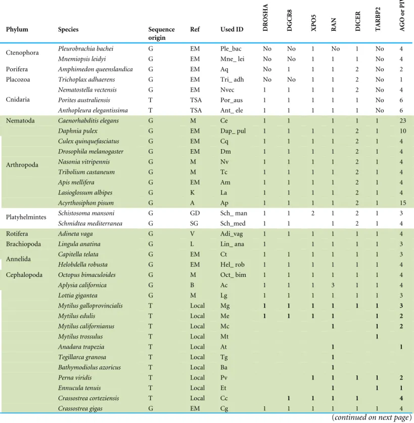
Sequences coding for proteins centrally involved in the miRNA pathway, namely DROSHA, DGCR8, XPO5, RAN, DICER, TARBP2, AGO and PIWI, have been methodically identified in the genomes and transcriptomes ofM. galloprovincialis(Mg) andC. gigas(Cg) as well as in other bivalve and non-bivalve species (66 species, listed inTable 1).
Sequence retrieval and analysis
The Mg WGS project (ID APJB000000000.1 (Nguyen, Hayes & Ingram, 2014)) and the Cg genome draft (GCA_000297895 (Zhang et al., 2012)) were retrieved from GenBank, whereas the oyster genome annotations were obtained from Ensembl Metazoa release 29 (http://metazoa.ensembl.org/Crassostrea_gigas/Info/Index). A Mg reference transcriptome was produced using 18,788 ESTs of mixed tissues previously obtained by Sanger sequencing (Venier et al., 2009) and 453 million reads obtained by paired-end (2 × 100 bp) Illumina Hiseq2000 sequencing of digestive gland
from North Adriatic Sea mussels (ID: PRJNA88481) (Gerdol et al., 2014), and from haemocytes, gills, mantle and muscle of Spanish mussels (ID: SRP033481) (Moreira et al., 2015). The quality of the sequencing readout was evaluated by the FastQC suite
(http://www.bioinformatics.babraham.ac.uk/projects/fastqc/) discarding the reads with
PHRED quality below 20 and presenting more than two ambiguous nucleotides. D e-novo assembly was performed with Trinity, release 2013-08-14 (Grabherr et al., 2011), setting the minimum contig length at 200 bp and using default settings. Subsequently,
Table 1 Organisms included in the present work.Phylum, organism name, sequence origin and reference, ID used in phylogenetic trees and iden-tified sequences are reported. Protostomia (green), Deuterostomia (orange) and novel protein sequences (numbers in bold) are well discernible.
Phylum Species Sequence
origin
Ref Used ID DROSHA DGCR8 XPO5 RAN DICER TARBP2 AGO
or
PIWI
Pleurobrachia bachei G EM Ple_bac No No 1 No 1 No 4
Ctenophora
Mnemiopsis leidyi G EM Mne_ lei No No 1 1 1 No 4
Porifera Amphimedon queenslandica G EM Aq No 1 1 1 2 No 2
Placozoa Trichoplax adhaerens G EM Tri_ adh No No 1 1 2 No 1
Nematostella vectensis G EM Nvec 1 1 1 1 2 No 4
Porites australiensis T TSA Por_aus 1 1 1 1 1 No 6
Cnidaria
Anthopleura elegantissima T TSA Ant_ ele 1 1 1 1 1 No 6
;Nematoda Caenorhabditis elegans G M Ce 1 1 1 1 1 23
; Daphnia pulex G EM Dap_ pul 1 1 1 1 2 1 10
; Culex quinquefasciatus G EM Cq 1 1 1 1 2 1 4
; Drosophila melanogaster G EM Dm 1 1 1 1 2 1 4
; Nasonia vitripennis G M Nv 1 1 1 1 2 1 4
; Tribolium castaneum G M Tc 1 1 1 1 2 1 4
; Apis mellifera G EM Am 1 1 1 1 2 1 4
; Lasioglossum albipes G K La 1 1 1 1 2 1 4
;
Arthropoda
Acyrthosiphon pisum G A Ap 1 1 1 1 2 1 15
Schistosoma mansoni G GD Sch_ man 1 1 2 1 2 1 3
Platyhelmintes
Schmidtea mediterranea G SG Sch_med 1 1 1 2 1 4
;Rotifera Adineta vaga G V Adi_vag 1 1 1 1 1 1 4
;Brachiopoda Lingula anatina G L Lin_ ana 1 1 1 1 1 3
; Capitella telata G EM Ct 1 1 1 1 1 1 3
;Annelida Helobdella robusta G EM Hel_ rob 1 1 1 1 1 1 4
;Cephalopoda Octopus bimaculoides G M Oct_ bim 1 1 1 1 1 1 4
; Aplysia californica G B Ac 1 1 1 3 1 1 4
; Lottia gigantea G M Lg 1 1 1 1 1 1 3
; Mytilus galloprovincialis T Local Mg 1 1 1 1 1 1 3
; Mytilus edulis T Local Me 1 1 1 1 1 2
; Mytilus californianus T Local Mc 1 1 2
; Mytilus trossulus T Local Mt 1
; Anadara trapezia T Local At 1 1
; Tegillarca granosa T Local Tg 1
; Bathymodiolus azoricus T Local Ba 1
; Perna viridis T Local Pv 1 1 1 1 2
; Ennucula tenuis T Local Et 1 1 1
; Crassostrea corteziensis T Local Cc 1 1 1 1 4
; Crassostrea gigas G EM Cg 1 1 1 1 1 1 4
(continued on next page)
Table 1(continued)
Phylum Species Sequence
origin
Ref Used ID DROSHA DGCR8 XPO5 RAN DICER TARBP2 AGO
or
PIWI
; Crassostrea hongkongensis T Local Ch 1 1 3
; Crassostrea virginica T Local Cv 1 4
; Crassostrea angulata T local Ca 1 1 1 1 2
; Ostrea chilensis T Local Oc 1
; Ostrea edulis T Local Oe 1 2
; Ostrea lurida T local Ol 1 1
; Ostreola stentina T Local Os 1
; Saccostrea glomerata T Local Sg 1
; Argopecten irradians T Local Ai 1 1
; Mizuhopecten yessoensis T Local My 1 1 1 1 2
; Pecten maximus T Local Pm 1 2
; Pinctada fucata G F Pf 1 1 1 1
; Solemya velum T Local Sv 1 1 1 1 1 3
; Elliptio complanata T Local Ec 1 1 1 1 1
; Pyganodon grandis T Local Pg 1 1 2
; Uniomerus tetralasmus T Local Ut 1 1 3
; Villosa lienosa T Local Vl 1 1
; Corbicula fluminea T local Cf 1 1
; Meretrix meretrix T local Mm 1 2
; Ruditapes decussatus T local Rd 1 1
; Mollusca
Ruditapes philippinarum T local Rp 1 1
;Echinodermata Strongylocentrotus purpuratus G M Sp 1 1 1 1 1 1 3
;Hemichordata Saccoglossus kowalevskii G M Sk 1 1 1 1 1 1 1
; Homo sapiens G M Hs 1 1 1 1 1 1 8
; Ciona intestinalis G M Ci 1 1 1 1 1 1 3
; Branchiostoma floridae G M Bf 1 1 1 1 1 1 7
; Oncorhynchus mykiss G O Om 1 1 1 1 1 1 5
;
Chordata
Danio rerio G M Dr 1 1 1 1 1 1 5
Arabidopsis thaliana G P At No No 1 1 4 No 10
Streptophyta
Populus trichocarpa G P Pt No No 1 1 5 No 11
Notes.
Abbreviations:: A, Aphidbase; B, broadinstitute.org/ftp/pub/assemblies/invertebrates/aplysia/; EM, Ensambl Metazoa v.29; F, Takeuchi et al. (2012) DNA Res. 19(2): 117– 130;; G, Genome; GD, GeneDB; K, Kocher et al. (2013) Genome Biology 14 (12): R142; L, Lou et al. (2015) Nat Commun. 8; 6:8301; M, Metazome v3.0; O, Berthelot et al. (2014) Nat Commun. 22; 5: 3657; P, Phytozome 11; SG, SmedGD; T, Transcriptome; TSA, NCBI Transcriptome shotgun assembly; V, Genoscope.
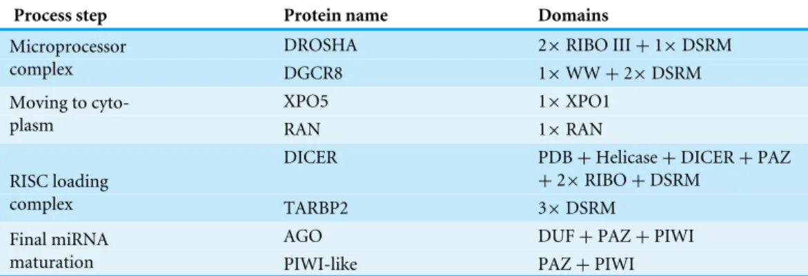
Table 2 Key proteins of the miRNA biogenesis with their structural domains.
Process step Protein name Domains
DROSHA 2×RIBO III+1×DSRM
Microprocessor
complex DGCR8 1×WW+2×DSRM
XPO5 1×XPO1
Moving to
cyto-plasm RAN 1×RAN
DICER PDB+Helicase+DICER+PAZ
+2×RIBO+DSRM RISC loading
complex TARBP2 3×DSRM
AGO DUF+PAZ+PIWI
Final miRNA
maturation PIWI-like PAZ+PIWI
protein coding sequences (cds) were predicted with Transdecoder (Grabherr et al., 2011). Transcriptomic reads of 30 bivalvespp. (Cg plus other 29 species) were retrieved from the SRA archive and assembled as described above (details inFile S1). The protein predictions of further 33 organisms were directly retrieved from public repositories or extracted from the corresponding genome releases. The NCBI transcriptome shotgun assembly (TSA) database was interrogated to retrieve hits for two additional cnidarians,Porites australiensis
andAnthopleura elegantissima(Table 1).
Protein domain searches
To investigate the presence of eight key proteins of miRNA biogenesis (DROSHA, DGCR8, XPO5, RAN, DICER, TARBP2, AGO and PIWI), we downloaded their predictive HMM from PFAM v.27 (listed inTable 2) and we scanned the sequence datasets with HMMer v3.1 (Eddy, 2011) applying a cut-off E-value of 0.01. To achieve a meaningful comparison of proteins from different organisms, we retained only hits presenting all diagnostic domains. Moreover, we identified several mussel transcripts related to protein interactions occurring in the miRNA biogenesis. To identify such proteins, we retrieved from PFAM the diagnostic domains of human homologs (listed inTable 3) and we scanned their presence in the Mg transcriptome as described above. Protein domain organization was reconstructed using SMART (Letunic, Doerks & Bork, 2012).
Gene structure analysis
We used the transcript sequences of DROSHA, DGCR8, XPO5, DICER and TARBP2 as blast queries against all Mg genomic contigs (blastn) in order to recover the related gene structures. Positive hits havinge-value lower than 10−20 were extracted and assembled on
the corresponding transcript, used as backbone. RNA-seq read mappings with adapted parameters (CLC Genomic Workbenchlarge gap mapping tool, with similarity and length fraction set at 0.9) allowed us to ascertain the correct gene assembly. Homolog gene structures were retrieved by interrogating genomic browsers, like Metazome v.3 (for C.intestinalis, B. floridae, D. rerio, S. kowalevskii, S. purpuratus, N. vectensis, T. castaneum, L. gigantea, O. bimaculoides, C. elegans andH. sapiens) and Ensembl Metazoa v.29 (for
C. gigas,C. quinquefasciatus, D. melanogaster, N. vitripennis, A. mellifera, A. queenslandica,
Table 3 miRNA biogenesis proteins ofMytilus galloprovincialis.Protein name, GenBank ID, transcript (bp) and protein length (aa), identified domains and annota-tion (first hit,e-value and percentage of similarity) are reported.
Protein name GenBank ID Transcript length (bp) Protein length (aa) Identified domain(s) Annotation (first hit) E-value (e∧)
Similarity (%)
MgDROSHA KT447251 4,384 1,377 2×RIBO III+1×DSRM Ribonuclease 3-like (Crassostrea gigas) 0 67
MgDGCR8 KT447252 2,483 728 1×WW+2×DSRM Microprocessor complex subunit DGCR8-like
(Crassostrea gigas)
0 50
MgXPO5 KT447259 3,875 1,201 XPO1 Exportin-5-like (Crassostrea gigas) 0 55
MgRAN KT447254 1,113 214 RAN GTP-binding nuclear protein Ran (Crassostrea gigas) −143 93
MgDICER KT447258 6,013 1,850 PDB+Helicase+DICER
+PAZ+2×RIBO+DSRM
Endoribonuclease Dicer-like (Crassostrea gigas) 0 58
MgTARBP2 KT447253 7,583 321 3×DSRM Probable RISC-loading complex subunit (Crassostrea
gigas)
−143 69
MgAGO KT447257 3,337 892 DUF+PAZ+PIWI Protein argonaute-2-like (Crassostrea gigas) 0 84
MgPIWIa KT447255 2,686 867 PAZ+PIWI Piwi-like protein 1 (Crassostrea gigas) 0 75
Key
miRNA
biogenesis
proteins
MgPIWIb KT447256 3,603 948 PAZ+PIWI Piwi-like protein 2 (Hydra vulgaris) 0 59
MgGW182 KT447250 3,825 1,274 UBA+RRM Trinucleotide repeat-containing gene 6C protein-like
(Crassostrea gigas)
0 45
MgCNOT1 KT694355 5,373 1,791 DUF3819+NOT1 CCR4-NOT transcription complex subunit 1-like
(Crassostrea gigas)
0 69
MgCNOT2 KT694357 864 288 NOT2_ 3_ 5 CCR4-NOT transcription complex subunit 2
(Pinctada fucata)
−156 82
MgCNOT3 KT694358 2,142 714 NOT3+NOT2_ 3_ 5 CCR4-NOT transcription complex subunit 3-like
(Crassostrea gigas)
0 97
MgCNOT6 KT694359 2,592 864 Exo_ endo_ phos Uncharacterized protein LOC105348954 isoform X1
(Crassostrea gigas)
0 71
MgCNOT7 KT694360 897 299 CAF1 CCR4-NOT transcription complex subunit 7-like
(Crassostrea gigas)
0 84
MgCNOT9 KT694361 927 309 RCD1 Cell differentiation protein RCD1 homolog
(Crassostrea gigas)
0 93
MgCNOT10 KT694356 2,133 711 TPR_ 1 CCR4-NOT transcription complex subunit 10-like
(Crassostrea gigas)
0 71
MgDDX5 KT694371 1,740 538 DEAD+Helic ATP-dependent RNA helicase DDX5 (Crassostrea
gigas)
0 75
MgDDX6 KT694372 1,332 443 DEAD+Helic ATP-dependent RNA helicase me31b (Crassostrea
gigas)
0 88
(continued on next page)
Table 3(continued) Protein name
GenBank ID
Transcript length (bp)
Protein length (aa)
Identified domain(s)
Annotation (first hit)
E-value (e∧)
Similarity (%)
MgDDX20 KT694373 1,836 612 DEAD+Helic ATP-dependent RNA helicase DDX20 (Crassostrea
gigas)
0 77
MgDDX42 KT694374 2,196 731 DEAD+Helic ATP-dependent RNA helicase DDX42 (Crassostrea
gigas)
0 72
MgPABP KT694365 1,881 627 4×RRM+PABP polyadenylate-binding protein 4 (Hydra vulgaris) 0 74
MgeIF4G KT694364 5,019 1,672 MIF4G+MA3+W2 eukaryotic translation initiation factor 4 gamma
(Crassostrea gigas)
0 57
MgPAN2 KT694367 3,606 1,202 UCH_ 1+RNase_T PAB-dependent poly(A)-specific ribonuclease
sub-unit PAN2 (Lingula anatina)
0 72
MgPAN3 KT694368 2,334 778 None PAB-dependent poly(A)-specific ribonuclease
sub-unit PAN3 (Lingula anatina)
0 67
MgDCP1 KT694362 1,611 536 DCP1 mRNA-decapping enzyme 1A-like (Crassostrea gigas) −126 73
MgDCP2 KT694363 1,313 385 DCP2+NUDIX m7GpppN-mRNA hydrolase (Lingula anatina) −117 67
MgPRMT5 KT694369 1,893 631 PRMT5 protein arginine N-methyltransferase 5-like
(Crassostrea gigas)
0 72
MgTudor-11 KT694370 2,682 894 4×SNc+TUDOR Hypothetical protein mRNA (Lottia gigantea) 0 73
Other
interacting
proteins
MgMaelstrom KT694366 1,321 404 HMG+MAEL Protein maelstrom (Crassostrea gigas) −155 62
R
osani
e
t
al.
(2016),
P
eerJ
,
DOI
10.7717/peerj.1763
P. bachei, M. leidyi, T. adhaerens, N. vectensis, D. pulex, S. mansoni, S. mediterranea, A. vaga, L. anatine, H. robustaandC. telata) or by localblastnagainst the downloaded genomes (A. pisumandL. albipes).
Phylogenetic analysis
The inferred protein sequences were aligned using MUSCLE, release 2014-05-29 (Edgar, 2004). Subsequently, the fasta alignments were analyzed using Gblocks v.0.91 (Castresana, 2000) to extract conserved positions (positions common to 51% of the locally aligned sequences). Trees were built using neighbor joining or maximus likelihood clustering methods with 1,000 bootstrap replicates. Bayesian phylogenies were reconstructed using MrBayes v.3.2.5 (Ronquist et al., 2012), with GTR substitution evolutionary model with gamma-distributed rate variation across sites, evaluating the convergence after 1,000,000 runs (0.5 was considered as cut-off value). Trees were visualized and edited with FigTree v1.4.2 (http://tree.bio.ed.ac.uk/software/figtree/).
Digital expression analysis
To analyze the expression of the selected genes in Cg and Mg RNA datasets, we retrieved all available RNA-seq samples from the NCBI SRA archive. For Cg, we analyzed 123 Illumina RNA-seq samples related to adult tissues or developmental stages. For Mg, we analyzed 13 RNA samples from gills (1), digestive gland (6), haemocytes (2), mantle (2) and muscle (2). Overall, we included in the expression analysis 2,271 and 453M reads for Cg and Mg, respectively (File S2). The trimmed reads were mapped to Cg and Mg genes using the CLC Genomics Workbench v.8.0 (Qiagen, Hilden, Germany) mapping tool, with length and similarity fractions set at 0.75 and 0.95, respectively, and mismatch/insertion/deletion penalties at 3/3/3. The number of uniquely mapped reads of each dataset were counted and used to calculate digital expression values as TPM (Transcripts Per Kilobase Million mapped reads) as described by (Wagner, Kin & Lynch, 2013), considering 3 TPMs as lower detection limit.
RESULTS
Mussel transcripts related to the miRNA biogenesis
We identifiedMytilus galloprovincialistranscripts involved in the miRNA biogenesis by systematic searches of diagnostic domains (Table 2) in a transcriptome assembly produced from 453 million Illumina reads. Thus, we recovered nine transcripts coding for DROSHA, DGCR8, XPO5, RAN, DICER, TARBP2 and for three Argonaute genes (one Ago and two Piwi-like proteins,Table 3). We also identified 21 mussel proteins expected to play a role in the miRNA maturation or involved in RNAi processes (File S3).
Figure 1 relates the general process of eukaryotic miRNA biogenesis to the mussel
proteins identified in this work. MgDROSHA and MgDGCR8 are expected to start the maturation of pri-miRNAs produced by RNA polymerase II. MgDROSHA codes for a 1,377 aa length protein containing all the canonical domains (2 RIBOc domains in positions 959–1,093 and 1,139–1,271 and one DSRM domain in position 1,278–1,351) whereas MgDGCR8 is a 728 aa length protein having one WW domain in position 229–258,
Figure 1 (A) Graphical reconstruction of mussel miRNA biogenesis process.(Modified fromKapinas & Delany, 2011). (B) Conserved domains of the mussel miRNA complements.
necessary for the interaction with DROSHA, and two DSRM domains (positions 472–536 and 578–642) necessary for pri-miRNA binding. MgXPO5 is expected to cooperate with MgRAN in the pre-miRNA cytoplasmic translocation. MgRAN encodes a 214 aa protein whereas MgXPO5 has a length of 1,201 aa and includes two 5’ conserved domains (IBN_N and Xpo1) and one conserved region necessary for the interaction with interleukin enhancer-binding factor 3 (position 525–562). In mussels, the RISC complex uploading pre-miRNAs is defined by the endoribonuclease MgDICER (1,850 aa) and MgTARBP2 (321 aa). Like in Lophotrocozoa, mussel DICER is encoded by a unique gene and contains the seven canonical domains, namely two helicase domains, one DICER-dimer domain, one PAZ, two RIBOc and a final DSRM domain. MgTARBP2 displays three DSRM domains in positions 9–73, 101–166 and 249–314. Moreover,
M. galloprovincialispossess three argonaute proteins ranging from 861 to 941 aa in length and representative of one AGO (DUF1785, PAZ and PIWI domains) and two PIWI-like proteins (PAZ and PIWI domains). We considered the above mentioned gene products as the key complement of the miRNA biogenesis.
Among the possible interacting proteins, we identified MgGW182, a transcript encoding a protein shorter than the human counterparts but holding all the features considered significant for its interaction with AGOs and the CCR4-NOT complex. In fact, MgGW182 possesses 19 N-terminal GW stretches, followed by one UBA domain, a Q-rich region (M domain) and a C-terminal RNA recognition motif (RRM domain). Moreover, we recognized a C-terminal conserved site known as PAM2 (Kozlov et al., 2010), expected to interact with the poly(A) binding protein 1 (MgPABPC1) through the MLLE motif and inhibit the mRNA translation by interfering with the mRNA circularization process (Piao et al., 2010;Van Kouwenhove, Kedde & Agami, 2011). In the mussel transcriptome, we also found putative homologs for a number of CNOT complex proteins (CNOT1, 2, 3, 6, 7, 9, and 10), for the eukaryotic translation initiation factor 4 gamma, 1 eIF4G, PAB-dependent poly(A)-specific ribonuclease subunits PAN2, PAN3, the decapping complex proteins DCP1 and DCP2, and several RNA helicases demonstrated to be crucial in the miRNA maturation (DDX5) and RNAi (DDX5- 6- 20 and 42). Finally, we recognized the putative mussel homologs of protein arginine methyltransferase 5 (MgPRMT5), tudor domain containing protein (MgTDRD-11) and maelstrom spermatogenic transposon silencer (MgMAEL).
Mussel genes related to the miRNA biogenesis
Taking advantage of mussel WGS data (Nguyen, Hayes & Ingram, 2014) we investigated the organization of the main genes involved in the mussel miRNA biogenesis. Fragmentation of the genomic mussel assembly (2.3 million contigs; 700 bp on average) and considerable dimension of the analyzed genes (9.6–17.6 kbp gene size in the case of Cg) prevented the recovery of the full gene sequences. Nevertheless, we can describe the complete gene structures of DROSHA, DGCR8, EXP5, DICER and TARBP2 (i.e., five of eight searched sequences) whose length varies between 7.5 and 27 kbp, confirmed by the back-mapping of 115,377 Illumina paired reads (Fig. 2,File S4). Moreover, these mussel genes showed
Figure 2 Mussel gene structures of DROSHA (A), DGCR8 (B), EXP5 (C), DICER (D) and TARBP2 (E).Green boxes represent exons, length is reported as base pair scale.
a remarkable conservation in terms of exon number when compared with a selection of homolog genes from deuterostome and protostome organisms (Table 4).
Transcripts related to the miRNA biogenesis in bivalve spp
To identify the miRNA biogenesis complements in marine mollusks, we used homologous genes retrieved from the genomes of C. gigas, L. giganteaandA. californica. Since the
C. gigasgenome includes annotations only for the cds regions, we exploited full-length transcripts obtained from a locally assembled oyster transcriptome to expand the genome annotations in this species. In particular, we updated the annotation of CgDGCR8 and CgDICER and we added new annotations for CgPIWI-1 (CGI_10008757: genomic contig JH815696, position 184178–187825) and CgTARBP2 (JH818440, 414703–419857).
Since many marine bivalvespp. do not have at present a sequenced genome, we used publicly available RNA-seq data to build 29 specie-specific transcriptome assemblies and retrieve the homologous sequences of interest. After domain searching, we carefully considered the high number of positive hits to retain only proteins including all the expected protein features. Thus, we retrieved 132 complete hits from marine mollusks: 10 DROSHAs, 9 DGCR8s, 14 XPO5s, 34 RANs, 7 DICERs, 13 TARBP2s and 45 Argonaute-like proteins, the latter classified in 13 AGO and 32 PIWI proteins by phylogenetic analysis
(Table 1,File S5).
Phylogenetic analysis of the miRNA biogenesis proteins
The inferred sequences of single miRNA biogenesis proteins were aligned together with those retrieved from 34 sequenced genomes. Here, we report the phylogenetic analysis of
Table 4 Number of exons of five key miRNA biogenesis genes.Metazome 3.0 and Ensembl Metazoa v.29 genome browsers were interrogated with the previously analyzed hits for each organism. La and Ap genomes were downloaded and analyzed locally. Mg gene structures were retrieved as de-scribed in Methods. In green are reported Protostomia; in orange Deuterostomia.
Species Homo sapiens Ciona intestinalis Branchiostoma floridae Danio rerio Saccoglossus kowalevskii Strongylocentrotus purpuratus Nematostella vectensis Amphimedon queenslandica Caenorhabditis elegans Capitella telata Culex quinquefasciatus Drosophila melanogaster Nasonia vitripennis Tribolium castaneum Apis mellifera Lasioglossus albipes Acyrthosiphon pisum Lottia gigantea Crassostrea gigas Mytilus galloprovincialis
ID Hs Ci Bf Dr Sk Sp Nvec Aq Ce Ct Cq Dm Nv Tc Am La Ap Lg Cg Mg
DROSHA 27 24 29 17 24 20 13 14 6 28 3 3 11 9 13 23 1 23 30 23
DGCR8 14 10 15 10 15 13 7 No 11 18 4 5 11 6 6 6 2 11 18 16
XPO5 32 4 8 21 29 31 2 28 No 30 9 2 11 10 9 10 1 32 34 31
DICER 27 23 26 17 14 19 12 10 26 13 7 8 5 9 29 33 19 16 19 18
TARBP2 9 1 6 9 6 2 No No 11 8 4 5 7 7 7 6 7 6 7 6
the five proteins centrally involved in the miRNA biogenesis, namely DROSHA, DGCR8, DICER, TARBP2 and AGOs (File S5includes all protein sequences). We back-traced the presence of a canonical DROSHA up to Cnidaria, although we found only incomplete hits in Porifera and Placozoa and the genomes ofCtenophora spp.lack of both DROSHA and DGCR8, as reported by other authors (Maxwell et al., 2012). The DROSHA sequences from Cnidaria’s appeared as general outgroup whereas those of Chordata clustered as outgroup of the other protostomes. DROSHAs from Mollusca and Arthropoda clustered consistently with the different taxawhereas those from Platyhelmintes, Rotifera, Brachiopoda and Annelida grouped together, with DROSHA fromCaenorhabditis elegans(Nematoda) being the most far-related (Fig. 3A). Contrary to DROSHA, we identified a complete DGCR8 also in thePorifera Amphimedon queenslandica, suggesting that also DROSHA should be present in this taxa. Following phylogenetic analysis, we highlighted Cnidaria and Porifera proteins as outgroup, with mollusks (and Annelida) clustering with Arthropoda and more distantlyPlatyhelmintesandRotiferahits. TheChordatasequences clustered as a separate group (Fig. 3B).
The finding of putative DICER sequences in Ctenophoraspp. supports the presence of this gene through the whole Opisthokonta evolution (Maxwell et al., 2012). Also plants possess DICER homologues which occur in different copy number among taxa: two genes in Porifera, Placozoa, Cnidaria, Platyhelminthes and Arthropoda (with the exception of
D. pulex that possess three genes); four genes in plants likeA. thaliana andP. trichocarpa
and one gene in Ctenophora, Rotifera, Cephalopoda Mollusca and Chordata. Moreover, the presence of DICER was reported in some Protozoa and fungi (Mukherjee, Campos & Kolaczkowski, 2013). Phylogenetic analyses, separate insect DICER-2,plant DICERs from DICER-1. DICER-1 clade shows a consistent clustering of Arthropoda, Mollusca
Figure 3 Phylogenetic relationships of four miRNA biogenesis proteins.(A) DROSHA, (B) DGCR8, (C) DICER and (D) TARBP2. Inferred protein sequences were aligned using MUSCLE, conserved positions were extracted using Gblocks and subjected to MrBayes analysis.
R
osani
e
t
al.
(2016),
P
eerJ
,
DOI
10.7717/peerj.1763
and Chordata hits, whereas some branches of basal metazoans and Platyhelminthes are not well resolved (Fig. 3C). Likewise, the phylogenetic tree regarding TARBP2 displays a clear cut-off between the proteins of mollusks, chordates and arthropods (Fig. 3D). We back-traced the miRNA cytoplasm export complex composed by RAN and XPO5 in all analyzed metazoans. Both RAN and XPO5 represent widely expressed sequences since we found them also in transcriptome assemblies, although with suboptimal sequence coverage.
Several AGO and Piwi proteins can be present in individual organisms and, in fact, we identified a total of 235 proteins. Whereas humans possess eight proteins, we found four proteins in the majority of the analyzed insectspp.(with the exception of 15 proteins in
A. pisum) and three or four different proteins in bivalvespp. Also, basal Metazoa possess Argonaute-like sequences: four in the genomes of Ctenophora and Cnidariaspp., one in the PlacozoaT. adhaerensand two inA. queenslandica. The case ofC. elegansis remarkable since it holds several Argonaute gene families and at least 24 proteins (Hoogstrate et al., 2014). In agreement with other phylogenetic studies (Swarts et al., 2014), the Argonaute proteins from plants and the majority of those from C. elegansformed distinct clades and, moreover, a clear separation was evident between AGO and PIWI proteins. Bivalve protein sequences clustered always separately forming one cluster for AGO-like hits and two clusters for PIWI-like proteins (Fig. 4).
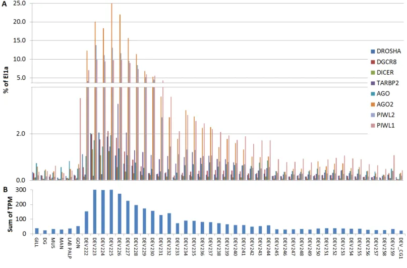
Digital expression analysis of mussel and oyster miRNA biogenesis genes
We used the 13 Mg and 124 Cg RNA-seq samples to evaluate the expression levels of miRNA biogenesis genes in different tissues and conditions. Based on total mapped reads, we computed TPM values and we used elongation factor 1α (El1α) as normalizer
housekeeping gene to compare the expression level of the different genes in each sample. For Mg, the sequence analysis indicated a scarce basal expression of the genes mentioned above in five adult tissues: gill, digestive gland, haemolymph, muscle and mantle (below 2% of El1α, except for DDX5, RAN and CNOT9). Mantle and muscle appeared the most
responsive tissues whereas haemolymph was the least responsive one. In particular, the genes that we considered as the core components of miRNA biogenesis were expressed at levels below 0.5% of El1α(File S6).
For Cg, we analyzed a considerable number of RNA-seq libraries representative of adult tissues (85) and developmental stages (39) (File S6). In adult oysters we observed low basal expression, as detected in the mussel samples. In fact, none of the experimental conditions reported for the analyzed RNA-seq samples influenced substantially the expression of the core miRNA pathway genes (expression levels below 2% of El1α), with the exception of the
high levels of CgPIWI-1 levels in male and female gonads (around 3.5%,Fig. 5). Conversely, most of the miRNA biogenesis genes were expressed at remarkable levels during the early stages of the oyster development: mainly from two cells to the rotary movement and, for some genes, also in the next developmental stages untilD-shapedlarvae, with no detectable signals afterward in spat and juveniles. Hence, these genes are particularly active in the early development, in particular one AGO (CGI_10020511) and two PIWI transcripts from the egg to trocophora (Fig. 5). In the same developmental stages we also noticed a
Figure 4 Phylogenetic relationships of Argonaute-like proteins.Proteins were aligned using MUSCLE and tree was generated using Neighbor Joining algorithm with 1,000 bootstrap replicates. Plant proteins are highlighted in green, whereasC. eleganshits are reported in grey. Blue lines rep-resent mollusk hits, red lines reprep-resent hits from basal metazoans.
Figure 5 Digital expression analysis in oyster.The expression of the 8 miRNA biogenesis genes were computed in tissue-specific RNA libraries and in RNA libraries from different developmental phases. (A) Expression values represented as percentage of El1α. (B) Cumulative TPM expres-sion values of the 8 genes in the same samples.
remarkable expression of the key miRNA genes, with the co-expression of DROSHA and DGCR8 evident in all the analyzed samples.
DISCUSSION
Small RNAs are important regulators of the gene expression, as recognized in various model and non-model organisms (Kim et al., 2014a; Kim et al., 2014b; Martini et al., 2014; Hussain & Asgari, 2014; Sahoo et al., 2014;Britton et al., 2014; Poole et al., 2014;
Solofoharivelo et al., 2014), including some bivalves (Jiao et al., 2014;Zhou et al., 2014). In addition to the identification of miRNAs, a general comprehension of the miRNA biogenesis in itself is also significant (Grimson et al., 2008;Wu et al., 2011;Moran et al., 2013). However, the main genes involved in miRNA formation in bivalves have not been described and characterized so far. In this study, we have provided an overview on the miRNA biogenesis complements in bivalves spp., with particular attention to
M. galloprovincialisandC. gigas. To the best of our knowledge, we report for the first
time the presence of a complete miRNA biogenesis pathway inM. galloprovincialis,the full-length transcript sequences of DICER, DGCR8, XPO5, RAN, DROSHA, TARBP2, three Argonaute genes and the identification of many other components that are candidate miRNA complement-interacting proteins such as MgGW182. By using local transcriptome assemblies, we identified these genes also in many other marine bivalves. The general low expression levels of these transcripts in the adult tissues of both M. galloprovincialis andC. gigas,and the considerable gene size, have probably prevented a previous identification of full-length sequences in not-well-covered bivalve transcriptomes. In fact, we obtained complete transcript sequences only from sequenced genomes or highly-covered transcriptomes whereas in other transcriptome assemblies we retrieved only few complete sequences. Overall, we have analyzed 523 miRNA complement sequences, 145 of them belonging to marine mollusks and displaying a consistent sequence clustering (OstreoidaandMytiloida proteins generated two distinct clades, located always as sister group of arthropods).
However, the copy number of Argonaute genes somewhat differs among bivalves, as
C. gigasandA. californicagenomes coding for four proteins (2 AGO and 2 PIWI proteins) whereasM. galloprovincialisandL. giganteapossess three proteins (1 AGO and 2 PIWIs). We also highlighted the over-expression of the miRNA biogenesis genes during the first phases of the oyster development. A genome protection mechanism based on piRNA expression during early developmental stages is well known in mammals (Malone & Hannon, 2009;Kim et al., 2014a;Kim et al., 2014b) but such mechanism has not been reported in bivalves and additional investigations are necessary.
Finally, the identification of several mussel proteins either necessary or cooperative in the miRNA biogenesis, supports the existence of a complete and functional miRNA pathway in mussels and, probably, in other bivalves. Up to now, protein–protein or protein-RNA interaction data are not available for bivalve spp. and these topics may represent a direction of work in the future. Meanwhile, the expression analyses of miRNA biogenesis genes coupled with the identification of the miRNAs expressed in naturally infected and laboratory-treated bivalves could provide both validation and new insights on these interesting processes.
ADDITIONAL INFORMATION AND DECLARATIONS
Funding
This work is supported by PRIN 2010-11 (20109XZEPR) and FP7-KBBE-2010-4-266157 (BIVALIFE). The funders had no role in study design, data collection and analysis, decision to publish, or preparation of the manuscript.
Grant Disclosures
The following grant information was disclosed by the authors: PRIN2010–11: 20109XZEPR.
BIVALIFE: FP7-KBBE-2010-4-266157.
Competing Interests
The authors declare there are no competing interests.
Author Contributions
• Umberto Rosani conceived and designed the experiments, performed the experiments,
analyzed the data, wrote the paper, prepared figures and/or tables.
• Alberto Pallavicini conceived and designed the experiments, wrote the paper, reviewed
drafts of the paper.
• Paola Venier conceived and designed the experiments, contributed
reagents/materials/-analysis tools, wrote the paper, reviewed drafts of the paper.
DNA Deposition
The following information was supplied regarding the deposition of DNA sequences: Genbank: KT447251, KT447252, KT447259, KT447254, KT447258, KT447253,
KT447257, KT447255, KT447256, KT447250, KT694355, KT694356, KT694357,
KT694358, KT694359, KT694360, KT694361, KT694362, KT694363, KT694364,
KT694365, KT694366, KT694367, KT694368, KT694369, KT694370, KT694371,
KT694372,KT694373,KT694374.
Data Availability
The following information was supplied regarding data availability: The research in this article did not generate any raw data.
Supplemental Information
Supplemental information for this article can be found online athttp://dx.doi.org/10.7717/
peerj.1763#supplemental-information.
REFERENCES
Ambros V. 2003.MicroRNA pathways in flies and worms: growth, death, fat, stress, and timing.Cell113(6):673–676DOI 10.1016/S0092-8674(03)00428-8.
Bartel DP. 2004.MicroRNAs: genomics, biogenesis, mechanism, and function.Cell 116(2):281–297DOI 10.1016/S0092-8674(04)00045-5.
Berezikov E, Guryev V, Van de Belt J, Wienholds E, Plasterk RHA, Cuppen E. 2005.
Phylogenetic shadowing and computational identification of human microRNA genes.Cell120(1):21–24DOI 10.1016/j.cell.2004.12.031.
Britton C, Winter AD, Gillan V, Devaney E. 2014.microRNAs of parasitic helminths— identification, characterization and potential as drug targets.International Journal for Parasitology: Drugs and Drug Resistance4(2):85–94DOI 10.1016/j.ijpddr.2014.03.001.
Carmell MA, Girard A, Van de Kant HJG, Bourc’his D, Bestor TH, De Rooij DG, Hannon GJ. 2007.MIWI2 is essential for spermatogenesis and repression of transposons in the mouse male germline.Developmental Cell12(4):503–514
DOI 10.1016/j.devcel.2007.03.001.
Carninci P, The FANTOM Consortium. 2005.The transcriptional landscape of the mammalian genome.Science309(5740):1559–1563DOI 10.1126/science.1112014.
Castresana J. 2000.Selection of conserved blocks from multiple alignments for their use in phylogenetic analysis.Molecular Biology and Evolution17(4):540–552
DOI 10.1093/oxfordjournals.molbev.a026334.
Chen G, Zhang C, Jiang F, Wang Y, Xu Z, Wang C. 2014.Bioinformatics analysis of hemocyte miRNAs of scallop chlamys farreri against acute viral necrobiotic virus (AVNV).Fish & Shellfish Immunology37(1):75–86DOI 10.1016/j.fsi.2014.01.002.
Denli AM, Tops BBJ, Plasterk RHA, Ketting RF, Hannon GJ. 2004.Processing of primary microRNAs by the microprocessor complex.Nature432(7014):231–235
DOI 10.1038/nature03049.
Diebel KW, Oko LM, Medina EM, Niemeyer BF, Warren CJ, Claypool DJ, Tibbetts SA, Cool CD, Clambey ET, Van Dyk LF. 2015.Gammaherpesvirus small noncoding RNAs are bifunctional elements that regulate infection and contribute to virulence
in vivo.mBio6(1):e01670–e01614DOI 10.1128/mBio.01670-14.
Eddy SR. 2011.Accelerated profile HMM searches.PLoS Computational Biology 7(10):e1002195DOI 10.1371/journal.pcbi.1002195.
Edgar RC. 2004.MUSCLE: multiple sequence alignment with high accuracy and high throughput.Nucleic Acids Research32(5):1792–1797DOI 10.1093/nar/gkh340.
Ender C, Meister G. 2010.Argonaute proteins at a glance.Journal of Cell Science 123(11):1819–1823DOI 10.1242/jcs.055210.
Fromm B, Billipp T, Peck LE, Johansen M, Tarver JE, King BL, Newcomb JM, Sempere LF, Flatmark K, Hovig E, Peterson KJ. 2015.A uniform system for the annotation of vertebrate microRNA genes and the evolution of the human microRNAome.Annual Review of Genetics49(1):213–242DOI 10.1146/annurev-genet-120213-092023.
Fabian MR, Sonenberg N. 2012.The mechanics of miRNA-mediated gene silenc-ing: a look under the hood of miRISC.Nature Structural & Molecular Biology 19(6):586–593DOI 10.1038/nsmb.2296.
Gammon DB, Mello CC. 2015.RNA interference-mediated antiviral defense in insects.
Current Opinion in Insect Science8(April):111–120DOI 10.1016/j.cois.2015.01.006.
Gao Z, Wang M, Blair D, Zheng Y, Dou Y. 2014.Phylogenetic analysis of the endori-bonuclease dicer family.PLoS ONE9(4):e95350DOI 10.1371/journal.pone.0095350.
Gerdol M, De Moro G, Manfrin C, Milandri A, Riccardi E, Beran A, Venier P, Pallavicini A. 2014.RNA Sequencing and de novo assembly of the digestive gland transcriptome inMytilus galloprovincialisfed with toxinogenic and non-toxic strains of alexandrium minutum.BMC Research Notes7:722DOI 10.1186/1756-0500-7-722.
Ghildiyal M, Zamore PD. 2009.Small silencing RNAs: an expanding universe.Nature Reviews Genetics10(2):94–108DOI 10.1038/nrg2504.
GIGA Community of Scientists. 2014.The global invertebrate genomics alliance (GIGA): developing community resources to study diverse invertebrate genomes.
The Journal of Heredity105(1):1–18DOI 10.1093/jhered/est084.
Grabherr MG, Haas BJ, Yassour M, Levin JZ, Thompson DA, Amit I, Adiconis X, Fan L, Raychowdhury R, Zeng Q, Chen Z, Mauceli E, Hacohen N, Gnirke A, Rhind N, Di Palma F, Birren BW, Nusbaum C, Lindblad-Toh K, Friedman N, Regev A. 2011.
Full-length transcriptome assembly from RNA-seq data without a reference genome.
Nature Biotechnology29(7):644–652DOI 10.1038/nbt.1883.
Grimson A, Srivastava M, Fahey B, Woodcroft BJ, Chiang HR, King N, Degnan BM, Rokhsar DS, Bartel DP. 2008.Early origins and evolution of microR-NAs and piwi-interacting RmicroR-NAs in animals.Nature455(7217):1193–1197
DOI 10.1038/nature07415.
Hong S, Noh H, Chen H, Padia R, Pan ZK, Su S-B, Jing Q, Ding H-F, Huang S. 2013.
Signaling by p38 MAPK stimulates nuclear localization of the microprocessor component p68 for processing of selected primary microRNAs.Science Signaling 6(266):ra16DOI 10.1126/scisignal.2003706.
Hoogstrate SW, Volkers RJM, Sterken MG, Kammenga JE, Snoek LB. 2014.Nematode endogenous small RNA pathways.Worm3(March)DOI 10.4161/worm.28234.
Huang J-T, Wang J, Srivastava V, Sen S, Liu S-M. 2014.MicroRNA machinery genes as novel biomarkers for cancer.Frontiers in Oncology 4:13
DOI 10.3389/fonc.2014.00113.
Huarte M. 2015.The emerging role of lncRNAs in cancer.Nature Medicine21(11): 1253–1261DOI 10.1038/nm.3981.
Hussain M, Asgari S. 2014.MicroRNAs as mediators of insect host–pathogen interac-tions and immunity.Journal of Insect Physiology70:151–158
DOI 10.1016/j.jinsphys.2014.08.003.
Iwasaki YW, Siomi MC, Siomi H. 2015.PIWI-interacting RNA: its biogenesis and functions.Annual Review of Biochemistry84:405–433
DOI 10.1146/annurev-biochem-060614-034258.
Jean-Philippe J, Paz S, Caputi M. 2013.hnRNP A1: the Swiss army knife of gene expression.International Journal of Molecular Sciences14(9):18999–19024
DOI 10.3390/ijms140918999.
Jiao Y, Zheng Z, Du X, Wang Q, Huang R, Deng Y, Shi S, Zhao X. 2014.Identification and characterization of MicroRNAs in pearl oyster pinctada martensii by solexa deep sequencing.Marine Biotechnology16(1):54–62DOI 10.1007/s10126-013-9528-x.
John B, Enright AJ, Aravin A, Tuschl T, Sander C, Marks DS. 2004.Human microRNA targets.PLoS Biology2(11):e363DOI 10.1371/journal.pbio.0020363.
Kapinas K, Delany AM. 2011.MicroRNA biogenesis and regulation of bone remodeling.
Arthritis Research & Therapy13(3):Article 220DOI 10.1186/ar3325.
Kenny NJ, Namigai EKO, Marlétaz F, Hui JHL, Shimeld SM. 2015.Draft genome assemblies and predicted microRNA complements of the intertidal lophotro-chozoans patella vulgata (Mollusca, Patellogastropoda) and spirobranchus (Pomatoceros) lamarcki (Annelida, Serpulida).Marine Genomics24:139–146
DOI 10.1016/j.margen.2015.07.004.
Kim S, Günesdogan U, Zylicz JJ, Hackett JA, Cougot D, Bao S, Lee C, Dietmann S, Allen GE, Sengupta R, Surani MA. 2014b.PRMT5 protects genomic integrity during global DNA demethylation in primordial germ cells and preimplantation embryos.
Molecular Cell56(4):564–579 DOI 10.1016/j.molcel.2014.10.003.
Kim M-C, Lee S-W, Ryu D-Y, Cui F-J, Bhak J, Kim Y. 2014a.Identification and char-acterization of microRNAs in normal equine tissues by next generation sequencing.
PLoS ONE9(4):e93662DOI 10.1371/journal.pone.0093662.
Kincaid RP, Sullivan CS. 2012.Virus-encoded microRNAs: an overview and a look to the future.PLoS Pathogens8(12):e1003018DOI 10.1371/journal.ppat.1003018.
Kozlov G, Safaee N, Rosenauer A, Gehring K. 2010.Structural basis of binding of P-body-associated proteins GW182 and Ataxin-2 by the Mlle domain of poly(A)-binding protein.The Journal of Biological Chemistry 285(18):13599–13606
DOI 10.1074/jbc.M109.089540.
Kozomara A, Griffiths-Jones S. 2014.miRBase: annotating high confidence mi-croRNAs using deep sequencing data.Nucleic Acids Research42:D68–D73
DOI 10.1093/nar/gkt1181.
Lau P-W, MacRae IJ. 2009.The molecular machines that mediate microRNA matura-tion.Journal of Cellular and Molecular Medicine13(1):54–60
DOI 10.1111/j.1582-4934.2008.00520.x.
Letunic I, Doerks T, Bork P. 2012.SMART 7: recent updates to the protein domain annotation resource.Nucleic Acids Research40(D1):D302–D305
DOI 10.1093/nar/gkr931.
Lucas K, Raikhel AS. 2013.Insect microRNAs: biogenesis, expression profiling and biological functions.Insect Biochemistry and Molecular Biology 43(1):24–38
DOI 10.1016/j.ibmb.2012.10.009.
MacRae IJ, Ma E, Zhou M, Robinson CV, Doudna JA. 2008.In vitroreconstitution of the human RISC-loading complex.Proceedings of the National Academy of Sciences of the United States of America105(2):512–517DOI 10.1073/pnas.0710869105.
Malone CD, Hannon GJ. 2009.Small RNAs as guardians of the genome.Cell 136(4):656–668DOI 10.1016/j.cell.2009.01.045.
Martín-Gómez L, Villalba A, Kerkhoven RH, Abollo E. 2014.Role of microRNAs in the immunity process of the flat oyster ostrea edulis against bonamiosis.Infection, Genetics and Evolution27(October):40–50DOI 10.1016/j.meegid.2014.06.026.
Martini P, Sales G, Brugiolo M, Gandaglia A, Naso F, De Pittà C, Spina M, Gerosa G, Chemello F, Romualdi C, Cagnin S, Lanfranchi G. 2014.Tissue-specific expression and regulatory networks of pig microRNAome.PLoS ONE9(4):e89755
DOI 10.1371/journal.pone.0089755.
Maxwell EK, Ryan JF, Schnitzler CE, Browne WE, Baxevanis AD. 2012.MicroRNAs and essential components of the microRNA processing machinery are not encoded in the genome of the ctenophoreMnemiopsis leidyi.BMC Genomics13:714
DOI 10.1186/1471-2164-13-714.
Mendell JT, Olson EN. 2012.MicroRNAs in stress signaling and human disease.Cell 148(6):1172–1187DOI 10.1016/j.cell.2012.02.005.
Min P-K, Chan SY. 2015.The biology of circulating microRNAs in cardiovascular dis-ease.European Journal of Clinical Investigation45:860–874DOI 10.1111/eci.12475.
Miyoshi K, Okada TN, Siomi H, Siomi MC. 2009.Characterization of the miRNA-RISC loading complex and miRNA-RISC formed in the Drosophila miRNA pathway.RNA 15(7):1282–1291DOI 10.1261/rna.1541209.
Moran Y, Praher D, Fredman D, Technau U. 2013.The evolution of microRNA pathway protein components in Cnidaria.Molecular Biology and Evolution30(12):2541–2552
DOI 10.1093/molbev/mst159.
Moreira R, Pereiro P, Canchaya C, Posada D, Figueras A, Novoa B. 2015.RNA-Seq in
mytilus galloprovincialis: comparative transcriptomics and expression profiles among different tissues.BMC Genomics16(1):728DOI 10.1186/s12864-015-1817-5.
Mortimer SA, Kidwell MA, Doudna JA. 2014.Insights into RNA structure and function from genome-wide studies.Nature Reviews. Genetics15(7):469–479
DOI 10.1038/nrg3681.
Mukherjee K, Campos H, Kolaczkowski B. 2013.Evolution of animal and plant dicers: early parallel duplications and recurrent adaptation of antiviral RNA binding in plants.Molecular Biology and Evolution30(3):627–641 DOI 10.1093/molbev/mss263.
Nguyen TTT, Hayes BJ, Ingram BA. 2014.Genetic parameters and response to selection in blue mussel (mytilus galloprovincialis) using a SNP-based pedigree.Aquaculture 420–421(January):295–301DOI 10.1016/j.aquaculture.2013.11.021.
Nottrott S, Simard MJ, Richter JD. 2006.Human Let-7a miRNA blocks protein production on actively translating polyribosomes.Nature Structural & Molecular Biology13(12):1108–1114DOI 10.1038/nsmb1173.
Piao X, Zhang X, Wu L, Belasco JG. 2010.CCR4-NOT deadenylates mRNA associated with RNA-induced silencing complexes in human cells.Molecular and Cellular Biology30(6):1486–1494DOI 10.1128/MCB.01481-09.
Poole CB, Gu W, Kumar S, Jin J, Davis PJ, Bauche D, McReynolds LA. 2014.Diversity and expression of microRNAs in the filarial parasite, brugia malayi.PLoS ONE9(5): e96498DOI 10.1371/journal.pone.0096498.
Qu S, Yang X, Li X, Wang J, Gao Y, Shang R, Sun W, Dou K, Li H. 2015.Circular RNA: a new star of noncoding RNAs.Cancer Letters365:141–148
DOI 10.1016/j.canlet.2015.06.003.
Resch AM, Palakodeti D. 2012.Small RNA pathways in schmidtea mediterranea.The International Journal of Developmental Biology56(1–3):67–74
DOI 10.1387/ijdb.113436ar.
Rhee S, Chae H, Kim S. 2015.PlantMirnaT: miRNA and mRNA integrated analysis fully utilizing characteristics of plant sequencing data.Methods83:80–87
DOI 10.1016/j.ymeth.2015.04.003.
Ronquist F, Teslenko M, Van der Mark P, Ayres D, Darling A, Höhna S, Larget B, Liu L, Suchard MA, Huelsenbeck JP. 2012.MrBayes 3.2: efficient Bayesian phylogenetic inference and model choice across a large model space.Systematic Biology61(3):539–542DOI 10.1093/sysbio/sys029.
Ruan X. 2015.Long noncoding RNA central of glucose homeostasis.Journal of Cellular BiochemistryEpub ahead of print Nov 26 2015DOI 10.1002/jcb.25427.
Sahoo G, Ansari MD, Dikhit M, Gupta N, Rana S, Das P. 2014.Computational identi-fication of microRNA-like elements in leishmania major.MicroRNA2(3):225–230
DOI 10.2174/2211536602666131203232422.
Sand M, Skrygan M, Georgas D, Sand D, Gambichler T, Altmeyer P, Bechara FG. 2012.
The miRNA machinery in primary cutaneous malignant melanoma, cutaneous malignant melanoma metastases and benign melanocytic nevi.Cell and Tissue Research350(1):119–126DOI 10.1007/s00441-012-1446-0.
Siomi MC, Sato K, Pezic D, Aravin AA. 2011.PIWI-interacting small RNAs: the vanguard of genome defence.Nature Reviews Molecular Cell Biology12(4):246–258
DOI 10.1038/nrm3089.
Sokolova OA, Iakushev EI, Stoliarenko AD, Mikhaleva EA, Gvozdev VA, Klenov MS. 2011.The interplay of transposon silencing genes in theDrosophila melanogaster
germline.Molekuliarnaia Biologiia45(4):633–641.
Solofoharivelo MC, Van der Walt AP, Stephan D, Burger JT, Murray SL. 2014.
MicroRNAs in fruit trees: discovery, diversity and future research directions.Plant Biology16(5):856–865DOI 10.1111/plb.12153.
Swarts DC, Makarova K, Wang Y, Nakanishi K, Ketting RF, Koonin EV, Patel DJ, Van der Oost J. 2014.The evolutionary journey of argonaute proteins.Nature Structural & Molecular Biology 21(9):743–753DOI 10.1038/nsmb.2879.
Tarver JE, Cormier A, Pinzón N, Taylor RS, Carré W, Strittmatter M, Seitz H, Coelho SM, Cock JM. 2015.microRNAs and the evolution of complex multicellularity: identification of a large, diverse complement of microRNAs in the brown alga ectocarpus.Nucleic Acids Research43:6384–6398DOI 10.1093/nar/gkv578.
Tarver JE, Donoghue PCJ, Peterson KJ. 2012.Do miRNAs have a deep evolutionary history?BioEssays34(10):857–866DOI 10.1002/bies.201200055.
Théron E, Dennis C, Brasset E, Vaury C. 2014.Distinct features of the piRNA Pathway in somatic and germ cells: from piRNA cluster transcription to piRNA processing and amplification.Mobile DNA5(1):28DOI 10.1186/s13100-014-0028-y.
Tolia NH, Joshua-Tor L. 2007.Slicer and the argonautes.Nature Chemical Biology 3(1):36–43DOI 10.1038/nchembio848.
Tomoyasu Y, Miller SC, Tomita S, Schoppmeier M, Grossmann D, Bucher G. 2008.
Exploring systemic RNA interference in insects: a genome-wide survey for RNAi genes in tribolium.Genome Biology9(1):Article R10DOI 10.1186/gb-2008-9-1-r10.
Van Kouwenhove M, Kedde M, Agami R. 2011.MicroRNA regulation by RNA-binding proteins and its implications for cancer.Nature Reviews Cancer11(9):644–656
DOI 10.1038/nrc3107.
Venier P, De Pittà C, Bernante F, Varotto L, De Nardi B, Bovo G, Roch P, Novoa B, Figueras A, Pallavicini A, Lanfranchi G. 2009.MytiBase: a knowledgebase of mussel (M. galloprovincialis) transcribed sequences.BMC Genomics10(February):72
DOI 10.1186/1471-2164-10-72.
Wagner GP, Kin K, Lynch VJ. 2013.A model based criterion for gene expression calls using RNA-seq data.Theory in Biosciences Theorie in Den Biowissenschaften 132(3):159–164DOI 10.1007/s12064-013-0178-3.
Wu B, Jin M, Gong J, Du X, Bai Z. 2011.Dynamic evolution of CIKS (TRAF3IP2/Act1) in metazoans.Developmental & Comparative Immunology35(11):1186–1192
DOI 10.1016/j.dci.2011.03.027.
Xu M-J, Qiu S-B, Nisbet AJ, Fu J-H, Shao C-C, Zhu X-Q. 2014.Global characteri-zation of microRNAs in trichomonas gallinae.Parasites & Vectors7:Article 99
DOI 10.1186/1756-3305-7-99.
Xu C, Zhang L, Li H, Liu Z, Duan L, Lu C. 2015.MiRNA-1469 promotes lung cancer cells apoptosis through targeting STAT5a.American Journal of Cancer Research 5(3):1180–1189.
Zhang G, Fang X, Guo X, Li L, Luo R, Xu F, Yang P, Zhang L, Wang X, Qi H, Xiong Z, Que H, Xie Y, Holland PWH, Paps J, Zhu Y, Wu F, Chen Y, Wang J, Peng C, Meng J, Yang L, Liu J, Wen B, Zhang N, Huang Z, Zhu Q, Feng Y, Mount A, Hedgecock D, Xu Z, Liu Y, Domazet-Lošo T, Du Y, Sun X, Zhang S, Liu B, Cheng P, Jiang X, Li J, Fan D, Wang W, Fu W, Wang T, Wang B, Zhang J, Peng Z, Li Y, Li N, Wang J, Chen M, He Y, Tan F, Song X, Zheng Q, Huang R, Yang H, Du X, Chen L, Yang M, Gaffney PM, Wang S, Luo L, She Z, Ming Y, Huang W, Zhang S, Huang B, Zhang Y, Qu T, Ni P, Miao G, Wang J, Wang Q, Steinberg CEW, Wang H, Li N, Qian L, Zhang G, Li Y, Yang H, Liu X, Wang J, Yin Y, Wang J. 2012.The oyster genome reveals stress adaptation and complexity of shell formation.Nature490(7418):49–54
DOI 10.1038/nature11413.
Zhou Z, Wang L, Song L, Liu R, Zhang H, Huang M, Chen H. 2014.The identification and characteristics of immune-related microRNAs in haemocytes of oyster Cras-sostrea gigas.PLoS ONE9(2):e88397DOI 10.1371/journal.pone.0088397.