Comparative Transcriptomic Analysis of
Primary Duck Hepatocytes Provides Insight
into Differential Susceptibility to DHBV
Infection
Liang Yan1☯, Su Qu1,2☯, Gang Liu3, Lei Liu3, Yao Yu3, Guohui Ding3,
Yanfeng Zhao4, Yixue Li3, Youhua Xie1*, Junqi Zhang1*, Di Qu1*
1Key Laboratory of Medical Molecular Virology, Ministry of Education and Health, School of Basic Medical Sciences, Fudan University, Shanghai, China,2Department of Biochemistry and Molecular Biology, School of Basic Medical Sciences, Fudan University, Shanghai, China,3Key Laboratory of Systems Biology, Shanghai Institutes for Biological Sciences, Chinese Academy of Sciences, Shanghai, China,4Department of Laboratory Medicine, the Second Affiliated Hospital of Nanjing Medical University, Nanjing, Jiangsu, China
☯These authors contributed equally to this work.
*[email protected](DQ);[email protected](JZ);[email protected](YX)
Abstract
Primary duck hepatocytes (PDH) displays differential susceptibility to duck hepatitis B virus when maintained in the media supplemented with fetal bovine serum or dimethyl sulfoxide (DMSO) which has been widely used for the maintenance of hepatocytes, and prolonging susceptibility to hepadnavirus. However the mechanism underlying maintenance of suscep-tibility to hepadnavirus by DMSO treatment remains unclear. In this study, a global tran-scriptome analysis of PDHs under different culture conditions was conducted for
investigating the effects of DMSO on maintenance of susceptibility of PDH to DHBVin vitro. The 384 differential expressed genes (DEGs) were identified by comparisons between each library pair (PDHs cultured with or without DMSO or fresh isolated PDH). We analyzed canonical pathways in which the DEGs were enriched in Hepatic Fibrosis / Hepatic Stellate Cell Activation, Bile Acid Biosynthesis and Tight Junction signaling. After re-annotation against human genome data, the 384 DEGs were pooled together with proteins belonging to hepatitis B pathway to construct a protein-protein interaction network. The combination of decreased expression of liver-specific genes (CYP3A4, CYP1E1, CFI, RELN and GSTA1 et al) with increased expression of hepatocyte-dedifferentiation-associated genes (PLA2G4A and PLCG1) suggested thatin vitroculture conditions results in the fading of hepatocyte phenotype in PDHs. The expression of seven DEGs associated with tight junc-tion formajunc-tion (JAM3, PPP2R2B, PRKAR1B, PPP2R2C, MAGI2, ACTA2 and ACTG2) was up-regulated after short-term culturein vitro, which was attenuated in the presence of DMSO. Those results could shed light on DHBV infection associated molecular events affected by DMSO.
a11111
OPEN ACCESS
Citation:Yan L, Qu S, Liu G, Liu L, Yu Y, Ding G, et al. (2016) Comparative Transcriptomic Analysis of Primary Duck Hepatocytes Provides Insight into Differential Susceptibility to DHBV Infection. PLoS ONE 11(2): e0149702. doi:10.1371/journal. pone.0149702
Editor:Haitao Guo, Indiana University, UNITED STATES
Received:September 29, 2015
Accepted:February 2, 2016
Published:February 22, 2016
Copyright:© 2016 Yan et al. This is an open access article distributed under the terms of theCreative Commons Attribution License, which permits unrestricted use, distribution, and reproduction in any medium, provided the original author and source are credited.
Data Availability Statement:The raw or processed transcript sequencing data for PDH cultured under different conditions were submitted and are available through the NCBI Sequence Read Archive under the accession numbers GSE66786 and GSE72868.
Introduction
Hepatitis induced by hepatitis B virus (HBV) infection remains a major public health problem with over 350 million chronically infected carriers worldwide [1]. The lack of an appropriate cell model supporting HBV infection has been a major limitation for the study of the pathogen-esis of hepadnavirus infection. NTCP has been demonstrated to be the functional receptor of HBV on the cell membrane recently [2]. However, more detailed interactions between hepad-navirus and hepatocytes need to be further investigated. Because of their susceptibility to DHBV infection, primary duck hepatocytes (PDH) serve as a valuable cell model to elucidate the key features of hepadnaviral infection [3–7]. A reverse transcribed RNA intermediate in the replication of hepadnavirus was first described in the DHBV-PDH infection model [3]. However, culturedin vitroover a period of time, PDH lose the hepatocyte phenotype and their susceptibility to DHBV infection rapidly diminishes [4,8]. Only 10%-20% cells retain suscepti-bility to DHBV infection 6 d after plating underin vitroculture condition. The rapid loss of susceptibility to DHBV infection has limited the application of PDH in hepadnavirus study [8].
Several modified culture methods have previously been developed for the maintenance of the primary hepatocyte, including the addition of a low concentration of DMSO to the culture medium [9,10], using plates pre-coated with rat-tail collagen [11], or supplementation with cell factors (epidermal growth factor or hepatocyte growth factor) [9]. Among these methods, the addition of DMSO has been widely used for the maintenance of hepatocytes, and prolonging susceptibility to hepadnavirus [12]. It shows differential susceptibility to DHBV infection between PDH maintained in culture media supplemented with 5% fetal bovine serum and 1.5% dimethyl sulfoxide (DMSO) [8]. It has been revealed that maintenance of polarization phenotypes and prevention of tight junction formation in primary human hepatocytes or HepaRG cells by DMSO is essential for the entry of the HBV underin vitroculture conditions [13]. However the mechanism underlying maintenance of susceptibility to hepadnavirus by DMSO remains to be elucidated.
Next-generation sequencing technology provides a useful tool to understand the activation patterns of the cellular response to external stimuli. Here we conducted a global transcriptome analysis of PDH under different culture conditions to investigate the effects of DMSO on main-tenance of susceptibility of PDH to DHBVin vitro. The differential expressed genes that were identified by comparison between PDHs cultured with or without DMSO, were pooled together with proteins belonging to hepatitis B pathway for the construction of a protein-pro-tein interaction (PPI) network. In the network, the expression of a cluster DEGs associated with tight junction formation was up-regulated after short-term culturein vitro, while the up-regulation was attenuated in the presence of DMSO. Supplementation with DMSO also relieved the declining of anti-virus associated DEGs expression in PDHsin vitro. Those results could shed light on DHBV infection associated molecular events affected by DMSO.
Results
Differential susceptibility to DHBV infection of primary duck hepatocytes
maintained under different conditions
In order to confirm susceptibility to DHBV infection, PDH isolated from Cherry Valley duck-lings were maintained in Leibovitz's L-15 Medium supplemented with 5% FBS for 1d (Con-PDH), and then followed by culture in media with 5% FBS (FBS-8d PDH) or 1.5% DMSO (DMSO-8d PDH) for another 7 days (Fig 1A). The PDH under different culture conditions were infected with DHBV at a multiplicity of genome equivalents of 100, and maintained in Foundation, Nanjing Department of Health
(YKK12198).
Competing Interests:The authors have declared
media with 5% FBS for another 5 days. DHBV in the supernatant of Con-PDH was 3.29×10⁵ copies/μl, 7.89×10⁴copies/μl in that of the DMSO-8d PDH, and only 6.72×10³copies/μl in that of FBS-8d PDH, determined by qPCR (Fig 1B). DHBV covalently closed circular DNA (cccDNA) and DHBV DNA in the core particles (core DNA) were extracted from the cells and analyzed by Southern blot assay. The highest level of cccDNA and core DNA were detected in Con-PDH, followed by DMSO-8d PDH. The lowest level was detected in FBS-8d PDH (Fig 1C and 1D). It confirmed that treatment with 1.5% DMSO could maintain the susceptibility of the PDH to DHBV infection [8].
Fig 1. Intracellular and extracellular viral DNA detection for PDH maintained in different culture media.(A) The Con-PDH, DMSO-8d PDH and FBS-8d PDH (Two lanes of FBS FBS-8d were transformed from a different gel.) were infected with DHBV at a multiplicities of genome equivalents (MGE) of 100 on the 1st or 8th d post-inoculation. (B) The supernatants from the PDH maintained in the different culture media were collected and DHBV DNA was extracted and quantified by qPCR. The cccDNA (C) and core DNA (D) were extracted and analyzed by Southern blot hybridization. RC, relaxed circular DNA; DSL, double-stranded linear DNA; SS, single-double-stranded DNA; cccDNA, covalently closed circular DNA.
Transcriptome analysis of primary duck hepatocytes under three culture
conditions
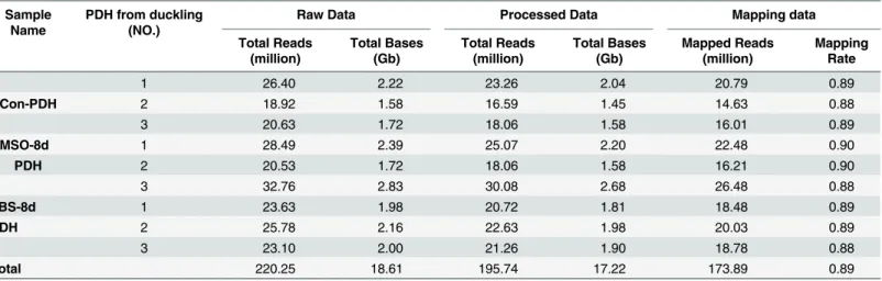
Transcriptomes of PDH with differential susceptibility to DHBV infection were analyzed using RNA-seq. The sequencing libraries were prepared in triplicates for PDH under three different culture conditions (Con-PDH, DMSO-8d PDH and FBS-8d PDH), and a total of 220.25 mil-lion raw reads were generated. After the removal of ambiguous and low-quality reads (Q20<20), 195.74 high-quality reads were obtained and 173.89 million were further mapped
onto theAnas platyrhynchosgenome(GenBank accession:PRJNA46621)(Table 1).
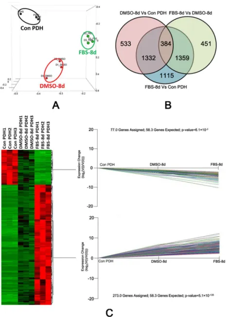
Principal component analysis (PCA) was applied to analyze the sample data from PDHs under the different culture conditions (3 samples for each condition). It showed that transcrip-tome profiles of the samples from the same culture condition were clustered together (Fig 2A). We further compared the differentially expressed gene of PDH under three different culture condition (Con-PDH, DMSO-8d and FBS-8d). A Gene with an FDR (False discovery rate) adjusted P value less than 0.05 (t-tests) and at least 2-fold change in transcript level were deemed to be differentially expressed. By three comparisons between each library pair (FBS-8d vs Con-PDH, DMSO-8d vs Con-PDH and FBS-8d vs DMSO-8d PDH), 4190, 2249 and 2194 genes were identified as DEGs, respectively. A total of 384 DEGs were overlapped, as illustrated by a Venn-diagram (Fig 2B). Of those overlapping DEGs, expression of 298 genes were up-reg-ulated and that of 86 genes down-regup-reg-ulated (S1 File). The overlapping DEGs were further ana-lyzed using K-mean clustering. The 273 up-regulated and 77 down-regulated DEGs (were assigned to two significant expression patterns as Con-PDH<DMSO-8d<FBS-8d (P value:
5.1×10−135, Fisher
’s exact test) and Con-PDH>DMSO-8d>FBS-8d (P value: 6.1×10⁻³, Fish-er’s exact test), respectively (Fig 2C).
Expression of five genes in PDH under different culture conditions were selected for valida-tion by quantitative reverse transcripvalida-tion PCR (RT-qPCR). Among them, two liver-specific expressed genes (albencoding albumin andambpfor alpha-1-microglobulin/bikunin precur-sor) were identified as DEGs by RNA-seq, and three other genes (cpd,gldcandfurinencoding carboxypeptidase D, glycine dehydrogenase and Furin, respectively) were not detected as DEGs but reported as proteins associated with DHBV infection [14–17]. Compared with that in FBS-8d PDH, PDH cultured under the culture condition supplemented with DMSO resulted in a 19-fold increase in expression of ALB and a 4-fold increase in AMBP determined by
RNA-Table 1. Summary of sequencing reads mapped to theAnas platyrhynchosreference genome.
Sample Name
PDH from duckling (NO.)
Raw Data Processed Data Mapping data
Total Reads (million)
Total Bases (Gb)
Total Reads (million)
Total Bases (Gb)
Mapped Reads (million)
Mapping Rate
1 26.40 2.22 23.26 2.04 20.79 0.89
Con-PDH 2 18.92 1.58 16.59 1.45 14.63 0.88
3 20.63 1.72 18.06 1.58 16.01 0.89
DMSO-8d 1 28.49 2.39 25.07 2.20 22.48 0.90
PDH 2 20.53 1.72 18.06 1.58 16.21 0.90
3 32.76 2.83 30.08 2.68 26.48 0.88
FBS-8d 1 23.63 1.98 20.72 1.81 18.48 0.89
PDH 2 25.78 2.16 22.63 1.98 20.03 0.89
3 23.10 2.00 21.26 1.90 18.78 0.88
Total 220.25 18.61 195.74 17.22 173.89 0.89
seq analysis (FPKM value), which was confirmed by RT-qPCR (Table 2). However,in vitro cul-ture of PDH led to dramatic reduction of ALB and AMBP expression, whether or not DMSO was applied. For the DHBV-infection-associated-genes (cpd,gldcandfurin), the presence of DMSO did not provide a distinct expression advantage over culture with FBS.
Fig 2. Expression pattern analysis of DEGs by PCA plot, Venn diagram and K-mean clustering.(A) PCA analysis of the transcriptomic data from RNA-seq. Single biological replicates are represented by dots, and PDH under different culture conditions are designated using separate colors. (B) Venn diagram of DEGs under different culture conditions. Three comparisons were performed between each library pair (Con-PDH, FBS-8d and DMSO-8d) and genes with an adjusted P value less than 0.05 and a change in transcription level of at least 2-fold were deemed to be differentially expressed. Among the comparisons, the number of overlapping genes is shown by the Venn diagram. (C) A heat map of the DEGs in PDH under various culture conditions. The columns represent triplicate samples of PDH under the three culture conditions and the relative expression level of genes are indicated by the color. Two significant K-mean clustering of the genes according to their expression pattern are shown.
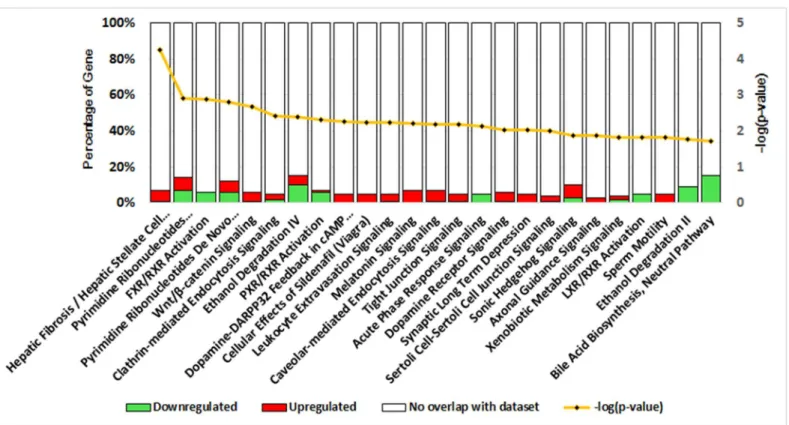
Functional analysis and biological enrichment of DEGs
To explore the biological behaviors of DEGs, DEGs were matched against the human genome using a BLAST search, with which 247 uncharacterized proteins in theAnas platyrhynchos
genome(GenBank accession:PRJNA46621)were further annotated. Ingenuity Pathway Analy-sis (IPA) software was employed to analyze the deregulated canonical pathways. A total of 44 pathways were indicated (S2 File) and the 25 most significant pathways are shown inFig 3.
Table 2. Expression of five genes validated by RT-qPCR.
Genes Functions of encoded proteins Ratio detected by qRT-PCR* Ratio detected by RNA-seq**
DMSO-8d/ Con
FBS-8d/ Con
DMSO-8d/ FBS-8d
DMSO-8d/ Con
FBS-8d/ Con
DMSO-8d/ FBS-8d
alb Main protein of human blood plasma specifically synthesized in hepatocytes.
0.02 0.01 8.00 0.05 0.00 19.24
ambp Belonging to the superfamily of lipocalin transport proteins and may play a role in the regulation of inflammatory processes.
0.09 0.02 4.50 0.50 0.12 4.30
cpd DHBV docking receptor. 0.53 0.69 0.77 0.77 1.20 0.64
gldc Involving in the membrane fusion of DHBV infection. 0.11 0.07 1.57 1.41 0.56 2.52 furin An enzyme in the endosome which can cleave DHBV large
surface protein.
0.95 0.46 2.07 0.53 0.72 0.73
*Relative expression of genes were calculated based on 2-ΔΔCTusing beta-actin as house-keeping gene.
**Ratio of FPKM value.
doi:10.1371/journal.pone.0149702.t002
Fig 3. DEGs-associated canonical pathways as indicated by IPA.Canonical pathways were analyzed by IPA software using genes that were differentially expressed in PDH under the different culture conditions. The significance (P value, yellow line) of the pathways was calculated using Fisher’s Exact Test, and the percentages of down-regulated (green bar) and up-regulated genes (red bar), respectively, in each pathway are indicated.
Among these pathways, Hepatic Fibrosis / Hepatic Stellate Cell Activation (P value: 5.75×10⁻⁵, Fisher’s exact test) was the most significant overrepresented pathway, followed by Pyrimidine Ribonucleotides Interconversion (P value: 1.25×10⁻³, Fisher
’s exact test) and FXR/RXR Activa-tion (P value: 1.28×10⁻³, Fisher
’s exact test). The ratios of DEGs in each canonical pathway ranged between 3.2% and 15.4%. Most importantly, up-regulated or down-regulated genes accounted for the majority of the DEGs in pathways including Hepatic Fibrosis / Hepatic Stel-late Cell Activation (up/down: 10/2, P value: 5.75×10⁻⁵), Ethanol Degradation II (up/down: 0/3, P value: 1.70×10⁻²) and Bile Acid Biosynthesis (up/down: 0/2, P value: 1.90×10⁻²). In addi-tion, DEGs in Tight junction signaling (up/down: 7/1, P value: 6.76×10⁻³), Caveolar-mediated Endocytosis Signaling (up/down: 4/1, P value: 6.46×10⁻³) and Clathrin-mediated Endocytosis Signaling (up/down: 6/3, P value: 3.80×10⁻³) indicated the potential association with PDH sus-ceptibility to DHBV.
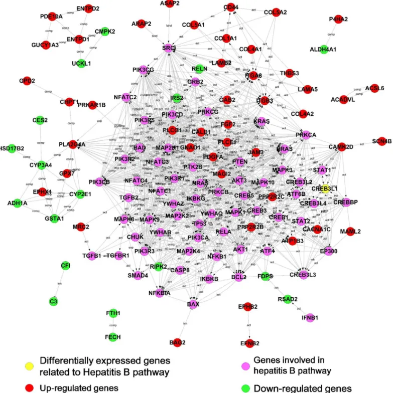
Key DEGs in DHBV infection revealed by a protein-protein interaction
network
Hepadnavirus infection in hepatocytes is a complex, multi-step process mediated by diverse proteins. To determine links between the DEGs and hepadnavirus infection, a protein-protein interaction (PPI) network was constructed based on the KEGG database. Due to the limited interaction information for duck proteins, a human protein interaction database was applied to construct the network. The proteins encoded by DEGs were pooled with 113 proteins in hepa-titis B (http://www.genome.jp/dbget-bin/get_linkdb?-t+orthology+path:map05161) as candi-dates of PPI network. The protein-protein interaction network illustrates 65 out of 384 proteins encoded by the DEGs and 48 out of 113 proteins encoded by genes belonging to hepa-titis B pathway. In the network, 49 proteins encoded by DEGs showed a direct or indirect inter-action with Hepatitis B pathway-associated proteins, and the others interact with each other among the DEGs. Most of the direct interactions were observed between proteins with similar alteration in expression under different culture conditions. Those up-regulated proteins inter-acted preferentially with proteins belonging to the Hepatitis B pathway. In the PPI network, 47 proteins encoded by DEGs were up-regulated (indicated as red nodes inFig 4) and 18 ones were down-regulated (as green nodes).
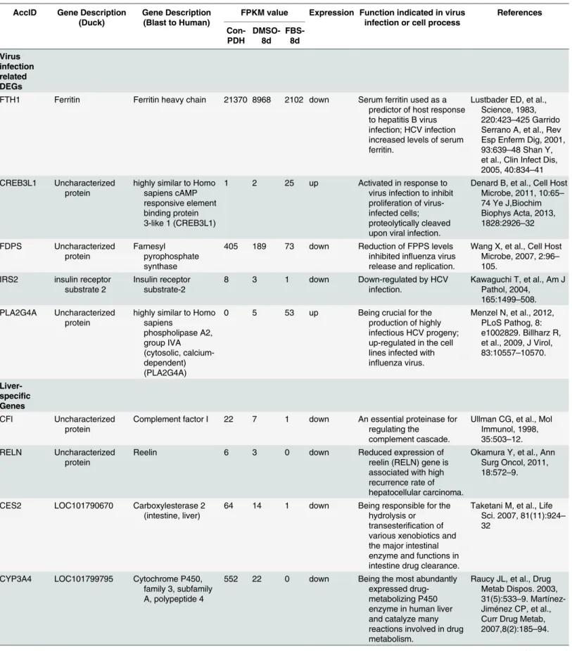
The combination of decreased expression of liver-specific genes (CYP3A4, CYP1E1, CFI, RELN and GSTA1 et al,Table 3) with increased expression of hepatocyte-dedifferentiation-associated genes (PLA2G4A and PLCG1) indicates thatin vitroculture conditions results in the fading of hepatocyte phenotype in PDHs. The presence of DMSO alleviated the hepatocyte phenotype fading and maintained the hepatocyte characters. The discrepancy of susceptibility of PDH to DHBV under different conditions was consistent with expression pattern of ferritin (FTH1) that was used as a predictor of host response to hepatitis B virus infection [18]. DEGs involving in virus infection were observed in the PPI network including CREB3L1, FDPS and IRS2 (Table 3), in which CREB3L1 was reported to be associated with iCell hepatocyte
(induced pluripotent stem cell derived hepatocytes from Cellular Dynamics International) sus-ceptibility to HBV infection [19]. It was unexpected that three genes (RSAD2, CMPK2, and RIPK2) playing roles in anti-viral immune responses (Table 3) showed down-regulated expres-sion in PDHs underin vitroculture.
Expression of seven DEGs associated with tight junction formation, including JAM3, PPP2R2B, PRKAR1B, PPP2R2C, MAGI2, ACTA2 and ACTG2, showed up-regulation pattern, which is in accordance with GO analysis of tight junction signaling pathway (P value:
1.25×10⁻³, Fisher
Fig 4. Interaction networks between DEGs and genes involved in the Hepatitis B pathway.The proteins encoded by DEGs (red, green or yellow nodes) or involved in the Hepatitis B pathway (purple nodes) in the KEGG database were extracted to construct a protein–protein interaction network. The lines in the network represent protein–protein interactions, including binding/association, phosphorylation, activation, and inhibition. Proteins encoded by up-regulated or down-up-regulated DEGs are indicated in red or green, respectively.
Table 3. Role of the differentially expressed genes in virus infection or cell process.
AccID Gene Description (Duck)
Gene Description (Blast to Human)
FPKM value Expression Function indicated in virus infection or cell process
References Con-PDH DMSO-8d FBS-8d Virus infection related DEGs
FTH1 Ferritin Ferritin heavy chain 21370 8968 2102 down Serum ferritin used as a predictor of host response to hepatitis B virus infection; HCV infection increased levels of serum ferritin.
Lustbader ED, et al., Science, 1983, 220:423–425 Garrido Serrano A, et al., Rev Esp Enferm Dig, 2001, 93:639–48 Shan Y, et al., Clin Infect Dis, 2005, 40:834–41 CREB3L1 Uncharacterized
protein
highly similar to Homo sapiens cAMP responsive element binding protein 3-like 1 (CREB3L1)
1 2 25 up Activated in response to virus infection to inhibit proliferation of virus-infected cells; proteolytically cleaved upon viral infection.
Denard B, et al., Cell Host Microbe, 2011, 10:65–
74 Ye J,Biochim Biophys Acta, 2013, 1828:2926–32
FDPS Uncharacterized protein
Farnesyl pyrophosphate synthase
405 189 73 down Reduction of FPPS levels inhibited influenza virus release and replication.
Wang X, et al., Cell Host Microbe, 2007, 2:96–
105. IRS2 insulin receptor
substrate 2
Insulin receptor substrate-2
8 3 1 down Down-regulated by HCV
infection.
Kawaguchi T, et al., Am J Pathol, 2004, 165:1499–508. PLA2G4A Uncharacterized
protein
highly similar to Homo sapiens phospholipase A2, group IVA (cytosolic, calcium-dependent) (PLA2G4A)
0 5 53 up Being crucial for the
production of highly infectious HCV progeny; up-regulated in the cell lines infected with influenza virus.
Menzel N, et al., 2012, PLoS Pathog, 8: e1002829. Billharz R, et al., 2009, J Virol, 83:10557–10570.
Liver-specific Genes
CFI Uncharacterized protein
Complement factor I 22 7 1 down An essential proteinase for regulating the
complement cascade.
Ullman CG, et al., Mol Immunol, 1998, 35:503–12. RELN Uncharacterized
protein
Reelin 6 3 0 down Reduced expression of
reelin (RELN) gene is associated with high recurrence rate of hepatocellular carcinoma.
Okamura Y, et al., Ann Surg Oncol, 2011, 18:572–9.
CES2 LOC101790670 Carboxylesterase 2 (intestine, liver)
64 14 1 down Being responsible for the hydrolysis or transesterification of various xenobiotics and the major intestinal enzyme and functions in intestine drug clearance.
Taketani M, et al., Life Sci. 2007, 81(11):924–
32
CYP3A4 LOC101799795 Cytochrome P450, family 3, subfamily A, polypeptide 4
552 22 0 down Being the most abundantly expressed drug-metabolizing P450 enzyme in human liver and catalyze many reactions involved in drug metabolism.
Raucy JL, et al., Drug Metab Dispos. 2003, 31(5):533–9. Martínez-Jiménez CP, et al., Curr Drug Metab, 2007,8(2):185–94.
Table 3. (Continued)
AccID Gene Description (Duck)
Gene Description (Blast to Human)
FPKM value Expression Function indicated in virus infection or cell process
References Con-PDH DMSO-8d FBS-8d ADH1A Uncharacterized protein Alcohol dehydrogenase 1A (class I), alpha polypeptide
132 60 11 down Catalyzing the oxidation of alcohols to aldehydes.
Lange LG, Biochemistry, 1976, 15(21): 4687–93.
GSTA1 LOC101797566 Glutathione S-transferase A1
601 209 9 down Function in the detoxification of electrophilic
compounds by conjugation with glutathione.
Karpusas M, et al., PLoS One. 2013, 8(2): e56337
CYP2E1 LOC101796596 Cytochrome P450 family 2 subfamily E polypeptide 1 (vase tunicate).
142 42 2 down Catalyzing many reactions involved in drug
metabolism and synthesis of cholesterol, steroids and other lipids.
Deka M, et al., World J Gastroenterol. 2010, 16 (38):4800–8.
Innate immune response related with anti-virus infection
RSAD2 Radical S-adenosyl methionine domain-containing protein 2
Highly similar to Homo sapiens viperin (cig5)
124 15 1 down A cellular protein which could inhibit many DNA and RNA viruses such as CHIKV, HCMV, HCV, DENV, WNV, SINV, influenza, HIV LAI strain.
Teng TS, et al., J Clin Invest, 2012, 122:4447–60; Cho H, et al., Nat Med. 2013;19(4):458–64; Thomas E, et al., Gastroenterology, 2012,142:978–88 CMPK2 Uncharacterized protein Mitochondrial cytidine monophosphate (UMP-CMP) kinase 2
75 22 2 down Antiviral ISGs in primary neurons induced by
IFN-βtreatment.
Li J, et al., Glia, 2011, 59:58–67
RIPK2 Uncharacterized protein
Highly similar to Receptor-interacting serine/threonine-protein kinase 2 (EC 2.7.11.1)
40 16 7 down Modulated by HBeAg which supporting the concept that HBeAg could impair both innate and adaptive immune responses to promote chronic HBV infection; NOD2-RIPK2 signaling in protection against virally triggered immunopathology.
Wu S, et al., J Infect Dis.2012, 206:415–20; Lupfer C, et al., Nat Immunol, 2013, 14: 480–488
Tight junction related DEGs ACTA2 Uncharacterized protein
Highly similar to Actin, aortic smooth muscle
13 93 819 up Playing a role in cell motility, structure and integrity.
Lee HW, et al., Clin Cancer Res, 2013, 19:5879–89. JAM3 Uncharacterized
protein
Junctional adhesion molecule C
0 2 9 up Interact with the
coxsackievirus and adenovirus receptor (CAR).
Mirza M, et al., Exp Cell Res. 2006, 312:817–
30.
than that in FBS-8d PDHs, suggesting that DMSO-8d PDH had less tight junction formation that serves a blockade of HBV infection [13].
DHBV attachment to PDH with or without DMSO was carried out. DHBV were inoculated into PDH maintained under the three different culture conditions, and incubated at 4°C for 2 h. Anti-Pres/s of DHBV was used as control, which could block DHBV attaching to PDH. After washing of free virus, the attached DHBV were detected by qPCR. A decrease of DHBV attachment was observed in the presence of anti-Dpres/s for Con PDH. Attached DHBV in FBS-8d PDH (2.94-fold decrease) were, lower than that in DMSO-8d PDH (1.37-fold decrease), compared with Con PDH (Fig 5).
Discussion
Hepadnaviruses exhibit a strong liver tropism and preferentially replicate in hepatocytes, the predominant liver cell type. Primary cultured hepatocytes remain the most important cell model used to elucidate the molecular pathogenesis underlying HBV infection. However, it
Table 3. (Continued)
AccID Gene Description (Duck)
Gene Description (Blast to Human)
FPKM value Expression Function indicated in virus infection or cell process
References Con-PDH DMSO-8d FBS-8d PPP2R2B Serine/threonine-protein phosphatase 2A 55 kDa regulatory subunit B Serine/threonine-protein phosphatase 2A 55 kDa
regulatory subunit B beta isoform
0 2 22 up Involved in the negative control of cell growth and division.
Cheng WT, et al., BMC Cell Biol, 2009,10:91
PRKAR1B Uncharacterized protein
Protein kinase, cAMP-dependent, regulatory, type I, beta, isoform CRA_b
1 10 63 up Controling many biochemical events in the cell including regulation of metabolism, ion transport, and gene transcription.
Elphinstone MS, et al., Clin Endocrinol (Oxf), 2004, 61:716–23.
PPP2R2C Serine/threonine-protein phosphatase 2A 55 kDa regulatory subunit B Serine/threonine-protein phosphatase 2A 55 kDa
regulatory subunit B gamma isoform
0 3 10 up Involved in the negative control of cell growth and division.
Backx L, et al., Eur J Med Genet, 2010, 53:239–
43.
MAGI2 Uncharacterized protein
Highly similar to WW andPDZ domain-containing protein 2
0 0 1 up Acting as scaffold molecule at synaptic junctions by assembling
neurotransmitter receptors and cell adhesion proteins.
Wapenaar MC, et al., Gut, 2008, 57:463–7
ACTG2 Actin, gamma-enteric smooth muscle
cDNA FLJ42347fis, clone
UTERU2003399
13 101 271 up Involved in various types of cell motility and in the maintenance of the cytoskeleton.
Citi S, et al., Ann N Y Acad Sci, 2009,1165:88–98
CLDN2 Claudin Claudin-2 16 4 1 down Expressed in an
organ-specific manner and regulate tissue-specific physiologic properties of tight junctions.
Wu F, et al., Exp Eye Res, 2008, 87:214–25; Rosenthal R et al., J Cell Sci,
2010,123:1913–21; Van Itallie CM, et al., Mol Biol Cell, 2009, 20:3930–40.
remains unclear why primary hepatocytes gradually lose susceptibility to DHBV infection underin vitroculture conditions. Supplementation with DMSO can prolong PDH susceptibil-ity to hepadnavirus [8]. In this study, DEGs were identified in PDH cultured under conditions with or without DMSO with RNA-seq analysis. Our results indicated that multiple factors were affected by DMSO treatment, and differential cell-cell junction may be one of the reasons lead-ing to differential susceptibility in PDHin vitro.
For screening the genes of interest, we compared transcriptome data from PDHs cultured with 1.5% DMSO or 5% FBS, and fresh isolated PDH was used as a control. To identify related DEGs associated with PDH susceptibility to DHBV infection, a Venn diagram was used to visualize overlapping DEGs across the three comparisons (FBS-8d/Con-PDH, FBS-8d/DMSO-8d, DMSO-8d/Con-PDH). Among the DEGs (fold change>2 and FDR adjusted p-value<0.05)
observed in the three comparisons, a total of 384 overlapping genes were obtained. Meanwhile, significant changes in 350 overlapping genes (273 up-regulated and 77 down-regulated genes) were enriched in two expression patterns (Con-PDH<DMSO-8d<FBS-8d and
Con-PDH>DMSO-8d>FBS-8d), which provided further confirmation of a relationship between
the DEGs and PDH susceptibility to DHBV infection. In another point of view, FPKM values of the down-regulated gene, which may be helpful in DHBV infection, are supposed to be higher than that of up-regulated ones. Therefore, FPKM values of the overlapped DEGs in Con PDH were compared by t-test. It indicated FPKM values of down-regulated gene were higher than that of up-regulated ones (p<0.0001,S1 Fig).
Fig 5. Differential DHBV attachment to PDH maintained under different conditions.PDH with differential susceptibility was pretreated with nocodazole and infected with DHBV at a MGE of 500. The DHBV attached to PDH were quantified by qPCR. Anti-Dpres/s or IgG were used as control. The asterisk represents a significant difference (t test, P value<0.05).
To obtain more protein information, DEGs were matched against the human genome using a BLAST search and 247 uncharacterized proteins were further annotated. Then Proteins encoded by overlapped DEGs and those from hepatitis B pathway were pooled together to con-struct a protein-protein interaction network. In the PPI network, a cluster of tight junction associated proteins (JAM3, PPP2R2B, PRKAR1B, PPP2R2C, MAGI2, ACTA2 and ACTG2) were up-regulated, suggesting that the formation of tight cell-cell junction between hepatocytes culturedin vitroover time. Since it has been assumed that tight junction formation creates a physical barrier preventing virus access to basolateral localized receptor(s), cell-cell junction alteration may contribute to differential susceptibility to DHBV between PDHs under different conditions. Under the culture condition with DMSO, tight junction formation between PDHs seemed to be retarded, evidenced by lower expression of tight junction genes than that in the absence of DMSO. The result will drive us to focus on molecular pathway of tight junction for-mation and virus entry in further investigation of influence of DMSO on hepadnavirus infection.
It is admitted that impacts of DMSO involve in more events of virus life cycle. Therefore, we analyzed the DHBV attachment, replication and release in PDH under different culture condi-tions. We have studied the effect of DMSO on DHBV attachment in PDHs (Fig 5) and virus replication in the LMH-D2 cells (S2 Fig). It showed that DHBV attachment and replication were increased in the presence of DMSO, indicating the extensive effect of DMSO on DHBV life cycle.
Differential susceptibility of PDH to DHBV also could be due to differential virus defense response to DHBV infection in PDHs under different culture condition. We observed down-regulated expression of anti-virus genes (RSAD2, CMPK2, and RIPK2), suggesting the collapse of cellular anti-virus immune concomitant with the loss of hepatic characters in PDHin vitro. It could rule out the possibility that weaken susceptibility of PDH to DHBV is the consequence of enhancement of cell defense against virus infection. In addition, expression of FTH1, a pre-dictor of host response to hepatitis B virus infectionin vivo[18] was down-regulated in FBS-8d PDH but partially recovered in DMSO-8d PDH. Expression of FTH1 could be used to predict availability of cell model for DHBV/HBV infection or to screen anti-virus infection chemicals. We also analyzed the expression of genes which play important roles in the DHBV infec-tion, such ascpd,gldcandfurin. CPD is the docking receptor and binds to DHBV particles attached to heparin sulfate and co-factors on the hepatocyte membrane [14]. DHBV particles that bind to CPD are internalized into the cytoplasm, forming endosomes where the large pro-tein on the DHBV membrane is cleaved by the Furin enzyme [15]. The cleaved large DHBV protein then binds to GLDC [16,17], followed by the membrane fusion of the DHBV particle and the endosome. NTCP is considered to be a functional receptor on the hepatocyte mem-brane and leads to the susceptibility of HepaG2 cell to HBV infection [2]. However, low FPKM value of NTCP was observed in PDH under different conditions (0.068, 0.36 and 0.28 in PDH-Con, DMSO-8d and FBS-8d, respectively) andcpd,gldc,furinand NTCP were not indi-cated as DEGs both by RNA-seq analysis and RT-qPCR.
Despite the candidate genes revealed in this study, further studies are still needed to investi-gate the association between these candidate genes and PDH susceptibility to DHBV infection. The mechanisms underlying the contribution of these genes to DHBV infection will be helpful for establishing new strategy against hepadnavirus infection.
Conclusion
network was constructed by pooling the DEGs with proteins belonging to hepatitis B pathway. The combination of decreased expression of liver-specific genes with increased expression of hepatocyte-dedifferentiation-associated genes suggested that the fading of hepatocyte pheno-type of PDHs were resulted byin vitroculture conditions. At the same time, the expression of seven DEGs associated with tight junction formation was up-regulated, which was attenuated by DMSO. The study paves the way to fully understand mechanism underlying DHBV life cycle and to establish new strategy against hepadnavirus infection.
Materials and Methods
Ethics statement
All animal experiments with Cherry Valley ducklings that we performed in this study were approved by the Institutional Animal Care and Use Committee (IACUC) of Fudan University (NO.20150305).
Experimental animals and cell lines
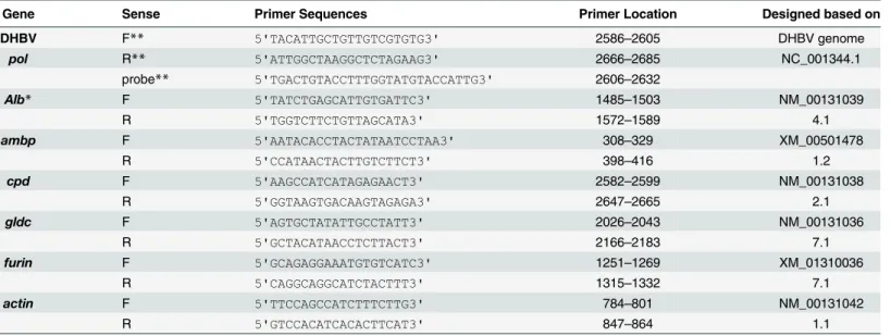
The 1-day-old Cherry Valley ducklings (Anas domesticus) were purchased from Breeding Center of Shanghai Institute of Veterinary Medical Sciences, Shanghai, China. The ducklings without of DHBV infection were used for PDH preparation, DHBV in the serum of ducklings were detected by qPCR with the specific primers (based on DHBV genome NC_001344.1), listed inTable 4.
LMH-D2 cells (a generous gift of Dr. William Mason, Institute for Cancer Research, Phila-delphia, Pa. USA), were maintained in DMEM F12 with 10% FBS and constitutively secreted DHBV virions into the supernatant [20].
PDH preparation and culture
The PDH were isolated from the livers of 1-day-old cherry valley ducklings as previously described [21]. Briefly, DHBV-negative ducklings were anesthetized with 0.75%
Table 4. Primers for detection of DHBV DNA and differential expressed genes.
Gene Sense Primer Sequences Primer Location Designed based on
DHBV F** 5'TACATTGCTGTTGTCGTGTG3' 2586–2605 DHBV genome
pol R** 5'ATTGGCTAAGGCTCTAGAAG3' 2666–2685 NC_001344.1
probe** 5'TGACTGTACCTTTGGTATGTACCATTG3' 2606–2632
Alb* F 5'TATCTGAGCATTGTGATTC3' 1485–1503 NM_00131039
R 5'TGGTCTTCTGTTAGCATA3' 1572–1589 4.1
ambp F 5'AATACACCTACTATAATCCTAA3' 308–329 XM_00501478
R 5'CCATAACTACTTGTCTTCT3' 398–416 1.2
cpd F 5'AAGCCATCATAGAGAACT3' 2582–2599 NM_00131038
R 5'GGTAAGTGACAAGTAGAGA3' 2647–2665 2.1
gldc F 5'AGTGCTATATTGCCTATT3' 2026–2043 NM_00131036
R 5'GCTACATAACCTCTTACT3' 2166–2183 7.1
furin F 5'GCAGAGGAAATGTGTCATC3' 1251–1269 XM_01310036
R 5'CAGGCAGGCATCTACTTT3' 1315–1332 7.1
actin F 5'TTCCAGCCATCTTTCTTG3' 784–801 NM_00131042
R 5'GTCCACATCACACTTCAT3' 847–864 1.1
*Designed based on assembled transcripts in RNA-seq
**F: forward primer, R: reverse primer, Probe: FAM™dye-labeled TaqMan MGB probe.
pelltobarbitalum natricum (10 mg/kg) and the portal vein was exposed. The liver was perfused with 80 ml of hepatocyte perfusion medium (Invitrogen, Carlsbad, CA, USA) and digested with collagenase IV (Sigma, St. Louis, MO, USA) via the portal vein. After removal of the blade, the liver was dispersed in hepatocyte wash medium (Invitrogen, Carlsbad, CA, USA), and the hepatocytes were separated from the non-parenchymal cells by centrifugation at 50×g. After washing, the hepatocytes were suspended in Leibovitz’s L-15 medium (Invitrogen, Carls-bad, CA, USA) supplemented with 5% FBS (Invitrogen, CarlsCarls-bad, CA, USA), 100 units/ml Pen-icillin-Streptomycin (Invitrogen, Carlsbad, CA, USA), 1 mg/L insulin (Sigma, St. Louis, MO, USA), and 10−5M hydrocortisone-hemisuccinate (Sigma, St. Louis, MO, USA), and seeded at a density of 1×10⁷cells per 10 cm dish. The PDH were maintained at 37°C without CO2. After 24
h post-inoculation, the PDH were maintained in L-15 medium supplemented with 5% FBS or 1.5% DMSO for 7 d.
Preparation and quantification of DHBV
For viral infection, virions of DHBV in the LMH D2 cell supernatants were precipitated with 10% PEG-8000 and dissolved in L-15 medium. The DHBV DNA in 200μl L-15 medium was extracted with a TIANamp Virus DNA/RNA Kit (Tiangen, Beijing, China), followed by quan-tification using a TaqMan probe-based qPCR assay, which carried out with the primers and probe listed inTable 4. The qPCR amplification was performed with the Takara Probe PCR Kit on an Eppendorf RealPlex 4, using 50 cycles of a 2-step PCR program.
DHBV infection and detection of viral cccDNA and core DNA
The PDH were maintained in Leibovitz's L-15 Medium supplemented with 5% FBS for 1 d (Con-PDH), and then followed by culture in media with 5% FBS (FBS-8d PDH) or 1.5% DMSO (DMSO-8d PDH) for 7 days. DHBV infection was carried out at a MGE of 100 by incu-bating overnight at 37°C. Then the inoculum was removed and L-15 medium supplemented with 5% FBS was added to the cells. On the 5thday post-DHBV infection, PDH and culture supernatants were collected for isolation of DHBV cccDNA, and core DNA.
To isolate DHBV core DNA, PDH in a 35-mm-diameter dish were lysed with 0.5 ml of lysis buffer (1% NP-40, 0.15M NaCl, 50mM Tris-Cl 8.0, 1mM EDTA, 1×Protease inhibitor). Fifteen units of Micro Nuclease (Roche Diagnostics, Basel, Switzerland) were added to the lysate, incu-bated for 45 min at 37°C. The supernatants were collected by centrifuging at 14,000 rpm for 1 min, and the DHBV core particles were concentrated by adding PEG-8000 to a final concentra-tion 10% (w/v). After centrifuging at 13,000 rpm for 5 min, the pellet was suspended in TNE lysis buffer (10 mM Tris-HCl, pH8.0, 100 mM NaCl, 1 mM EDTA, 0.5% SDS, 0.6μg/μl pro-teinase K (Invitrogen, Carlsbad, CA, USA)), and incubated at 37°C for 1 h. The DHBV core DNA was extracted twice with Tris-buffered phenol/chloroform (pH 7.4), followed by one extraction with chloroform and precipitation by adding 3 volumes of 100% ethanol. The extracted DHBV core DNA was suspended in 20μl of TE.
To isolate DHBV cccDNA, PDH were lysed in 1 ml of cccDNA extraction buffer containing 50 mM Tris-HCl, pH 8.0, 150 mM NaCl, 10 mM EDTA and 1% SDS. Then 250μl of 2.5 M KCl was added to the lysate, rotated overnight at 4°C and centrifuged at 18,000 ×gfor 30 min at 4°C. The DHBV cccDNA was extracted from the supernatant 3 times with phenol and once with chloroform. After adding 2.2 volumes of absolute ethanol and incubating at -80°C over-night, precipitated DHBV cccDNA was centrifuged at 18,000 ×gfor 20 min, washed 3 times with 75% Ethanol and dissolved in 35μl of TE buffer.
p32-labeled probes which were synthesized with a random-primed DNA Labeling Kit (Roche Diagnostics, Basel, Switzerland); the signals were exposed to a Phosphor screen for 24 h, and visualized by a PhosphorImager (Fujifilm, Kanagawa, Japan).
Detection of DHBV attachment to PDH
The detection of DHBV attachment to PDH was described by Funk A et al [22]. In brief, PDH in 6-well plate were pretreated with 16.5μM nocodazole for 1 h. The DHBV was inoculated at a MGE of 500 and incubated at 4°C for 2 h. DHBV premixed with anti-Dpres/s monoclonal antibody (1:1000) (a generous gift of Dr. William Mason in Fox Chase Cancer Center) or IgG (1:1000) were used as controls. After incubation with DHBV, the cells were washed with cold PBS for 5 times and lyzed with 200μl lysis buffer (50 mM KCl, 10 mM Tris-HCl [pH 8.3], 0.45% Tween 20, 0.45% Nonidet P-40, 0.1 mg/ml proteinase K). The lysates were centrifuged at 16100 g for 10 min followed by DHBV DNA extraction with TIANamp Virus DNA/RNA Kit. Then DHBV DNA was quantified by qPCR.
Preparation of the PDH cDNA library, and sequencing
For transcriptome analysis, cDNA libraries were established for the PDH samples. The total RNA was extracted from PDH under three culture conditions (in Leibovitz's L-15 Medium supplemented with 5% FBS for 1 d, followed by culturing in media with 5% FBS or 1.5% DMSO for 7 d) using Trizol reagent following the manufacturer’s protocol (Invitrogen, Carls-bad, CA, USA). Then, poly-A mRNA was isolated from 10μg total RNA using a TruSeq1RNA Sample Preparation v2 kit (Illumina Inc., San Diego, CA, USA) and was fragmented at 94°C for 8 min. The first and second cDNA strands were synthesized successively, followed by the purification of double-stranded cDNA using AMPure XP beads (Beckman Coulter, Brea, CA, USA). After end repair, cDNA was adenylated at the 30end, ligated to the sequencing adapters and amplified by PCR. The quality and quantity of the cDNA library was determined with an Agilent DNA 1000 Kit and an Agilent 2100 Bioanalyzer (Agilent Technologies, Palo Alto, CA, USA). PDH cDNA libraries were sequenced on the Illumina Solexa platform using the pair-end approach and the Illumina data processing pipeline.
Processing and mapping of Illumina reads
In order to rule out sequencing errors, all raw reads obtained from the Illumina platform were pre-processed prior to the next step. Clean reads were obtained by removing the adaptor sequences, reads with>5% ambiguous bases (noted as N) and low-quality reads containing
more than 20 percent of bases with qualities of<20. High-quality reads were mapped to the
Anas platyrhynchosgenome and transcripts were downloaded from ENSEMBL (http://www. ensembl.org) with TopHat v2.0.12 software (http://ccb.jhu.edu/software/tophat/index.shtml). TopHat allows multiple alignments per read and the default parameters were used. Cufflinks v2.2.1 software (http://cufflinks.cbcb.umd.edu/) was later used for analyses that included tran-script assembly and FPKM value calculations to quantify gene expression; this program was also run with default parameters. Genes with an FDR adjusted P value less than 0.05 and a change in levels of at least 2-fold were considered to be differentially expressed.
Validation of DEGs by real-time PCR
manufacturer’s protocols (Takara, Dalian, China) on a Mastercycler ep realplex (Eppendorf, Hamburg, Germany). The specific primers for each gene were designed using Beacon designer 7.5 software (Palo Alto, CA, USA), and are listed inTable 4.
Canonical pathway analysis by IPA
The differentially expressed genes with corresponding expression values were uploaded to the IPA Software, and analyzed by canonical pathway. We used Fisher’s exact test to select the sig-nificant pathways and the significance threshold was defined by a P value of 0.05. The signifi-cance of the pathway was indicated by the ratio of the number of genes form the dataset that mapped to the pathway to the total number of genes present in the canonical pathway.
Construction of protein-protein interaction network
A protein-protein interaction network was constructed using Cytoscape software. The candidate proteins are encoded by DEGs in the present study and from proteins involved in hepatitis B pathway in KEGG (http://www.genome.jp/dbget-bin/get_linkdb?-t+orthology+path:map05161). A total number of 113 genes which were reported involved in hepatitis B pathway were extracted. Due to the limited interaction information for duck proteins, DEGs identified by RNA-seq analy-sis were matched against the human genome using BLAST method, and matched ones were pooled together as candidate genes. The protein–protein interaction network was constructed according to the functional relationships annotated in the KEGG database. In the network, lines were used to represent protein–protein interactions including binding/association, phosphoryla-tion, activaphosphoryla-tion, and inhibition. Proteins encoded by up-regulated or down-regulated DEGs were indicated in red or green, respectively, while purple nodes were used to present proteins involved in the hepatitis B pathway.
Statistical analysis
T test was used to filter the differentially expressed genes, after the significant analysis and FDR analysis under the following criteria: i) Fold Change>2 or<0.5; ii) FDR<0.05. Fisher’s exact
test was used in K-means clustering and Canonical pathway analysis. A P value<0.05 was
con-sidered to be statistically significant.
Supporting Information
S1 Fig. Comparison of FPKM values of up- and down-regulated DEGs in Con PDH.The FPKM values overlapped up- and down-regulated genes in Con PDH were compared by t-test. (TIF)
S2 Fig. Effects of DMSO on DHBV replication in LMH D2 cell.The LMH D2 cell were cul-tured in DMEM-F12 medium with 0.5% or 1.5% DMSO. LMH D2 cell under normal culture conditions were used as control. The cccDNA and core DNA were extracted on the 1st, 3rd, 5th and 7th day post DMSO treatment and detected by Southern Blot. D: DMSO group; C: Control group.
(TIF)
S1 File. Differential expressed genes associated with PDH susceptibility to DHBV infec-tion.
(XLSX)
Acknowledgments
We gratefully acknowledge Professor Zonghou Shen (Department of Biochemistry and Molec-ular Biology, Fudan University) for suggestions on analyzing the DGEs associated with PDH susceptibility to DHBV infection and Professor Jianming Hu (Department of Microbiology and Immunology, Pennsylvania State University) for helping on DHBV cccDNA and core DNA detection by Southern Blot.
Author Contributions
Conceived and designed the experiments: DQ YX JZ. Performed the experiments: LY SQ. Ana-lyzed the data: GL LL YY. Contributed reagents/materials/analysis tools: YZ GD. Wrote the paper: LY SQ DQ YX YL.
References
1. Lavanchy D (2005) Worldwide epidemiology of HBV infection, disease burden, and vaccine prevention. J Clin Virol 34 Suppl 1: S1–3. PMID:16461208
2. Yan H, Zhong G, Xu G, He W, Jing Z, Gao Z, et al. (2012) Sodium taurocholate cotransporting polypep-tide is a functional receptor for human hepatitis B and D virus. Elife 1: e00049. doi:10.7554/eLife. 00049PMID:23150796
3. Summers J, Mason WS (1982) Replication of the genome of a hepatitis B—like virus by reverse tran-scription of an RNA intermediate. Cell 29: 403–415. PMID:6180831
4. Tuttleman JS, Pugh JC, Summers JW (1986) In vitro experimental infection of primary duck hepatocyte cultures with duck hepatitis B virus. J Virol 58: 17–25. PMID:3512855
5. Tohidi-Esfahani R, Vickery K, Cossart Y (2010) The early host innate immune response to duck hepati-tis B virus infection. J Gen Virol 91: 509–520. doi:10.1099/vir.0.015529-0PMID:19846670
6. Qiao M, Scougall CA, Duszynski A, Burrell CJ (1999) Kinetics of early molecular events in duck hepati-tis B virus replication in primary duck hepatocytes. J Gen Virol 80 (Pt 8): 2127–2135. PMID:10466812
7. Funk A, Mhamdi M, Hohenberg H, Will H, Sirma H (2006) pH-independent entry and sequential endo-somal sorting are major determinants of hepadnaviral infection in primary hepatocytes. Hepatology 44: 685–693. PMID:16941679
8. Pugh JC, Summers JW (1989) Infection and uptake of duck hepatitis B virus by duck hepatocytes main-tained in the presence of dimethyl sulfoxide. Virology 172: 564–572. PMID:2678730
9. Kost DP, Michalopoulos GK (1991) Effect of 2% dimethyl sulfoxide on the mitogenic properties of epi-dermal growth factor and hepatocyte growth factor in primary hepatocyte culture. J Cell Physiol 147: 274–280. PMID:1828251
10. Gripon P, Diot C, Theze N, Fourel I, Loreal O, Brechot C, et al. (1988) Hepatitis B virus infection of adult human hepatocytes cultured in the presence of dimethyl sulfoxide. J Virol 62: 4136–4143. PMID:
3172341
11. Ezzell RM, Toner M, Hendricks K, Dunn JC, Tompkins RG, Yarmush ML (1993) Effect of collagen gel configuration on the cytoskeleton in cultured rat hepatocytes. Exp Cell Res 208: 442–452. PMID:
8375473
12. Pugh JC, Di Q, Mason WS, Simmons H (1995) Susceptibility to duck hepatitis B virus infection is asso-ciated with the presence of cell surface receptor sites that efficiently bind viral particles. J Virol 69: 4814–4822. PMID:7609048
13. Schulze A, Mills K, Weiss TS, Urban S (2012) Hepatocyte polarization is essential for the productive entry of the hepatitis B virus. Hepatology 55: 373–383. doi:10.1002/hep.24707PMID:21953491
14. Locarnini S, Warner N (2013) The new front-line in hepatitis B/D research: Identification and blocking of a functional receptor. Hepatology.
15. Tong Y, Tong S, Zhao X, Wang J, Jun J, Park J, et al. (2010) Initiation of duck hepatitis B virus infection requires cleavage by a furin-like protease. J Virol 84: 4569–4578. doi:10.1128/JVI.02281-09PMID:
20181690
16. Li J, Tong S, Wands JR (1999) Identification and expression of glycine decarboxylase (p120) as a duck hepatitis B virus pre-S envelope-binding protein. J Biol Chem 274: 27658–27665. PMID:10488106
18. Lustbader ED, Hann HW, Blumberg BS (1983) Serum ferritin as a predictor of host response to hepati-tis B virus infection. Science 220: 423–425. PMID:6301008
19. Chiao E, Hamilton MM, Kameoka S, Leonard B, Triyatni M (2014) Compounds for improved stem cell differentiation into hepatocytes. Google Patents.
20. Condreay LD, Aldrich CE, Coates L, Mason WS, Wu TT (1990) Efficient duck hepatitis B virus produc-tion by an avian liver tumor cell line. J Virol 64: 3249–3258. PMID:2352324
21. Zhao Y, Ben H, Qu S, Zhou X, Yan L, Xu B, et al. (2010) Proteomic analysis of primary duck hepato-cytes infected with duck hepatitis B virus. Proteome Sci 8: 28. doi:10.1186/1477-5956-8-28PMID:
20529248