ISSN 1553-345X
© 2009 Science Publications
Corresponding Author: Shaon RayChaudhuri, Department of Biotechnology, West Bengal University of Technology, BF-142, Sector 1, Salt Lake, Calcutta-700064, India Tel: 00919831034236 Fax: 009133233441030
Radiation Induced DNA Double Strand Break Studies of a Metal Sensitive
Novel Bacterial Isolate from East Calcutta Wetland
1
Sanhita Chowdhury,
2Anindita Chakraborty,
3Ashoke Ranjan Thakur
and
1Shaon Ray Chaudhuri
1Department of Biotechnology, West Bengal University of Technology,
BF-142, Sector 1, Salt Lake, Calcutta-700064, India
2
UGC-DAE Consortium for Scientific Research, Calcutta Centre,
III/LB-8, Bidhan Nagar, Calcutta-700064, India
3
West Bengal State University, (Barasat North 24 Parganas),
Barasat Government College (Annexe Building), 10KNC Road, Kolkata-00124, India
Abstract: Problem statement: This study was an attempt to isolate anaerobic microbes with potential for DNA double strand break repair using methanogen specific medium (DSMZ 120) from East Calcutta Wetland in India. It also intended to verify the specificity of the medium for isolation of the desired family of microbe. Approach: Culture based technique was used to obtain the pure isolate that was further characterized in details. For double strand break repair studies, isolate was irradiated with different doses of 60Co gamma rays and its subsequent repair was observed using pulse field gel electrophoresis and asymmetric field inversion gel electrophoresis. Inhibitor was used to predict the mechanism of repair. Results: In this study we isolated and characterized a metal sensitive anaerobic microbial strain obtained using methanogen specific medium (DSMZ 120) from East Calcutta Wetland in India. The strain was one of the members of the group of uncultivated bacterium as evident from phylogenetic analysis, thus indicating the successful cultivation of an as yet uncultivable novel microbe (GenBank Acc. No. FJ 930097) and also the non-specific growth of microbes in prescribed medium. It was a Gram positive Bacilli, member of Fermicutes with optimum growth at 25°C and pH-7. The growth curve analysis showed a lag phase up to 24 h, log phase from 24-48 h, an early stationary phase from 96 h onwards. The strain could repair the DNA double strand break caused by irradiation with 60Co γ rays. The dose profile study revealed maximum repair at 60 Grays and thereafter a drop in repair ability with increase in irradiation dose. The time required for repair showed an essential incubation period of 4 h. The DNA polymerase inhibitor, Arabinose CTP inhibited the repair indicating the involvement of polymerase in the repair process and thus pointing towards homologous recombination as the underlying mechanism. Conclusion: In this study we were able to cultivate an as yet uncultivable anaerobic bacterial isolate and predict the growth conditions for the isolate. On irradiation with 60Co γ rays the isolate showed maximum repair following 60 Gray damage. DNA polymerase inhibitor arabinose CTP inhibited the repair mechanism completely. This indicated that DNA polymerase took active part in repair process and thus the mechanism was that of homologous recombination repair.
Key words: DNA double strand break repair, East Calcutta Wetland, Methanogen specific media, Homologous recombination
INTRODUCTION
DNA molecules can be damaged in many ways. Spontaneous damage may occur due to replication errors, deamination, depurination and oxidation. DNA molecules may also get damaged by the effects of
of damage which unless repaired leads to cell death. The DNA repair ability of a cell is vital to the integrity of its genome and thus to the organism. DSB can be repaired by two different types of mechanism. One type takes advantage of proteins that promote homologous recombination to obtain instructions from the sister or homologous chromosome for proper repair of breaks. The other type permits joining of ends even if there is no sequence similarity between them. The latter process is called non-homologous end joining[1]. In bacteria and yeast homologous recombination is the process for repairing the DNA double strand break, while vertebrates mostly repair the double strand breaks in DNA by Non-Homologous End Joining (NHEJ)[2,3].
Methanogen is a single celled archaea that produce methane as a metabolic end product in an anaerobic environment. They are commonly founds in wetland, paddy fields and marshlands. Methanogen are used worldwide to reduce and detoxify agricultural, industrial and urban waste to generate methane from waste biomass to be used as fuel. Thus they are very important from the point of view of biotechnology and bioremediation. They also play an important role in interspecies H2 transfer in anaerobic ecosystem
[4]
. They are involved in anaerobic 2-propane degradation in anoxic paddy soil. Some are able to synthesize vitamin B12[5]. Their being strict anaerobe, improves the chances of avoiding cultivation of pathogens during their isolation from environmental samples. Methanogens are closer to bacteria in their morphology while being closer to their eukaryotic counterpart in the information processing systems[2]. The methanogens can be distinguished from other members of the domain Archaea in that they have coenzyme M, which are essential for methane synthesis in the final step of methanogenesis[6 ].
East Calcutta Wetland (ECW) is located at the eastern edge of Calcutta and lies approximately between 22°25'-22°40' latitude north and 88°20' to 88°35' longitude east[7]. It is well known for its integrated resource recovery[7-14]. It acts as a natural sewage treatment plant for the city where usage of city sewage for traditional practices of pisciculture and agriculture are being observed[7,12,13]. It receives effluents from domestic activities, industries, tanneries, battery manufacturing units as well as health sectors. The hot and humid climate prevailing throughout the year favors this site to act as an incubator for growth of diverse groups of microbes[7]. A microbial strain was obtained in methanogen specific medic from East Calcutta Wetland. In this study we have characterized the isolate studying the DNA-DSB repair mechanism in it.
MATERIALS AND METHODS
Isolation: For isolation, water samples from Captain bheri of ECW were collected from 4 cm below the surface of water. The water was inoculated (5 mL/vial with 50 mL media) in anaerobic media DSMZ 120 (specific for methanogen as formulated by DSMZ) and incubated under anaerobic condition (80%N2+
20%CO2) at 37°C. Sub-culturing was done after every
7days. After getting nearly pure culture through serial dilution, they were plated in agar plates and kept in anaerobic jar for 7 days. Isolated single colonies were picked up and maintained in broth.
Characterization of isolates:
Morphological characterization: The cellular morphology was determined by bright field microscopy of a Gram stained preparation at 100 × magnification on a Zeiss Axiostar Plus microscope.
Physiological characterization of the isolate:
Optimum temperature for growth: The strain was inoculated (1%) in DSMZ media 120 and incubated at different temperatures (4, 20, 25, 30, 37 and 45°C). After 7 days of growth the cell concentration was determined by measuring optical density at 660 nm.
Optimum pH for growth: DSMZ media of different pH (pH 2-10.5) were prepared. Strain was inoculated and incubated at 25°C for 7 days followed by determination of cell growth by measuring the optical density at 660 nm.
Growth profile: From confluent culture 1% inoculum was given in DSMZ media and allowed to grow at 25°C. The growth was monitored at regular intervals through direct counting of the cell number using haemocytometer.
rifampicin (15 µg), doxcycline hydrochloride (30 µg), cloxacillin (10 µg), trimethoprim (30 µg) and metronidazole (4 g). The sensitivity and resistance profile was determined based on the diameter of the zone of inhibition and evaluation was done according to National Committee for Clinical Laboratory Standard's (NCCLS) chart provided with the antibiotic discs by Himedia.
Metal tolerance: Whether the strain could tolerate metal salts was checked. For this study, complex salts of metals like Al(NO3)3.9H2O, CuSO4.5H2O, AgCl,
Pb(NO3), NiCl2.6H2O, HgCl2, CrO3, FeSO4, 7H2O,
CoCl.6H2O and CdCl3 were used. Molar stock solutions
of these salts were prepared and each of them was added as supplement in 1 mM concentration to the DSMZ medium which was seeded with 1% inoculum. For AgCl, the saturated solution was used. The tolerance was checked on the basis of observation of growth by turbidimetric method within 7 days.
Biochemical characterization: Biochemical characterization was performed for the following enzymatic assays: protease, lipase, DNase, catalase and oxidase. Commercially available ready-made media were used for detecting the presence of enzymes like DNAse and lipase (Media No M482 for DNase, M157 for lipase from HiMedia Laboratories Pvt Limited). For oxidase test isolated single colonies from plate were picked up using a tooth pick and were gently scratched on the oxidase disks (DD 018, HiMedia Laboratories Pvt Limited). For catalase assay on isolated single colonies, 1% H2O2 (Merck chemicals) was dropped
using a glass capillary tube and the appearance of effervescence demonstrates the presence of enzyme. For protease assay solid milk medium (0.3% yeast extract, 10% milk and 1.5% agar) was used. From the overnight cultures streaking was done on the respective medium and plates were incubated at 25°C for 7 days in an anaerobic jar. The results were assessed as per manufacturer's instructions. The detailed protocols for the different areas of characterization were as reported by Adarsh et al.[8].
Checking the growth in presence of Bromoethanesulfonate (BES): Since the isolate was found to grow in methanogen specific media so its survival in presence of methanogenesis inhibitor Bromoethanesulfonate (BES) was checked. About 1 M stock solution of BES was prepared and added in 1 mM concentration to the DSMZ 120 medium which was seeded with 1% inoculum. The tolerance of the strain to BES was checked on the basis of observation of growth by turbidimetric method within 7 days.
Molecular characterization of isolate through 16S rDNA amplification: Genomic DNA was isolated from the strain by modified alkali lysis method[15] with some modification like 40 µ g mL−1 proteinase K and 20 µg mL−1 lysozyme was added for protein degradation and better lysis of cells respectively; polyethylene glycol and sodium chloride aided precipitation of the cell lysate was avoided. PCR amplification of the 16S rDNA gene fragment was done using universal 16S rDNA primers (Forward primer-5'
TGACTGACTGAGAGCTCTACCTTGTTACGM 3'
and Reverse
primer-5'TGACTGACTGAGTGCCAGCMGCCGCGG 3')
followed by sequencing of the gene. The sequence obtained was subjected to nucleotide BLAST analysis. The sequence was submitted to GenBank. Phylogenetic analysis was done by neighbor joining method.
DNA-double strand break repair study:
Irradiation by 60Co γ rays: Irradiations with 60Co γ rays were done in 50 mL glass vials. After irradiation at desired dose cells in vials were immediately placed in ice. Aliquots containing 108 cells were taken, centrifuged at 10000 g for 10 min, resuspended in 30 µL PBS and formed into plugs by adding 70 µL of 1% low gelling agarose in PBS. For repair studies, plugs were formed after incubation of cells in media under anaerobic growth condition at 25°C for different time intervals. The plugs were then lysed overnight under aerobic condition at 55°C in the lysis buffer consisting of 10 mMTris, 0.5 M EDTA, 1% Lauryl sarcosine and 500 µg mL−1 Proteinase K. Post lysis the plugs were washed extensively in TE to remove any trace of detergent.
Dose profile: To check the extent of tolerance of irradiation the cells were irradiated at different doses. (20 Gray-100 Gray). After incubation of 4hrs following irradiation, plugs were formed, lysed and analyzed on an Asymmetric Field Inversion Gel Eletrophoresis (AFIGE) system (BIORAD) on 1% agarose gel with 0.5X TBE buffer at 10°C. The pulse program was set with ramping progression of 0.1-2.0 s, at 180 V forward voltage and 120 V reverse voltage for 12 h. The gel was stained with ethidium bromide (0.01 mg mL−1), scanned and the percentage FAR (Fraction of Activity Released) was calculated from the densitometric scanning data as:
= [(Amount of DNA in the lane)/ (Amount of DNA in lane + Percentage FAR groove) × 100]
plating method dilution plating was done with control, damaged and repaired cells. They were grown in strict anaerobic condition inside anaerobic jar at 25°C and incubated for 7 days. After that colonies were counted.
Time course of repair: For studying the kinetics of repair, the culture was irradiated at 40 Gray and following irradiation incubated at 25°C under growth condition. After time intervals of 1 h, 2, 3, 4 and 5 h the plugs were made and analysed on PFGE system on a 1% agarose gel with 0.5 X TBE buffer at 5°C with 68 volts potential difference for 48 h. The pulse program was divided in to 6 phases; phase 1-5 sec for both N/S and E/W for 8 h, phase 2-25 sec for both N/S and E/W for 8 h, phase 3-50 sec for both N/S and E/W for 8 h, phase 4-75 sec for both N/S and E/W for 8 h, phase 5-100 sec for both N/S and E/W for 8 h, phase 6-120 sec for both N/S and E/W for 8h. Percentage FAR was calculated as in case of dose profile.
Prediction of the mechanism of repair by using inhibitor: Arabinosine CTP, a polymerase inhibitor had been used to check the involvement of polymerase in the repair process as recombination repair would be affected by polymerase inhibitor while NHEJ would not be affected. For this study 1, 2 and 4 mM Ara-C was added to culture 2 h prior to irradiation and cell was irradiated at a dose of 30 Gy followed by incubation at 25°C for 4 h. Then plugs were formed and analyzed through AFIGE using 12 h run under the same conditions stated above followed by % FAR calculation.
RESULTS
Isolation: Microbial growth was obtained in methanogen specific media from water of Captain bheri (shallow waste water fed fish pond) of East Calcutta Wetland.
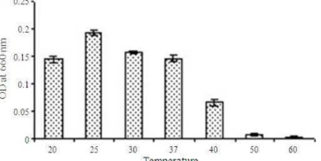
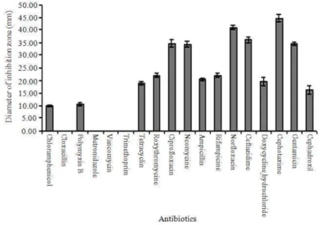
Characterization of the isolates: The characterization study revealed the isolate to be a gram positive bacilli (Fig. 1), which grew in a temperature range of 20o-50°C with optimum growth at 25°C (Fig. 2). It could tolerate pH in the range of 3-10 with optimum pH at 7 (Fig. 3). Its growth curve shows a lag period of 24 h, an early log phase up to 36 h followed by a slowed growth between 36-96 h and a death phase after 96 h (Fig. 4). It showed resistance against Chloramphenicol, Cloxacillin, Polymyxin B, Metronidazole, Vancomycin and Trimethoprim. It was sensitive to Tetracyclin, Roxythromycine, Ciprofloxacin, Neomycine, Ampicillin, Rifampicine, Norfloxacin, Ceftazidime, Doxycycline
hydrochloride, Cephotaxime and Gentamicin. It showed intermediate sensitivity to Cephadroxil (Fig. 5).
Fig. 1: Gram positive bacillus isolated from East Calcutta Wetland using methanogen specific medium at 100X magnification on a Zeiss Axiostar Plus microscope
Fig. 2: Graph representing the effect of temperatures on growth of the isolate. Temperature profile of the isolate was plotted with the OD 660 nm on Y axis and the corresponding temperature on the X axis. The result showed that the optimum temperature for growth was 25°C
Fig. 4: Growth curve of the isolate obtained by plotting the cell number on the Y axis and the time in hours on the X axis
Fig. 5: Bar diagram representing the sensitivity of the isolate to different antibiotics. The diameters of the zone of inhibitions around the antibiotic disks were plotted on the Y axis while X axis represented the names of the antibiotics
It could not grow in presence of 1mM concentration of elements like Cu (copper sulphate), Pb (lead nitrate), Cr (chromium trioxide), Ni (nickel chloride), Fe (ferrous sulphate), Co (cobaltous nitrate), Al (aluminum nitrate), Ag (silver chloride), Hg (mercuric chloride), Zn (zinc sulphate), Cd (cadmium chloride) indicating metal sensitivity (Table 1). It does not produce DNase, lipase, oxidase, protease and catalase.
Checking the growth in presence of Bromo Ethane Sulfonate (BES): Methanogenesis inhibitor BES fails to inhibit the growth of the microbe indicating the absence of methyl co-enzyme M reductase in the metabolic pathway of the microbe and indirectly indicating the isolate to be a non methanogen.
Table 1: The growth of the isolate in response to metal stress
Metals OD at 660 nm
Control 0.190
Copper sulphate 0.002
Lead nitrate 0.002
Chromium trioxide 0.003
Nickel chloride 0.002
Ferrous sulphate 0.003
Cobaltous nitrate 0.002
Aluminum nitrate 0.002
Silver chloride 0.002
Mercuric chloride 0.002
Zinc sulphate 0.002
Cadmium chloride 0.003
Fig. 6: Tree of 16S rRNA based phylogenetic analysis of isolate constructed using neighbor joining method. The isolate was indicated as WBUT 87 in the tree.
Molecular characterization of isolate through 16S rRNA amplification: Results of Blast analysis of the 16SrDNA sequence revealed identity of the isolate to be closer to as yet uncultivated microbes. The isolate was found to be a novel one and the sequence was submitted to GenBank (Accn. No. FJ930097). The phylogenetic position was predicted according to the neighbor joining method (Fig. 6).
DNA-double strand break repair study: Irradiation with 60Co γ rays showed induction of DSB and its subsequent repair as visualized through AFIGE (Fig. 7). The densitometric scanning data showed maximum repair following 60 Gray damage and thereafter a drop in repair ability with increase in irradiation dose (Fig. 8). This finding of reparability was also supported by the survival plating data [control unirradiated = 3.825×10⁸ CFU mL−1, Damage = 1.15×10⁸ CFU mL−1, Repair after 4 h = 2.65×10⁸ CFU mL−1, Control unirradiated after 4 h = 3.85×10⁸ CFU mL−1; at 40 Gray].
Fig. 7: Photograph of ethidium bromide stained 1% agarose gel showing the effect of different doses of 60Co γ ray on the isolate
Fig. 8: Bar diagram representing the dose profile of the isolate. Percentage FAR was plotted on Y axis while the X axis represented the doses in gray. It showed maximum repair post 60 Gray damage and thereafter a drop in reparability with increase in irradiation dose
Fig. 9: Gel photograph showing the kinetics of repair. The cells were irradiated at 40 Gy and reparability at different time intervals were measured to observe optimum repair after 4 hours of incubation
Arabinose CTP inhibited the repair of the isolate (Fig. 11). This finding proves that DNA polymerase was involved in the repair process and thus the repair takes place through homologous recombination.
Fig. 10: Bar diagram representing the kinetics of repair. % FAR was plotted on Y axis and time was plotted on the X axis. The time course of repair shows 4 h to be the optimum time for incubation following damage
Fig. 11: Bar diagram representing the effect of various doses of Arabinosine CTP on DSB repair. It showed that Ara C inhibited the repair. %FAR was plotted on the Y axis while the X axis represented the concentration of arabinose CTP
DISCUSSION
DSB which increased with increasing dose. The analysis of densitometric scanning data post PFGE analysis indicated maximum repair following 60 Gray damage after which there was a drop in reparability with increase in irradiation dose. This may be due to the fact that the damage induced at higher doses were of the non repairable type. For the lower doses the damage was of repairable type and so with increasing damage there was associated repair. It is at par with the finding of Raychaudhuri et al.[2] in case of methanogen. The time course of repair showed 4 h to be the optimum time required for repair. DNA polymerase inhibitor Arabinose CTP inhibited repair mechanism completely. This indicates that DNA polymerase took active part in repair process and thus the mechanism was that of homologous recombination.
CONCLUSION
In this study we report for the first time the isolation of a novel bacterial isolate with potential for DNA DSB repair through homologous recombination. It is a metal sensitive bacilli having maximum identity with the uncultivable members of Fermicutes despite being grown in methanogen specific medium.
ACKNOWLEDGMENT
The researchers would like to thank IUC-DAE under the CRS scheme for the financial assistance for the work. The authors would like to thank Dr. Asiti K. Sanyal of the Inter university Accelerator Center for availing the AFIGE facility at IUAC, New Delhi.
REFERENCES
1. Jackson Stephen, P., 2002. Sensing and repairing DNA double strand breaks. Carcinogenesis, 23: 687-696.
http://carcin.oxfordjournals.org/cgi/content/full/23/ 5/687
2. Ray Chaudhuri, S., P. Karmakar, D. Choudhary, A. Sarma and A.R. Thakur, 2003. Effect of heavy ion irradiation on DNA DSB repair in Methanosarcina barkeri. Anaerobe, 9: 15-21. DOI:
10.1016/S1075-9964(03)00027-1
3. Ray Chaudhuri, S., P. Karmaker and A.R. Thakur, 2000. γ-ray induced DNA damage and repair in Methanosarcina barkeri. Anaerobe, 6: 325-331. DOI: 10.1006/anae.2000.0359
4. Achtnich, C., A. Schuhmann, T. Wind and R. Conrad, 2006. Role of interspecies H2 transfer to sulfate and
ferric iron-reducing bacteria in acetate consumption in anoxic paddy soil. FEMS. Microbiol. Ecol., 16: 61-70. DOI: 10.1111/j.1574-6941.1995.tb00269.x
5. Zhenya, Z., Q. Taisheng, L. Pomin, Z. Yansheng, S. Norio and M. Takaaki, 2004. Study on methane fermentation and production of vitamin B12 from alcohol waste slurry. Applied Biochem.
Biotechnol., 115: 1033-1039.
http://www.ncbi.nlm.nih.gov/pubmed/15054251 6. Hales, B.A., E. Clive, D.A. Ritchie, G. Hall,
R.W. Pickup and R. Jon, 1996. Saunders isolation and identification of methanogen-specific DNA from blanket bog peat by PCR amplification and sequence analysis. Applied Environ. Microbiol., 62: 668-675.
http://aem.highwire.org/cgi/content/abstract/62/2/6 68
7. Raychaudhuri, S. and A.R. Thakur, 2006. Microbial genetic resource mapping of East Calcutta Wetland. Curr. Sci., 91: 212-217. http://www.ias.ac.in/currsci/jul252006/212.pdf 8. Adarsh, V.K., M. Mishra, S. Chowdhury, M. Sudarshan,
A.R. Thakur and S. Ray Chaudhuri, 2007. Studies on metal microbe interaction of three bacterial isolates from East Calcutta Wetland. J. Biol. Sci., 7: 80-88.
http://www.scipub.org/fulltext/ojbs/ojbs7280-88.pdf
9. Chowdhury, S., M. Mishra, V.K. Adarsh and A. Mukherjee et al., 2008. Novel metal accumulator and protease secretor microbes from East Calcutta Wetland, Am. J. Biochem. Biotech., 4: 255-264.
http://www.scipub.org/fulltext/ajbb/ajbb43255-264.pdf
10. Pradhan, A., P. Bhaumik, S. Das, M. Mishra and S. Khanam et al., 2008. Phytoplankton diversity as indicator of water quality for fish cultivation. Am. J. Environ. Sci., 4: 271-276.
http://www.scipub.org/fulltext/ajes/ajes44406-411.pdf
12. Raychaudhuri, S., M. Mishra, S. Salodkar, M. Sudarshan and A.R. Thakur, 2008. Traditional aquaculture practice at East Calcutta Wetland: The safety assessment. Am. J. Environ. Sci., 4: 140-144. http://www.scipub.org/fulltext/ajes/ajes42173-177.pdf
13. Ray Chaudhuri, S., S. Salodkar, M. Sudarshan and A.R. Thakur, 2007. Integrated resource recovery at East Calcutta Wetland-how safe is these? Am. J.
Agric. Biol. Sci., 2: 75-80.
http://www.scipub.org/fulltext/AJAB/AJAB22
75-80.pdf
14. Ray Chaudhuri, S., S. Salodkar, M. Sudarshan, I. Mukherjee and A.R. Thakur, 2008. Role of water hyacinth mediated phytoremediation in waste water purification at East Calcutta Wetland. Environ. Sci., 5: 53-62.
http://www.informaworld.com/smpp/content~conte nt=a790738169~db=all~jumptype=rss