Members of the
RAD52
Epistasis Group
Contribute to Mitochondrial Homologous
Recombination and Double-Strand Break
Repair in
Saccharomyces cerevisiae
Alexis Stein, Lidza Kalifa, Elaine A. Sia*
Department of Biology, University of Rochester, Rochester, New York, United States of America
Abstract
Mitochondria contain an independently maintained genome that encodes several proteins required for cellular respiration. Deletions in the mitochondrial genome have been identified that cause several maternally inherited diseases and are associated with certain cancers and neurological disorders. The majority of these deletions in human cells are flanked by short, repetitive sequences, suggesting that these deletions may result from recombination events. Our current understanding of the maintenance and repair of mtDNA is quite limited compared to our understanding of similar events in the nucleus. Many nuclear DNA repair proteins are now known to also localize to mitochondria, but their function and the mecha-nism of their action remain largely unknown. This study investigated the contribution of the nuclear double-strand break repair (DSBR) proteins Rad51p, Rad52p and Rad59p in mtDNA repair. We have determined that both Rad51p and Rad59p are localized to the matrix of the mitochondria and that Rad51p binds directly to mitochondrial DNA. In addition, a mitochondrially-targeted restriction endonuclease (mtLS-KpnI) was used to produce a unique double-strand break (DSB) in the mitochondrial genome, which allowed direct analy-sis of DSB repairin vivoinSaccharomyces cerevisiae. We find that loss of these three pro-teins significantly decreases the rate of spontaneous deletion events and the loss of Rad51p and Rad59p impairs the repair of induced mtDNA DSBs.
Author Summary
Mitochondria are the powerhouse of the cell, providing energy in the form of ATP. Several proteins required for the production of ATP are encoded in the mitochondrial genome, which is maintained independently from the nuclear genome. Mutations in mitochondrial DNA are responsible for several inherited diseases and are associated with certain cancers, neurological disorders, and aging. In human mtDNA, 90% of deletions, a specific type of mutation, are flanked by repetitive sequences. Deletions in this context are generally pro-duced through homologous recombination, specifically single-strand annealing. For the
a11111
OPEN ACCESS
Citation:Stein A, Kalifa L, Sia EA (2015) Members of theRAD52Epistasis Group Contribute to
Mitochondrial Homologous Recombination and Double-Strand Break Repair inSaccharomyces cerevisiae. PLoS Genet 11(11): e1005664. doi:10.1371/journal.pgen.1005664
Editor:Sue Jinks-Robertson, Duke University, UNITED STATES
Received:June 10, 2015
Accepted:October 22, 2015
Published:November 5, 2015
Copyright:© 2015 Stein et al. This is an open access article distributed under the terms of the
Creative Commons Attribution License, which permits unrestricted use, distribution, and reproduction in any medium, provided the original author and source are credited.
Data Availability Statement:All relevant data are within the paper and its Supporting Information files.
Funding:This work was funded by the National Science Foundation (grants MCB0841857 and MCB1243428 to EAS). The funders had no role in study design, data collection and analysis, decision to publish, or preparation of the manuscript.
first time, in this study we show that the proteins involved in nuclear homologous recom-bination, Rad51p, Rad52p, and Rad59p, also contribute to the formation of mitochondrial deletions. Our data also suggest a novel mitochondrial-specific role for Rad51p in the gen-eration of this type of deletion. Further study of these repair pathways allows us to garner a better understanding of these processes that are involved in disease pathologies and aging.
Introduction
Mitochondria are dynamic organelles that function as the primary source of ATP. Mitochon-dria contain an autonomously replicating genome that is packaged into DNA-protein struc-tures called nucleoids. This multi-copy genome encodes subunits of electron transport chain (ETC) complexes and the rRNAs and tRNAs that are specifically required for the expression of these proteins. The number and types of proteins encoded by the mitochondrial genome are highly conserved among eukaryotes, even though the overall size and structure of the genome varies widely between species. The remaining proteins required for the ETC, as well as those required for maintenance and repair of the mitochondrial genome are encoded in the nucleus and imported into mitochondria from the cytoplasm.
Stability of the mitochondrial genome is required for the survival of most eukaryotic cells. Mutations in the mitochondrial genome cause metabolic defects that underlie multiple dis-eases, both inherited and spontaneous [1–6]. Accumulation of both point mutations and dele-tions in mtDNA are also proposed to contribute to the normal aging process [7]. Consistent with this hypothesis, transgenic mice expressing a proofreading-defective Polγ, the replicative mitochondrial polymerase, display accelerated aging phenotypes due to the accumulation of somatic mtDNA mutations [8]. Despite its profound importance to metabolic control and cell maintenance, the mechanisms of mitochondrial mutagenesis and repair are poorly understood, particularly double-strand break repair (DSBR) [9]. Though homologous recombination (HR) in yeast, plant and mammalian mtDNA has been reported [10–15], few specific proteins involved in mitochondrial recombination have been identified [16–22]. For those proteins that have been implicated in mitochondrial HR, the inability to generate and visualize specific, con-trolled mitochondrial double-strand breaks has precluded a direct analysis of their role in DSBR.
Deletions have been observed in yeast, plants, flies, and mammalian mtDNA [10,11,31,32], and often these deletions involve sequences originally flanked by direct repeats. For example, in human cells nearly 90% of deletions in mtDNA are flanked by either perfect or imperfect repeats. This suggests that recombination is a possible mechanism for the generation of mtDNA deletions, but the exact mechanisms and proteins involved are currently unknown. [33,34].
Recent studies have localized members of theRAD52epistasis group to mitochondria. In plants, a mitochondrial-specific isoform of Rad52p has been identified, and demonstrated to promote annealing of complementary DNA sequencesin vitro, consistent with a role in recom-bination in mitochondria [35]. In human cell lines, Rad51, and two of its paralogs Rad51C and XRCC3 were found to localize to mitochondria and promote mtDNA replication upon oxida-tive stress [36,37]. These studies strongly suggest that at least a subset of the nuclear recombi-nation proteins can localize to mitochondria and that once there, they may facilitate processes that require homologous sequences such as HR. Mammalian systems do not lend themselves to mechanistic studies because these organisms require intact mtDNA molecules to survive. In addition, it is not currently possible to introduce exogenous DNA reportersin vivoin these sys-tems.Saccharomyces cerevisiaeis an ideal model system for these studies due to the fact that these repair proteins are highly conserved, mtDNA is not required for cell survival, and it is possible to introduce exogenous reporter constructs directly into the mitochondrial genome.
We previously developed a reporter system for quantitatively measuring the occurrence of direct repeat mediated deletions (DRMD) in mtDNA [38–40]. This reporter introduces a uniqueKpnI restriction recognition site between direct repeats. By expressing mitochondrially-targetedKpnI, induction of a directed mtDNA DSB can be precisely controlled. In the work presented here, we use a modified version of this endonuclease fusion to demonstrate that loss Rad51p, Rad52p and Rad59p significantly decreases the generation of both spontaneous and DSB induced mitochondrial DRMDs. Improved control of DSB induction has also allowed us to study the kinetics of mtDNA DSBR using both genetic and molecular methods. Southern blot analysis confirms that loss of these proteins leads to delays in break processing and/or gen-eration of deletion products. The localization of Rad51p and Rad59p to yeast mitochondria
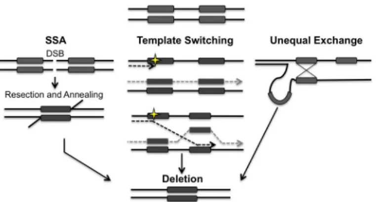
Fig 1. Models for the generation of deletions between directly repeated sequences.Repair of a double-strand break (DSB) located between directly repeated sequences (grey boxes) can result in the deletion of one of the repeats and all of the intervening sequence. 5’to 3’resection at the DSB reveals the direct repeats. Rad52p alone or in conjunction with Rad59p, promotes the annealing of the repeats. Template switching can occur if a lesion (yellow star) is encountered during replication. At a stalled fork, the nascent strand may invade at the incorrect repeat, leading to the generation of a deletion. Unequal exchange can occur when HR occurs between misaligned repeats leading to a deletion. Both template switching and unequal exchange can happen intramolecularly or intermolecularly. Arrowheads indicate 3’end.
argue that these effects are direct. Analysis of double mutants indicates that the contribution of these proteins to the repair process differs between the mitochondrial and the nuclear
genomes.
Results
Rad51p and Rad59p localize to the mitochondrial matrix
Previous studies have shown that Rad51, Rad51C and XRCC3 are found in the mitochondria of human cells [36]. In order to determine if this dual localization of Rad51 to both the nucleus and mitochondria is conserved in yeast, mitochondria were purified from cells expressing a C-terminal Rad51-HA tag expressed from the endogenous locus. We were able to detect at least four Rad51-HA specific bands in whole cell extracts and associated with the mitochondrial fraction (Fig 2A). These bands are specific to the strain expressing the Rad51-HA as they can-not be detected in whole cell extracts or mitochondrial extracts from an untagged control strain (S3 Fig). In order to determine the precise mitochondrial localization a protease protection assay was performed. Isolated mitochondria were subjected to proteinase K treatment in the presence or absence of 20mM Hepes, pH 7.4 to remove the outer mitochondrial membrane, or Triton X-100 to rupture both membranes. The inter membrane space protein, cytochrome b (Cob), is sensitive to digestion when the mitochondria are treated with 20mM Hepes (Fig 2A). The ~ 55 kDa band of Rad51p is also sensitive to digestion when the mitochondria are treated with 20mM Hepes suggesting this isoform is also found in the inter membrane space (Fig 2A, lanes 1 and 2). It is possible this isoform is also found in the matrix but is not detectable with our current system. Citrate synthase (Cit1p), a matrix protein (Fig 2A) and the ~ 60 kDa band of Rad51 remains protected from proteinase K unless treated with Triton X-100, confirming Rad51p’s localization in the matrix of the mitochondria in yeast (Fig 2A, lane 4). The specific modification(s) responsible for the larger Rad51p mitochondrial isoform(s) is currently unknown. Mitochondria were also purified in the same manner from cells expressing
Rad59-HA from the endogenous locus. A protease protection assay confirmed the presence of Rad59 in the matrix of the mitochondria (Fig 2AandS3 Fig).
In order to further confirm the mitochondrial localization of Rad51p and to determine if it binds directly to mtDNA, chromatin immunoprecipitation (ChIP) was performed using an antibody to the native untagged Rad51 protein. Using primers that anneal to a region previ-ously demonstrated to be near a recombination hotspot in the yeast mitochondrial genome, we were able to detect a significant 4-fold (p= 0.008) increase in mtDNA signal in the Rad51p IP compared to the mock IP (Fig 2B) [41]. The western blot and ChIP data together clearly dem-onstrate the Rad51p is localized to the mitochondria ofS.cerevisiae.
Rad51p, Rad52p, and Rad59p promote spontaneous mitochondrial
deletions
Rad51p, Rad52p, and Rad59p participate in spontaneous DRMD events and the repair of induced DSBs in the nucleus [26]. To determine whether they also contribute to spontaneous mitochondrial DRMD, we generated deletions of these genes both individually and in combi-nation, in yeast strains containing genetic reporters for nuclear and mitochondrial DRMD events [38–40].
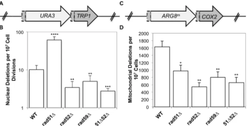
flankingARG8m. Strains that contain an intact reporter are phenotypically Arg+and respira-tion deficient. When a mitochondrial DRMD event occurs, the strain becomes respirarespira-tion pro-ficient and will become Arg-following sorting to homoplasmy, which occurs rapidly in yeast. The nuclear reporter is similarly constructed, with theURA3gene inserted 99 bp into theTRP1 gene, followed by the entireTRP1gene, generating 96bp of directly repeated sequence (Fig 3A) [39]. Strains with an intact reporter are phenotypically Ura+and Trp-. If a nuclear DRMD event occurs, the strain becomes Ura-and Trp+. Plating of cells on the appropriate media allows selection of both mitochondrial and nuclear deletions from the same culture.
As we have previously shown, the mitochondrial deletion rate is approximately160-fold greater than the nuclear rate for similar repeats in wild-type cells. It is important to note that nuclear and mitochondrial rates should not be compared directly, since the multicopy nature of the mitochondrial genome, and its subsequent replication and segregation into daughter cells will impact the rate. Rates within each compartment are compared between wild-type and
Fig 2. Rad51p and Rad59p localize to the mitochondrial matrix.(A) Intact mitochondria, mitoplasts, or Triton X-100 lysed mitochondria were treated with proteinase K and subjected to immunoblot analysis with the indicated antibodies. Por1p, outer membrane protein; Cob, intermembrane space protein; Cit1p, matrix protein. (B) Rad51p physically interacts with mtDNA. DNA samples (3 biological replicates) were
immunoprecipitated with antibodies against endogenous Rad51p or V5 epitope tag (mock). Samples were evaluated by qPCR and changes in fold enrichment were compared to the mock IP. Error bar indicates SD. Asterisks indicate significant differences between the Rad51p IP and the mock IP (**=p0.01).
mutant strains. Deletion ofRAD52orRAD59resulted in similar decreases of both nuclear and mitochondrial DRMDs (Fig 3C and 3D). Spontaneous nuclear and mitochondrial DRMDs decreased 3-fold in therad52-Δstrain (p= 0.001, in each case) and 2-fold in therad59-Δstrain (nuclearp= 0.001, mitochondrialp= 0.002) relative to wild-type. This suggests that the contri-bution of Rad52p and Rad59p to the generation of spontaneous deletions is equally as impor-tant in both the nucleus and mitochondria. In contrast,RAD51deletion resulted in different nuclear and mitochondrial phenotypes, with a 6-fold increase in nuclear DRMD (p= 2 x 10⁻⁸) and an approximately 2-fold decrease in mitochondrial DRMD (p= 0.01) (Fig 3C and 3D). The nuclear spontaneous DRMD results for this strain are consistent with the current model of SSA, however, the decrease measured for mitochondrial DRMD is not. According to the cur-rent nuclear model, SSA is dependent on Rad52p and Rad59p and independent of Rad51p. Deletions generated by SSA increase in the absence of accurate repair mediated by Rad51p [28,43]. Thus, the observed decreases in nuclear DRMD events in therad52-Δ,rad59-Δ, and rad51-Δrad52-Δstrains, and the increase in nuclear DRMD events inrad51-Δstrains were as expected [27,28]. The decrease in mitochondrial DRMD events observed in therad51null mutants indicates that in contrast to the nuclear DRMD events, spontaneous mitochondrial DRMDs are at least partially dependent on Rad51p, suggesting a unique functional role for Rad51p in mitochondria.
To confirm that the decreases in mitochondrial DRMD rates in theradmutants are due to specific deficiencies in mitochondrial pathways leading to deletions, and not due to general mitochondrial genome instability, the spontaneous frequency ofpetiteswas measured in each of the above mutants. Spontaneously arisingpetitecolonies (ρ-) are non-respiring, usually due
to large-scale deletions and rearrangements of mtDNA, and do not grow on non-fermentable carbon sources such as glycerol [10]. The mechanism by which these deletions and rearrange-ments occur has not been defined, although homologous recombination has been proposed [10]. None of the deletions we tested exhibited any significant change from wild-type in the fre-quency of respiration loss (S2 Table). This suggests that the phenotypes we observe do not
Fig 3. Spontaneous nuclear and mitochondrial repeat mediated deletions.(A) The nuclear DRMD reporter. TheURA3gene was inserted 99 bp into theTRP1sequence, and is followed directly by the entire
TRP1gene lacking the start codon, resulting in a 96 bp direct repeat flanking theURA3marker. Spontaneous deletions are selected on medium lacking tryptophan, and rates were determined using the method of the median. (B) The mitochondrial DRMD reporter. TheARG8mgene was inserted 99 bp into theCOX2
sequence, and is followed directly by the entireCOX2gene lacking the start codon, resulting in a 96 bp direct repeat flanking theARG8mmarker. Spontaneous deletions are selected on medium containing glycerol as the sole carbon source, and rates were determined using the method of the median. (C) Average rates of nuclear DRMD. (D) Average rates of mitochondrial DRMD. Error bars indicate SD. Asterisks indicate significant differences between the mutant and wild-type rates (*=p0.05,**=p0.01,***=p0.001,
****=p0.0001).
result from a general increase in mtDNA instability, and these proteins are not involved in the mechanism that generates spontaneouspetitecolonies.
Specific mitochondrial double-strand breaks increase the frequency of
mtDNA deletions
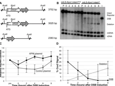
TheARG8msequence used in our mitochondrial reporter contains a uniqueKpnI restriction site. Cleavage at this site generates a specific DSB 370 bp downstream of theARG8mstart codon, with 4 bp 3’single-stranded complementary overhangs at the broken ends (Fig 4A). We previously reported that the galactose-inducible promoter regulating the expression of our mtLS-KpnI was leaky, which stimulated the generation of deletion products prior to the addi-tion of galactose [40]. In order to ensure that we did not have premature expression ofKpnI endonuclease, a temperature sensitive intein was inserted into the active site ofKpnI [44]. This intein self-cleaves at 20°C generating an activeKpnI enzyme. A control strain was also con-structed utilizing a nonfunctional intein (intdead) that cannot self-cleave at any temperature [44].
This modification allows tight control of the activity of mtLS-KpnI. Southern blot analysis shows no detectible digestion at the mitochondrialKpnI recognition sequence, and no evidence of deletion product prior to induction of mtLS-KpnI-intts(Fig 4B, lane P).KpnI cleavage is achieved by inducing mtLS-KpnI-inttsexpression with galactose and reducing the incubation temperature to 20°C (Fig 4B, lane 0). Furthermore, the frequency of DSB-induced DRMDs increases linearly with extended incubation time at 20°C prior to assaying at 30°C, suggesting thatKpnI activity is largely restricted to 20°C (S1 Fig). Longer incubations at 20°C generated a larger proportion of DSBs, but never exceeded 60% of the totalCOX2signal. This result may suggest that some genomes may not be accessible to theKpnI enzyme or that alternative repair processes are rapidly repairing the break (S4 Fig). In subsequent experiments, 16–18 hours incubation at 20°C was used to ensure significant visible DSBs were generated.
Quantification ofCOX2-hybridizing species from independent experiments reveals that an average of 30.2 ± 16.7% of theCOX2signal appears as a specificKpnI digested fragment after 16–18 hours incubation at 20°C, and no measurable deletions are observed (Fig 4B and 4D). Two hours after cultures are returned to 30°C, deletion products can be observed, and their concentration continues to increase over the course of the experiment to represent, on average, 16.2% of theCOX2signal (Fig 4B and 4D). Thus, the percent of theCOX2signal that hybrid-izes to DRMD products after 8 hours at 30°C is approximately equivalent to two-thirds of the amount observed in the DSB at time 0. By 8 hours after shift to 30°C the cultures have reached saturation, and no further increase in deletion product was seen when cultures were monitored for an additional 4 hours (S4 Fig), suggesting that repair is complete by 8 hours in the wild-type strain. In addition, while breaks are stimulated at 20°C, the bulk of DRMD events occur after the cells are shifted back to 30°C, since deletions are not detected by Southern blot analysis at time of shift back to 30°C. In control cultures, expressing the mtLS-KpnI-intdead, no stimula-tion of DRMD was observed, indicating that no significant cleavage of the intdeadoccurs.
mtLS-KpnI::inttsexpressing strains recovered significantly faster than the control strain, how-ever, both the control and mtLS-KpnI::inttsstrains fully recovered to pre-induction levels of mtDNA after eight hours of growth at 30°C (Fig 4C).
It has been reported that mitochondrial DNA can move to the nucleus, although this occurs rarely [45]. In order to ensure that the signal we are detecting in the Southern blots is originat-ing from the mitochondrial genome, both pre- and post-induction wild type cultures were treated with ethidium bromide. Growth of yeast in culture medium in the presence of ethidium bromide has been demonstrated to suppress mtDNA replication [46]. Southern blot analysis confirms that greater than 98% of the signal from theCOX2and the 21S rRNA probes is lost when the cells are subjected to ethidium bromide treatment. (S2 Fig).
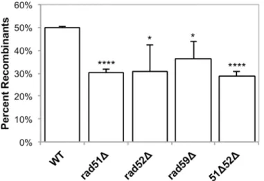
Eight hours after shifting to 30°C, approximately 16% of the mtDNA detected contains a deletion of theARG8mreporter (Fig 4B and 4D). However, the distribution of the 16% of deleted genomes within the population of cells is unclear. For example, many deletions may be concentrated in a small subset of the cell population, or distributed throughout, with most cells containing one or more mitochondrial genomes with deletions. By assaying for respiring colo-nies, we can directly distinguish between these possibilities. Cells from time 0 in the above experiments (Fig 4B) were plated on non-selective medium, allowed to grow, and individual colonies were tested for the presence of the intact reporter (Arg+) or the deletion (respiration proficient) by replica plating. We found that 50% of the population is respiration competent, consistent with a wide distribution of mitochondrial genomes containing deletions (Fig 5). The majority of these colonies also contain Arg+cells, indicating heteroplasmy at the time of
Fig 4. Inducing a specific mitochondrial DSB.(A) The mitochondrial DRMD reporter, with restriction recognition sequences indicated. Horizontal line beneathCOX2indicates where theCOX2probe anneals. (B) Representative Southern blot ofAvaII-digested DNA extracted from EAS748 containing pEAS115
(mtLS-KpnI-inteindead) or pEAS114 (mtLS-KpnI-inteints). Both strains were grown in SRaffinose-Arg-Ura until OD 600
= 0.150. Galactose was added to a final concentration of 2%, and the strains were shifted to 20°C to induce the either mtLS-KpnI-inteintsor mtLS-KpnI-inteindead. Genomic DNA in lanes labeled P are from preinduced
cultures. Cultures were incubated for 16–18 hours at 20°C and samples were taken for the time 0 timepoint. The cultures then were shifted to 30°C to allow repair. Lane I contains DNA from the t = 0 timepoint of the mtLS-KpnI-inteints-containing strain digestedin vitrowithKpnI andAvaII to demonstrate the migration of DNA
with a DSB. The 21S rRNA gene was probed to detect total mtDNA. The 25S rRNA gene was probed to detect total nuclear DNA. (C) Relative mtDNA content was determined by taking the ratio of the 21S rRNA signal and the 25S rRNA signal, then normalizing to the t = 0 sample for each strain. (D) The intensity of the
COX2-hybridizing bands were measured using Image Lab software (www.biorad.com) The proportions of the totalCOX2signal present in the DSB and deletion products were calculated as a percent of the totalCOX2
signal.
plating. Significantly, we did not observe DSB stimulation ofρ-orρ0(lacking mtDNA)petites,
which would be both Arg-and non-respiring. After cutting, 1.5% of the colonies from wild-type cells were both Arg-and respiration deficient. This is comparable to the percentage of spontaneouspetitesseen in wild-type cultures prior toKpnI-induced mtDNA DSBs, indicating the induction of the DSB does not lead to general mitochondrial genome instability (S3 Table).
Rad51p, Rad52p, and Rad59p influence mitochondrial DSBR
To determine the effect of Rad51p, Rad52p, and Rad59p on the repair of mitochondrial DSBs, plasmids expressing either mtLS-KpnI::intts(pEAS114) or mtLS-KpnI::intdead(pEAS115) were introduced into the mutant strains. Mitochondrial DSBs were induced as described above, and Southern blots reveal that the level of cutting by mtLSKpnI-inttsat the time of shift to 30°C in all of the mutant strains is equivalent to wild-type. (Table 1).
In our genetic assay for induced deletions, 50% of colonies from wild-type cells had under-gone at least one selectable deletion event (Fig 5), and each of theRADsingle mutants exhibited a modest, yet significant, decrease in selectable deletion events when compared to wild-type. In therad51-Δ,rad52-Δ,rad59-Δstrains 30% (p= 4.2 x 10⁻⁵), 31% (p= 0.03), and 36% (p= 0.03)
Table 1. Average level of cutting after 16 hours of mtLSKpnI-inttsinduction.
Average level of cutting p-valuea
Wild-type (EAS930) 30.2±16.7%
-rad51-Δ(ASY113) 31.5±3.0% 0.90
rad52-Δ(ASY115) 40.6±3.7% 0.21
rad59-Δ(ASY117) 24.9±8.3% 0.52
rad51-Δrad52-Δ(ASY119) 29.8±2.0% 1.0
apvalues were calculated were calculated by un-paired, two-tailed t-tests.
doi:10.1371/journal.pgen.1005664.t001
Fig 5. Induced mitochondrial repeat mediated deletions.Strains were grown in SRaffinose-Arg-Ura until OD600= 0.150. Galactose was added to a final concentration of 2%, and the strains were shifted to 20°C for
16–18 hours in order to induce DSBs via mtLS-KpnI. Appropriate dilutions were plated on YPD and allowed to grow for 3 days at 30°C. The colonies were then replica plated to YPG and SD-Arg. Growth on YPG was scored in order to determine the percent of colonies that had at least some cells that had undergone a deletion after non-selective growth. Error bars indicate SD. Asterisks indicate significant differences between the mutant and wild-type rates (*=p0.05,****=p0.0001).
of the cells were able to give rise to respiring colonies respectively, after 16–18 hours of induc-ing DSBs (Fig 4). As with the wild-type strain, non-respiring and Arg-petitesdid not signifi-cantly increase after DSB induction in these strains (S3 Table).
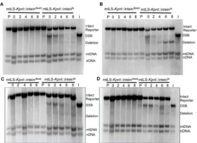
Southern blots analysis of the mtDNA inrad51-Δandrad59-Δstrains revealed that the same proportions of the mitochondrial genomes are being cut (31% and 25%, respectively) compared to the wild-type strain (Table 1). However, consistent with the genetic data, these cut genomes are not converted to deletion products to the same extent as wild-type (Figs6A and 6Cand7). In therad51-Δstrain after 8 hours of recovery only 14.4% ± 7.9 (p= 0.001) of the cutCOX2signal had been converted to a deletion product compared to 66.8% ± 7.4 in the wild-type strain. In therad59-Δstrain, 36.7% ± 14.9 (p= 0.01) of the cutCOX2signal is con-verted to a deletion product. The cut band is completely lost in all mutant and wild-type strains after 8 hours of recovery, suggesting that DSB processing is not being eliminated but the path-way choice is altered in the absence of Rad51p or Rad59p.
Analysis of therad52-Δstrain revealed no significant change in cutCOX2signal at time 0, 40.6% ± 2 (p= 1.0) compared to wild-type (Table 1). Surprisingly, in contrast to the genetic data, the proportion of the cutCOX2signal converted to deletion product, 55.9% ± 26.3 (p= 0.5) is equivalent to wild-type. We know that the distribution or maintenance of these genomes in the population of cells is not equivalent to wild-type because only 31% of the colo-nies contain respiring cells, while 50% of wild-type colocolo-nies contain respiration-competent cells after 3 days growth on solid medium.
Therad51-Δrad52-Δdouble mutant exhibited the same decrease in deletion events as the rad51-Δandrad52-Δsingle mutants, with 29% (p= 4.5 x 10⁻⁵) of colonies capable of respira-tion after inducrespira-tion of mtLS-KpnI::intts(Fig 5). Interestingly, the mtDNA in this mutant dis-played a similar significant decrease in the proportion of cutCOX2signal converted to deletion product, 7.6% ± 5.7 (p= 0.0004), as therad51-Δstrain (p= 0.3) (Figs6Dand7). This would suggest that during the initial generation of deletion products after induction of a DSB that RAD51is epistatic toRAD52in the mitochondria.
We considered the possibility that the relative levels of the differentCOX2-hybridizing spe-cies in all cases might be affected by differences in growth rates of the mutant and wild-type strains under the conditions of the experiment. Monitoring the growth of the cultures by opti-cal density and total cell counts throughout the protocol revealed that therad52-Δandrad59-Δ
strains displayed a 20% increase in doubling time at 20°C, while therad51-Δmutant strain was comparable to the wild-type. All strains undergo one population doubling during the eight-hour recovery period. It is not likely then that the wild-type levels of deletion product generated in therad52-Δstrain are dependent on this growth defect, considering therad59-Δstrain also has inhibited growth at 20°C but clearly shows impaired deletion product formation. We believe there is no significant correlation of differences in growth rate with differences in DSBs or deletions (Fig 6B).
Single-stranded DNA is present at break after induction of
Kpn
I
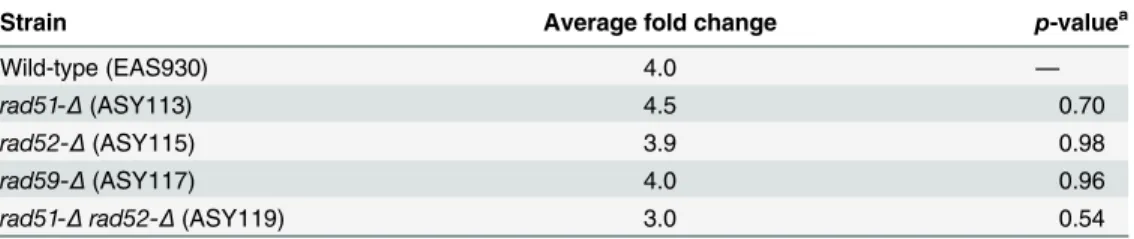
rad52-Δstrains is also consistent with HR models that show these proteins function in the repair of DSBs after resection has occurred (Table 2).
Discussion
The faithful maintenance of the mitochondrial genome is required for eukaryotic cells to have a functional electron transport chain. Mutations and deletions in the mitochondrial genome can compromise energy production for the cell. However, studying mtDNA repair has been difficult, due to the necessity for intact mitochondrial genomes in most eukaryotic systems. Thus, the extent of mtDNA repair has remained unclear. For years it was believed that since there are multiple copies of the mitochondrial genome, the damaged mtDNA was simply degraded in lieu of repair [47]. While there is evidence that mitochondrial genomes are degraded under high levels of chronic damage, it is unclear if this is the response under more moderate damaging conditions [48].
Mitochondrial base excision repair has been more extensively studied [9,49,50], indicating that at least some highly conserved repair pathways exist in mitochondria. In addition, there is evidence for homologous recombination (HR) repair pathways in mitochondria of several dif-ferent species including yeast,Drosophila, and mice [40,51,52]. In this study, we have shown that the HR proteins Rad51p and Rad59p localize to the matrix of the mitochondria. We have also demonstrated that Rad51p, Rad52p and Rad59p impact the rate of spontaneous mitochon-drial DNA deletions and the repair of induced mitochonmitochon-drial DSBs in yeast. This is the first study to demonstrate that Rad51p, Rad52p, and Rad59p impact the repair of specific mtDNA lesions.
In the present study, we have greatly improved our reporter system to introduce a maxi-mum of a single DSB per mitochondrial genome to identify the proteins involved in the repair of these breaks. We had previously generated a plasmid expressing a galactose-inducible,
Fig 6. Repair of induced mitochondrial DSBs in HR single mutants.All samples were obtained after 16–
18 hours of mtLS-KpnI induction. DNA was extracted from the appropriate strains and digested withAvaII. Samples in lanes labeled P are from pre-induced cultures. Lane I is the t = 0 sample from the mtLS-Kpn I-inteintsexpressing strain digested withKpnI andAvaII to demonstrate the migration of the reporter with a DSB
in relation to the otherCOX2bands. (A) Representative Southern blot ofrad51-Δstrains ASY114 (inteindead)
and ASY113 (inteints). (B) Representative Southern blot of
rad52-Δstrains ASY116 (inteindead) and ASY115
(inteints). (C) Representative Southern blot ofrad59-Δstrains ASY118 (inteindead) and ASY117 (inteints). (D) A representative Southern blot ofrad51-Δrad52-Δstrains, ASY120 (inteindead) and ASY119 (inteints).
mitochondrially-targetedKpnI endonuclease that showed evidence of expression under unin-duced conditions [40]. To add an additional level of control over endonuclease activity, we have inserted a temperature-sensitive intein into the active site of theKpnI enzyme. This modi-fied expression construct does not result in efficient cleavage of the mitochondrial genome, since we must incubate at the permissive temperature for 16–18 hours to observe digestion of approximately 30% of the mtDNA by Southern blot analysis (Fig 3). However, it is likely that a larger proportion of breaks are present, but these breaks are not detectible due to the end resec-tion that occurs at the site of DSBs. We have demonstrated that 3’single-stranded ends are present at the site of the break following cleavage. The heterogeneous nature of these interme-diates would render them undetectable by our current Southern blot techniques. We would also be unable to detect any breaks that occurred and were subsequently ligated with fidelity.
Fig 7. Conversion of DSBs to deletion product is impaired in HR mutants.Experiments were performed as inFig 4. The ratios of the amount ofCOX2signal in the cut band at t = 0 and the amount ofCOX2signal in the deletion product at t = 8 were calculated. The intensity of theCOX2-hybridizing bands was measured using Image Lab software (www.biorad.com). The proportions of the totalCOX2signal present in the DSB and deletion products were calculated as a percent of the totalCOX2signal. Error bars indicate SD. Asterisks indicate significant differences between the mutant and wild-type rates (*=p0.05,***=p0.001).
doi:10.1371/journal.pgen.1005664.g007
Table 2. Average fold change of amount of 3’ssDNA atKpnI cleavage site after induction.
Strain Average fold change p-valuea
Wild-type (EAS930) 4.0 —
rad51-Δ(ASY113) 4.5 0.70
rad52-Δ(ASY115) 3.9 0.98
rad59-Δ(ASY117) 4.0 0.96
rad51-Δrad52-Δ(ASY119) 3.0 0.54
apvalues were calculated were calculated by un-paired, two-tailed t-tests.
This system is an improvement over our previously published mtLS-KpnI construct, as there is no molecular or genetic evidence of mtDNA cleavage prior to induction, nor is there selection for cleavage-resistant genomes in our cultures. In addition, this level of digestion is sufficient to observe DNA repair over the course of 8 hours after shifting back to the non-permissive tem-perature for intein cleavage.
It bears noting that deletion products do not appear after extensive incubation at the per-missive temperature for intein cleavage, although they are clearly visible by 2 hours after shift back to the non-permissive temperature. It is unlikely that this delay simply results from slow repair, because no deletions are visible on a Southern blot after 24 hours of incubation at the permissive temperature (S1 Fig). Rather, this observation suggests that at least one step of the deletion process is inefficient or non-functional at 20°C. Deletion formation does not appear to be the only temperature-dependent process. We observed a slight decrease in relative mtDNA copy number upon shifting the cultures to 20°C. However, a decrease was also seen in our con-trols expressing mtLS-KpnI-intdead, leading us to conclude that temperature shift alone is cause of this temporary decrease in mtDNA copy number (Fig 4C). Consistent with this hypothesis, both strains recover by 8 hours after shift back to 30°C. We conclude that in our system, repair-able DSBs do not stimulate mtDNA loss from the population of cells. Our genetic assay sup-ports this observation, as induction of a specific mitochondrial DSB does not stimulate an increase in the loss of functional mtDNA. Therefore, mechanisms exist in cells to maintain functional mtDNA. It is important to note that this selection cannot be applied at the level of respiratory function, or ATP production, as the genomes containing the intact DRMD reporter confer an Arg+phenotype, and have active mitochondrial protein synthesis, but do not assem-ble a functional electron transport chain. In yeast, there is evidence for a mitochondrial G1-to-S phase checkpoint that is activated in response to the loss of mtDNA and not linked to respira-tory capacity [53], suggesting that the cell has a mechanism for sensing mtDNA. Clearly, although mtDNA can be eliminated by treatment of cells with ethidium bromide and sponta-neouspetitemutants arise in yeast cultures [10] this should not be taken as evidence that mech-anisms do not exist to maintain an intact genome.
Our results demonstrate that Rad51p, Rad52p, and Rad59p are involved in a portion of DSBR events. In the nucleus, SSA is the primary HR pathway that generates DSB-induced dele-tions between repeated sequences [26], and it seems likely to be an important pathway for spontaneous events at direct repeats as well. This mutagenic DSB repair pathway leads to the deletion of one of the repeats and any intervening sequence. In the nucleus, SSA is independent of Rad51p, but requires Rad52p and Rad59p [43]. Previous studies have shown [26,27], and this study confirms, that in the nucleus, deletion ofRAD51leads to an increase in deletions generated by a Rad52p-dependent pathway, likely SSA (Fig 3B). In contrast, in mitochondria, the deletion ofRAD51caused a significant decrease in the frequency of spontaneous deletions and those generated after inducing a DSB. Genetically, the contribution of Rad51p to the gen-eration of DRMDs was equivalent to Rad52p and Rad59p (Figs3Dand5). This would imply that both compartments use at least some of the same proteins to generate these deletions, but that the pathways these proteins are participating in are unique compared to the nucleus. In the absence of the full suite of nuclear HR proteins in the mitochondria we speculate that HR proteins, such as Rad51, have been co-opted to perform their HR functions in contexts that are not necessary in the nucleus.
increased dependence on Rad51p for deletion formation, is that the AT-rich mitochondrial genome requires further stabilization of the annealed intermediate and thus utilizes Rad51p uniquely in this manner. However, at this time, we cannot rule out the existence of a novel Rad51p dependent mechanism that generates deletions solely in the mitochondria.
The localization of Rad51p and Rad59p to mitochondria strongly suggests that the effects we observe on deletion product formation in the mutant strains are direct, for several reasons. First, we demonstrated that mitochondrial DSB repair and homology-dependent deletions were specifically affected, while mitochondrial genomes were stably maintained, as indicated by the maintenance of wild-type copy number and the lack of additionalρ-in our mutant
strains. In addition, the epistatic relationships betweenRAD51,RAD52,RAD59with respect to mitochondrial phenotypes were not the same as the nuclear phenotypes, most notably in the case of Rad51p, which had a similar impact on mitochondrial DRMD’s as both Rad52p and Rad59p. Finally, their action at DSBs is consistent with their known function. Identification of mitochondrial-specific factors that interact either physically or genetically with Rad51p, Rad52p, and Rad59p will help to elucidate the mitochondrial specific repair pathways or path-way components.
Materials and Methods
Media and strains
Media used in this study were previously described [39].S.cerevisiaestrains used in this study are listed inS1 Tableand are isogenic with DFS188 except NPY66, which is derived from DFS160 [38,54]. Single-deletion strains were constructed by one-step gene replacement of the wild-type gene with akanMXmarker using standard protocols [55]. Strains containing two gene replacements were constructed by mating isogenic haploid strains, dissecting diploids, and screening by PCR for the desired genotypes. The spontaneous DRMD reporter strain (LKY196) was constructed previously [39]. Gene deletions in this strain were constructed by mating and dissection. The desired genotypes were screened for by PCR. These strains were then cured of their mtDNA by treatment with ethidium bromide, and cytoduced with the mitochondrial reporter from NPY66 [56].
Mitochondrially localized, V5-tagged mtLS-KpnI was constructed by amplifying the bacte-rialKpnI gene from pETRK [57], using a 5’primer containing the mitochondrial localization signal ofCOX4 5’-GGATCCATGCTGAGCCTGCGCCAGAGCATTCGCTTTTTTAAACCG GCGACCCGCACCCTGTGCAGCAGCCGCTATCTGGTGATGGTGATGATGGATGTCT TTGATAAAGTTTATAG-3’and 3’primer 5’- TCTTTTCAATCCATAATAATTCAA-3’. The PCR product was cloned into pYES2.1 using the TOPO TA cloning kit (Invitrogen) then trans-formed into Top10F’bacterial cells containing pACMK+, a plasmid that carries theKpnI meth-ylase [57]. Transformants were screened by digestion with restriction endonucleasesBamHI andXbaI. A positive clone was confirmed by sequencing and named pEAS101.
the cysteine at position 198 ofKpnI. Transformants were selected on SD-Ura medium. Geno-mic DNA was collected from the transformants and plasmids were rescued by the transforma-tion ofE.coliTop10F’cells and selection on LB+ampicillin. Plasmids containing the intein were screened by digestion withBamHI andBglI and confirmed by sequencing. pEAS114 con-tains mtLS-KpnI+inteints303. pEAS115 contained mtLS-KpnI+inteindead.
Mitochondrial isolation and protease protection assay
Mitochondria were isolated as previously described in Diekertet al., 2001 [58]. The protease protection assay was performed as previously described in Diekertet al., 2001 with the follow-ing modification. 200μg of isolated mitochondria were used in each protease protection assay
for both the ASY092 (Rad51-HA) and ASY127 (Rad59-HA) strains. 100μg of isolated
mito-chondria were used in the protease protection assay for DFS188 (WT). Samples were subjected to western blot analysis on 10% SDS-PAGE gels. Antibodies used for detection were as follows: 1:2500 dilution of anti-HA-HRP (Santa Cruz Biotechnology), 1:4000 anti-Cytb2 (generous gift from Dr. Tom Fox), 1:4000 Cit1 (generous gift from Dr. Tom Fox), and 1:25,000 anti-POR1 (Invitrogen).
Chromatin immunoprecipitation
A 150 ml culture (RCY248) was grown to OD600= 0.800 in YPD and crosslinked in 1%
formal-dehyde for 20 minutes at room temperature. Cells were pelleted and washed twice in 1X TBS. Pellets were resuspended in 1ml of lysis buffer (50 mM Hepes-KOH, pH7.5, 150 mM NaCl, 1 mM EDTA, 1% TritonX-100, 0.1% sodium deoxycholate, 0.5% SDS) and 0.4 g of glass beads. Cellular lysates were obtained by bead beating at 4°C for 2.5 minutes. The lysates were then sonicated to generate fragments between 400-700bp in size. Lysates were precleared by incu-bating with 20μl of Protein A/G magnetic beads (Thermo Scientific). One milliliter of lysate
was incubated over night with either 1μg of anti-Rad51 antibody (Abcam) or anti-V5 (mock)
(Invitrogen) rocking at 4°C. Immune complexes were precipitated by incubating with 20μl of
Protein A/G magnetic beads (Thermo Scientific) for 1 hour at 4°C. Beads were washed twice with lysis buffer, once with LiCl buffer (, and twice with 1X TE. Immune complexes were eluted twice with 150μl of elution buffer (1% SDS, 0.1M NaHCO3). Crosslinks were reversed by
add-ing 0.2M NaCl and heatadd-ing overnight at 65°C. DNA was purified and equal amounts of DNA were analyzed by qPCR using SYBR green mix (Biorad) with the following primer pair: 5’ -GGTAAATAGCAGCCTTATTATG-3’, 5’-CGATCTATCTAATTACAGTAAAGC-3’.
Measuring nuclear and mitochondrial DRMDs and respiration loss
The rates of nuclear and mitochondrial spontaneous DRMDs and the average frequency of res-piration loss were determined as previously described [39,40]. Fluctuation analyses consisted of 10–20 independent samples each. Rates were determined using the method of the median [59], and each experiment was performed a minimum of 3 times. Induced DRMDs were mea-sured by growing strains to saturation in SRaffinose-Arg-Ura. These cultures were then diluted back to an OD600~ 0.08 and grown to OD600of 0.15–0.20. A pre-induction sample was
Southern blot analysis
DNA for Southern blot analysis was prepared from cells grown as stated above. A pre-induc-tion sample was obtained prior to inducpre-induc-tion with 2% galactose and shifting to 20°C. After 16–
18 hours of cutting, the cultures were shifted back to 30°C and samples were collected after 0, 2, 4, 6, and 8 hours. Total cellular DNA was extracted using standard procedures and trans-ferred to Amersham Hybond-XL (GE Healthcare) nylon membranes following standard proto-cols. DNA was obtained from three independent experiments for each strain. Representative blots are shown in Figs3,5and6. TheCOX2, nuclear 25S rRNA, and mitochondrial 21S rRNA probes were constructed as previously described [40]. Quantification of Southern blots was performed using Image Lab software (www.biorad.com). The percentage of theCOX2signal represented by deletion product and break product was calculated by measuring the intensity of each band and dividing by the total signal for all 3COX2-hybridizing bands.
In order to confirm that the signal from theCOX2probe was originating from the mtDNA the mitochondrial DNA from both pre-induction and time 0 cultures were diluted 1:1000 into minimal media containing 0.125mg of ethidium bromide and grown to satura-tion [46]. The saturated cultures were diluted 1:1000 into minimal media containing 0.125mg of ethidium bromide and grown to saturation. DNA was obtained from these cul-tures using standard protocol. The ethidium bromide treated culcul-tures as well as DNA col-lected from these cultures prior to the ethidium bromide treatment were analyzed by Southern blot as described above.
Slot blot analysis of 3
’
ssDNA
Samples collected at time 0 were also subjected to slot blot analysis in order to detect 3’-ssDNA near theKpnI cleavage site. Total cellular DNA was extracted using standard procedures. Sam-ples were prepared as previously described [60] with the following modifications. Native geno-mic DNA (1μg) was prepared in 10X SSC and denatured samples (0.15μg) were prepared in
0.4N NaOH for 5 minutes. The samples were neutralized by the addition of 3M sodium acetate. Native and denatured samples were transferred to Amersham Hybond-XL (GE Healthcare) nylon membranes with a slot blot manifold following the manufacturers suggested protocol (Schleicher & Schuell). The DNA was UV cross-linked to the membrane. The membrane was prehybridized for a minimum of 1 hour in prehybridization solution (6X SSC, 5X Denhardt’s, 0.05% sodium pyrophosphate, 3 mg boiled fish sperm) at 37°C. The membrane was probed with a small synthetic end-labeled oligo (5’CAGCAATACCAGCTGTGAAATCG 3’) over-night at 48°C. This probe anneals 213 nucleotides up stream of theKpnI cleavage site. The membrane was washed in 6X SSC, 0.05% sodium pyrophosphate at room temperature. Quanti-fication of slot blots was performed using Image J software (http://rsbweb.nih.gov/ij/). The native samples were normalized to the appropriate denatured control.p-values were deter-mined by un-paired, two-tailed t-tests using Microsoft Excel for Macintosh 2011. Three inde-pendent experiments were performed for each strain.
Supporting Information
S2 Fig.COX2signal is dependent on the presence of mtDNA.All samples labeled pre were obtained prior to the induction of mtLS-KpnIts. All samples labeled 0 were obtained after 16 hours of induction of mtLS-KpnIts. All samples labeled + EtBr were grown to saturation in ethidium bromide twice in order to remove the mtDNA. Lane I contains DNA from the wild-type t = 0 timepoint of the mtLS-KpnI-inteints-containing strain digestedin vitrowithKpnI andAvaII to demonstrate the migration of DNA with a DSB.
(TIFF)
S3 Fig. HA signal is specific to RAD51-HA and RAD59-HA strains.Immunoblot analysis of
whole cell extracts and mitochondrial extracts from RAD51-HA, RAD59-HA tagged strains and wild-type untagged strain with HA antibody.
(TIF)
S4 Fig. Percentage of detectable DSB maximized after 24 hours at 20°C.(A) Induction of
DSBs after 24 hours at 20°C reaches ~60% of the total COX2 signal. (B) After induction of mitochondrial DSBs, the amount of COX2 signal found in the recombinant product does not increase after 8 hours of recovery at 30°C.
(TIF)
S1 Table. List of strains used in this study.
(DOCX)
S2 Table. Spontaneous respiration loss frequency in theradmutants.The median frequency
of spontaneous respiration loss was determined for each strain using 10–20 independent cul-tures. The average median frequencies for at least three experiments are presented. Mutant fre-quencies are compared to wild-type using unpaired two-tailed t-tests, andp-values are
provided. (DOCX)
S3 Table. The average difference inpetitefrequency following double-strand break
induc-tion.The samples analyzed inFig 4were also assessed forpetites(Arg-, non-respiring colo-nies). For each experiment, we calculated the difference in the frequency ofpetitesfrom the same culture before and after DSB induction. The average difference for all experiments is shown. The unpaired two-tailed t-tests were used to compare the frequencies for each strain before and after break induction, andp-values are presented.
(DOCX)
Author Contributions
Conceived and designed the experiments: AS LK EAS. Performed the experiments: AS LK EAS. Analyzed the data: AS EAS. Contributed reagents/materials/analysis tools: EAS. Wrote the paper: AS EAS.
References
1. Schapira AH (2012) Mitochondrial diseases. Lancet 379: 1825–1834. doi:10.1016/S0140-6736(11) 61305-6PMID:22482939
2. Ylikallio E, Suomalainen A (2012) Mechanisms of mitochondrial diseases. Ann Med 44: 41–59. doi:10. 3109/07853890.2011.598547PMID:21806499
3. Yang JL, Weissman L, Bohr VA, Mattson MP (2008) Mitochondrial DNA damage and repair in neurode-generative disorders. DNA Repair (Amst) 7: 1110–1120.
5. Milone M (2012) Mitochondria, diabetes, and Alzheimer's disease. Diabetes 61: 991–992. doi:10. 2337/db12-0209PMID:22517655
6. Federico A, Cardaioli E, Da Pozzo P, Formichi P, Gallus GN, et al. (2012) Mitochondria, oxidative stress and neurodegeneration. J Neurol Sci 322: 254–262. doi:10.1016/j.jns.2012.05.030PMID:22669122
7. Ma YS, Wu SB, Lee WY, Cheng JS, Wei YH (2009) Response to the increase of oxidative stress and mutation of mitochondrial DNA in aging. Biochim Biophys Acta 1790: 1021–1029. doi:10.1016/j. bbagen.2009.04.012PMID:19397952
8. Trifunovic A, Wredenberg A, Falkenberg M, Spelbrink JN, Rovio AT, et al. (2004) Premature ageing in mice expressing defective mitochondrial DNA polymerase. Nature 429: 417–423. PMID:15164064
9. Kazak L, Reyes A, Holt IJ (2012) Minimizing the damage: repair pathways keep mitochondrial DNA intact. Nat Rev Mol Cell Biol 13: 659–671. doi:10.1038/nrm3439PMID:22992591
10. Dujon B (1981) Mitochondrial genetics and functions. In: Jones EW, Broach JR, editors. The Molecular Biology of the Yeast Saccharomyces: Life Cycle and Inheritance. Cold Spring Harbor, NY: Cold Spring Harbor Laboratory. pp. 505–592.
11. Poulton J, Deadman ME, Bindoff L, Morten K, Land J, et al. (1993) Families of mtDNA re-arrangements can be detected in patients with mtDNA deletions: duplications may be a transient intermediate form. Human Molecular Genetics 2: 23–30. PMID:8490619
12. Thyagarajan B, Padua RA, Campbell C (1996) Mammalian mitochondria possess homologous DNA recombination activity. Journal of Biological Chemistry 271: 27536–27543. PMID:8910339
13. Lakshmipathy U, Campbell C (1999) Double strand break rejoining by mammalian mitochondrial extracts. Nucleic Acids Research 27: 1198–1204. PMID:9927756
14. Kajander OA, Karhunen PJ, Holt IJ, Jacobs HT (2001) Prominent mitochondrial DNA recombination intermediates in human heart muscle. EMBO Rep 2: 1007–1012. PMID:11713192
15. Kraytsberg Y, Schwartz M, Brown TA, Ebralidse K, Kunz WS, et al. (2004) Recombination of human mitochondrial DNA. Science 304: 981. PMID:15143273
16. Ling F, Makishima F, Morishima N, Shibata T (1995) A nuclear mutation defective in mitochondrial recombination in yeast. EMBO J 14: 4090–4101. PMID:7664749
17. Ling F, Morioka H, Ohtsuka E, Shibata T (2000) A role for MHR1, a gene required for mitochondrial genetic recombination, in the repair of damage spontaneously introduced in yeast mtDNA. Nucleic Acids Res 28: 4956–4963. PMID:11121487
18. Ling F, Shibata T (2002) Recombination-dependent mtDNA partitioning: in vivo role of Mhr1p to pro-mote pairing of homologous DNA. EMBO J 21: 4730–4740. PMID:12198175
19. MacAlpine DM, Perlman PS, Butow RA (1998) The high mobility group protein Abf2p influences the level of yeast mitochondrial DNA recombination intermediates in vivo. Proc Natl Acad Sci U S A 95: 6739–6743. PMID:9618482
20. Sembongi H, Di Re M, Bokori-Brown M, Holt IJ (2007) The yeast Holliday junction resolvase, CCE1, can restore wild-type mitochondrial DNA to human cells carrying rearranged mitochondrial DNA. Hum Mol Genet 16: 2306–2314. PMID:17666405
21. Ezekiel UR, Zassenhaus HP (1993) Localization of a cruciform cutting endonuclease to yeast mito-chondria. Mol Gen Genet 240: 414–418. PMID:8413191
22. Mookerjee SA, Sia EA (2006) Overlapping contributions of Msh1p and putative recombination proteins Cce1p, Din7p, and Mhr1p in large-scale recombination and genome sorting events in the mitochondrial genome of Saccharomyces cerevisiae. Mutat Res 595: 91–106. PMID:16337661
23. McIlwraith MJ, Van Dyck E, Masson JY, Stasiak AZ, Stasiak A, et al. (2000) Reconstitution of the strand invasion step of double-strand break repair using human Rad51 Rad52 and RPA proteins. J Mol Biol 304: 151–164. PMID:11080452
24. Davis AP, Symington LS (2004)RAD51-dependent break-induced replication in yeast. Mol Cell Biol 24: 2344–2351. PMID:14993274
25. Krogh BO, Symington LS (2004) Recombination proteins in yeast. Annu Rev Genet 38: 233–271. PMID:15568977
26. Symington LS (2002) Role ofRAD52epistasis group genes in homologous recombination and double-strand break repair. Microbiol Mol Biol Rev 66: 630–670, table of contents. PMID:12456786
27. Kagawa W, Kurumizaka H, Ikawa S, Yokoyama S, Shibata T (2001) Homologous pairing promoted by the human Rad52 protein. J Biol Chem 276: 35201–35208. PMID:11454867
29. Prado F, Cortes-Ledesma F, Huertas P, Aguilera A (2003) Mitotic recombination in Saccharomyces cerevisiae. Curr Genet 42: 185–198. PMID:12589470
30. Branzei D, Foiani M (2010) Maintaining genome stability at the replication fork. Nat Rev Mol Cell Biol 11: 208–219. doi:10.1038/nrm2852PMID:20177396
31. Newton KJ, Gabay-Laughan S, De Paepe R (2004) Mitochondrial mutations in plants. In: Day DA, Millar AH, Whelan J, editors. Plant Mitochondria: From Genome to Function. Great Britain: Kluwer Academic Publishers. pp. 121–142.
32. Yui R, Ohno Y, Matsuura ET (2003) Accumulation of deleted mitochondrial DNA in agingDrosophila melanogaster. Genes Genet Syst 78: 245–251. PMID:12893966
33. Bianchi NO, Bianchi MS, Richard SM (2001) Mitochondrial genome instability in human cancers. Mutat Res 488: 9–23. PMID:11223402
34. Krishnan KJ, Reeve AK, Samuels DC, Chinnery PF, Blackwood JK, et al. (2008) What causes mito-chondrial DNA deletions in human cells? Nat Genet 40: 275–279. doi:10.1038/ng.f.94PMID: 18305478
35. Samach A, Melamed-Bessudo C, Avivi-Ragolski N, Pietrokovski S, Levy AA (2011) Identification of plantRAD52homologs and characterization of theArabidopsis thaliana RAD52-like genes. Plant Cell 23: 4266–4279. doi:10.1105/tpc.111.091744PMID:22202891
36. Sage JM, Gildemeister OS, Knight KL (2010) Discovery of a Novel Function for Human Rad51: Mainte-nance of the mitochondrial genome. Journal of Biological Chemistry 285: 18984–18990. doi:10.1074/ jbc.M109.099846PMID:20413593
37. Sage JM, Knight KL (2013) Human Rad51 promotes mitochondrial DNA synthesis under conditions of increased replication stress. Mitochondrion 13: 350–356. doi:10.1016/j.mito.2013.04.004PMID: 23591384
38. Phadnis N, Sia RA, Sia EA (2005) Analysis of repeat-mediated deletions in the mitochondrial genome ofSaccharomyces cerevisiae. Genetics 171: 1549–1559. PMID:16157666
39. Kalifa L, Beutner G, Phadnis N, Sheu SS, Sia EA (2009) Evidence for a role ofFEN1in maintaining mitochondrial DNA integrity. DNA Repair (Amst) 8: 1242–1249.
40. Kalifa L, Quintana DF, Schiraldi LK, Phadnis N, Coles GL, et al. (2012) Mitochondrial genome mainte-nance: roles for nuclear nonhomologous end-joining proteins inSaccharomyces cerevisiae. Genetics 190: 951–964. doi:10.1534/genetics.111.138214PMID:22214610
41. Fritsch ES, Chabbert CD, Klaus B, Steinmetz LM (2014) A genome-wide map of mitochondrial DNA recombination in yeast. Genetics 198: 755–771. doi:10.1534/genetics.114.166637PMID:25081569
42. Steele DF, Butler CA, Fox TD (1996) Expression of a recoded nuclear gene inserted into yeast mito-chondrial DNA is limited by mRNA-specific translational activation. Proceedings of the National Acad-emy of Sciences 93: 5253–5257.
43. Ivanov EL, Sugawara N, Fishman-Lobell J, Haber JE (1996) Genetic requirements for the single-strand annealing pathway of double-strand break repair inSaccharomyces cerevisiae. Genetics 142: 693–
704. PMID:8849880
44. Tan G, Chen M, Foote C, Tan C (2009) Temperature-sensitive mutations made easy: generating condi-tional mutations by using temperature-sensitive inteins that function within different temperature ranges. Genetics 183: 13–22. doi:10.1534/genetics.109.104794PMID:19596904
45. Thorsness PE, Fox TD (1990) Escape of DNA from mitochondria to the nucleus in Saccharomyces cer-evisiae. Nature 346: 376–379. PMID:2165219
46. Goldring ES, Grossman LI, Krupnick D, Cryer DR, Marmur J (1970) The petite mutation in yeast. Loss of mitochondrial deoxyribonucleic acid during induction of petites with ethidium bromide. J Mol Biol 52: 323–335. PMID:5485912
47. Liu P, Demple B (2010) DNA repair in mammalian mitochondria: Much more than we thought? Environ Mol Mutagen 51: 417–426. doi:10.1002/em.20576PMID:20544882
48. Shokolenko IN, Wilson GL, Alexeyev MF (2013) Persistent damage induces mitochondrial DNA degra-dation. DNA Repair (Amst) 12: 488–499.
49. de Souza-Pinto NC, Eide L, Hogue BA, Thybo T, Stevnsner T, et al. (2001) Repair of oxodeoxygua-nosine lesions in mitochondrial dna depends on the oxoguanine dna glycosylase (OGG1) gene and 8-oxoguanine accumulates in the mitochondrial dna of OGG1-defective mice. Cancer Res 61: 5378–
5381. PMID:11454679
51. Bacman SR, Williams SL, Moraes CT (2009) Intra- and inter-molecular recombination of mitochondrial DNA after in vivo induction of multiple double-strand breaks. Nucleic Acids Research 37: 4218–4226. doi:10.1093/nar/gkp348PMID:19435881
52. Morel F, Renoux M, Lachaume P, Alziari S (2008) Bleomycin-induced double-strand breaks in mito-chondrial DNA ofDrosophilacells are repaired. Mutat Res 637: 111–117. PMID:17825327
53. Crider DG, Garcia-Rodriguez LJ, Srivastava P, Peraza-Reyes L, Upadhyaya K, et al. (2012) Rad53 is essential for a mitochondrial DNA inheritance checkpoint regulating G1 to S progression. J Cell Biol 198: 793–798. doi:10.1083/jcb.201205193PMID:22927468
54. Sia EA, Butler CA, Dominska M, Greenwell P, Fox TD, et al. (2000) Analysis of microsatellite mutations in the mitochondrial DNA ofSaccharomyces cerevisiae. Proc Natl Acad Sci U S A 97: 250–255. PMID: 10618404
55. Adams A, Gottschling DE, Stearns T, Kaiser CA (1997) Methods in Yeast Genetics, 1997. A Cold Spring Harbor Laboratory Course Manual. Cold Spring Harbor, NY: Cold Spring Harbor Laboratory Press.
56. Fox TD, Folley LS, Mulero JJ, McMullin TW, Thorsness PE, et al. (1991) Analysis and manipulation of yeast mitochondrial genes. In: Guthrie C, Fink GR, editors. Guide to Yeast Genetics and Molecular Biol-ogy. San Diego: Academic Press. pp. 149–165.
57. Saravanan M, Bujnicki JM, Cymerman IA, Rao DN, Nagaraja V (2004) Type II restriction endonuclease R. KpnI is a member of the HNH nuclease superfamily. Nucleic Acids Res 32: 6129–6135. PMID: 15562004
58. Diekert KdK, A. I. P. M; Kispal G.; Lill R. (2001) Isolation and Subfractionation of Mitochondria from the YeastSaccharomyces cerevisiae. In: Pon LAS E. A., editor. Methods in Cell Biology. New York, NY: Academic Press. pp. 37–51. PMID:11381604
59. Lea DE, Coulson CA (1949) The distribution of the number of mutants in bacterial populations. J Genet 49: 264–284. PMID:24536673