Cryptococcus neoformans
and
C. gattii
, the Two Etiologic
Agents of Cryptococcosis
Popchai Ngamskulrungroj1,2, Yun Chang1, Jamin Roh1, Kyung J. Kwon-Chung1*
1Molecular Microbiology Section, Laboratory of Clinical Infectious Diseases, National Institute of Allergy and Infectious Diseases, National Institutes of Health, Bethesda, Maryland, United States of America,2Department of Microbiology, Faculty of Medicine Siriraj Hospital, Mahidol University, Bangkok, Thailand
Abstract
Two members of the Cryptococcus neoformans-gattii species complex, the etiologic agents of cryptococcosis, can be differentiated by biological, biochemical, serological and molecular typing techniques. Based on their differences in carbon and nitrogen utilization patterns, cost effective and very specific diagnostic tests using D-proline and canvanine-glycine-bromthymol blue (CGB) media have been formulated and are widely used for identification of the two species. However, these methods have yet to be tested for strains with confirmed molecular types to assess the degree of specificity for each molecular type in the two species. We collected global isolates of every major molecular type available and tested their patterns of nitrogen utilization. We confirmed specificity of the CGB test to be 100% regardless of molecular type while the D-proline test yielded 8–38% false negative results in three of the fourC. gattiimolecular types, VGI–VGIII. The utilization pattern of a new set of amino acids: D-alanine, L-tryptophan and L-phenylalanine, showed species specificity comparable to that of D-proline. We discovered that the transcription factor Gat1 (Are1) regulates the utilization of nitrogen differently between C. neoformans and C. gattii strains. Unlike in C. neoformans, expression of the genes encoding glycine decarboxylase complex in C. gatti was only partially suppressed by nitrogen catabolite repression in the presence of ammonium.GAT1inC. neoformanscontrolled the induction of three of the four genes encoding the glycine decarboxylase complex when glycine was used as the sole nitrogen source while inC. gattiiits regulation of these genes was less stringent. Moreover, while virulence ofC. neoformansstrains in mice was not affected by Gat1, the transcription factor positively influenced the virulence ofC. gattiistrain.
Citation:Ngamskulrungroj P, Chang Y, Roh J, Kwon-Chung KJ (2012) Differences in Nitrogen Metabolism betweenCryptococcus neoformansandC. gattii, the Two Etiologic Agents of Cryptococcosis. PLoS ONE 7(3): e34258. doi:10.1371/journal.pone.0034258
Editor:Kirsten Nielsen, University of Minnesota, United States of America
ReceivedJanuary 18, 2012;AcceptedFebruary 24, 2012;PublishedMarch 27, 2012
This is an open-access article, free of all copyright, and may be freely reproduced, distributed, transmitted, modified, built upon, or otherwise used by anyone for any lawful purpose. The work is made available under the Creative Commons CC0 public domain dedication.
Funding:This study was supported by funds from the intramural program of the National Institute of Allergy and Infectious Diseases, National Institutes of Health. The funders had no role in study design, data collection and analysis, decision to publish, or preparation of the manuscript.
Competing Interests:The authors have declared that no competing interests exist.
* E-mail: [email protected]
Introduction
Two members ofCryptococcus neoformans-gattiispecies complex (Csc) are basidiomycetous yeasts that causes cryptococcosis in humans and animals world-wide [1]. Two closely related sister species,C. neoformans and C. gattii each composed of various subtypes are recognized within the complex [2]. Based on M13 DNA-fingerprinting [3], Amplified Fragment Length Polymorphism (AFLP) [4] and Multilocus Sequence Typing (MLST) [5,6,7],Csc can be separated into 8 molecular types, VNI-IV and VGI-IV that correlate with four serotypes [8] and 3 varieties [9,10]. They are,C. neoformans: VNI and VNII – serotype A, var.grubii; VNIII – serotype AD; VNIV – serotype D, var.neoformans andC. gattii: VGI-IV – serotype B or C, which was raised to species level in 2002 [11]. In addition, a novel molecular type inC. neoformans, VNB, was recently discovered as a unique cryptococcal population in Botswana [12]. Though very closely related, the two species are different in many aspects. C. neoformansmainly causes meningoencephalitis in HIV infected patients worldwide [1] except for countries in Far East Asia where cryptococcosis is more common among non-HIV patients [13,14]. C. gattii causes diseases more commonly in non-HIV patients and has gained its importance recently as the causative
agent of the cryptococcosis outbreak on Vancouver Island in Canada [15,16] and the northwest region of USA [17,18]. The environmental source ofC. neoformansreported world-wide is usually associated with pigeon guano [10].C. gattii, on the other hand, is known to be associated with a variety of trees, especially Eucalyptus trees [19] and was thought to be restricted to tropical and subtropical areas [20]. Extensive surveys in North and South America, however, revealed that the ecological niche ofC. gattiihas been expanded to areas with temperate climates such as the Pacific Northwest region of USA and Canada [17,18] or high mountain regions in Colombia [21].
was also found to differ between the two species and D-proline medium was reported to be highly specific for the differentiation of the two species [24,25]. The enzymatic mechanism of D-proline metabolism, however, has not been studied.C. gattiiis known to utilize glycine and dicarboxylic acids as carbon sources more effectively thanC. neoformans[22,26]. While 88% ofC. gattiistrains utilized glycine, only 20% ofC. neoformansstrains were reported to utilize glycine as a carbon source [26]. A combination of L-canavanine and glycine in the medium allowed 100% differenti-ation between the two species among the 101 strains of C. neoformansand the 70 strains ofC. gattiitested [27]. A further study from Brazil confirmed the high specificity of the canavanine glycine medium for the differentiation of the two species; only 1 out of 233 strains tested produced an ambiguous result [25].
Utilization of limited nitrogen sources in the host environment is crucial for growth of any pathogenic organism. In order to efficiently regulate the use of nitrogen when abundant, nitrogen catabolite repression (NCR) or nitrogen metabolite repression is induced. NCR ensures the use of preferred nitrogen sources first by suppressing the degradative pathways for secondary nitrogen sources until the readily assimilable nitrogen sources have been exhausted [28,29]. GATA transcription factors are known to play an important role in regulating the expression of nitrogen-regulated genes [28]. The GATA transcription factor, AreA, Nit2 and Gln3 were among the first and most important transcription factors to be characterized in ascomycetous fungi [28,29,30,31,32]. It mediates NCR and is required for utilization of secondary nitrogen sources. Apart from its role in nitrogen-regulation,GAT1also contributes to the virulence of pathogenic fungi in both humans [32,33] and plants [34]. Disruption ofGAT1 significantly reduced the virulence of Candida albicans [32] and Aspergillus fumigatus[33] in a mice model. InC. neoformans, the role of GAT1was extensively characterized where a relationship was observed between the transcription factor and the genes that are associated with cryptococcal virulence [35,36]. GAT1 has been shown to regulate melanin synthesis, capsule production, mating in vitro and virulence in either invertebrate and vertebrate hosts [35]. In spite of the apparent differences betweenC. neoformansand C. gattiiin the utilization of several nitrogen sources, the role of GAT1in regulation of nitrogen assimilation has not been studied. Although cryptococcosis caused by both species was discovered more than 100 years ago [2,37], most studies have focused onC. neformans and relatively little attention has been paid toC. gattii. Comparative biological studies between the two species were mostly carried out prior to the advent of AIDS epidemics [22,27,38,39,40]. However, after the cryptococcosis outbreak caused byC. gattiion Vancouver Island Canada in 1999 [15],C. gattii has gained prominence as a primary pathogen. Genetic characterizations of C. gattii have been conducted mostly to decipher the virulence traits of the species [41,42,43,44,45] and not its ability to utilize nitrogen or carbon. Since these biochemical features are crucial for understanding the differences in pathobi-ology between the two species, we compared theGAT1regulation of the amino acid utilization and its effect on virulence.
Results
Utilization of nitrogen sources
Initial screening of nitrogen utilization was performed with H99 [46] and R265 [15] as the reference strains ofC. neoformansandC. gattii, respectively, since these two genome sequenced strains are typically used for molecular genetics and pathogenesis studies [1]. Nitrogen sources used in the study included 23 amino acids (see Fig. 1) and ammonium sulfate. Yeast peptone glucose (YPD) agar
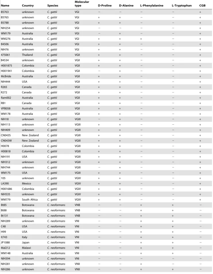
was used as a positive control for growth and yeast nitrogen base (YNB) with 2% glucose but without amino acids and ammonium sulfate (Fig. 1, Blank) was used as a negative control. As shown in Figure 1, utilization of the nitrogen sources was comparable between the two species except for L-phenylalanine, L-tryptophan, D-proline and D-alanine. Although threonine utilization was also different between the two strains, the difference was not as robust as with the aforementioned four amino acids (Fig. 1). While D-proline and D-alanine were utilized exclusively by R265, L-phenylalanine and L-tryptophan were utilized by H99 significantly better than R265. We randomly selected 67 strains of all the known major molecular types (8 VNI, 3 VNII, 3 VNB, 6 VNIII, 11 VNIV, 8 VGI, 12 VGII, 11 VGIII and 5 VGIV). The molecular types of the strains were confirmed byURA5-RFLP and then the strains were tested for utilization of the four amino acids as the source of nitrogen (Table 1). The results were compared with the standard CGB test (Table 1) and the species specificity of the amino acid utilization was summarized (Table 2). The CGB test was species specific, regardless of molecular type. Interestingly, utilization of D-alanine was found to be more species specific than D-proline which has been the second most widely used reagent to differentiate between the two species. While the test involving utilization of D-proline did not yield any false positive results for theC. neoformansstrains, six of the 36 (17%)C. gattiistrains were false negative. False negatives were obtained for three of the four C. gattiimolecular types (VGI–VGIII) with the highest frequency observed among strains of molecular type VGI (38%) followed by VGIII (18%) and VGII (8%). No false negative results were obtained with VGIV strains. As is the case with D-proline, noC. neoformansstrains utilized D-alanine (Table 2) while 94% (34/36) of theC. gattiistrains were able to utilize D-alanine as the sole source of nitrogen. Interestingly, false negatives for D-alanine were only obtained for strains of the VGI molecular type (25%). Among the C. gattistrains tested, only 6% could utilize L-Phenylalanine while none could utilize L-Tryptophan as a nitrogen source (Tables 1 and 2). Interestingly, the ability to utilize Phenylalanine or L-Tryptophan as a nitrogen source was specific to only the VNI, VNII, VNB (serotype A/var.grubii; 93% for L-Phenylalanine and 79% for L-tryptophan) and VNIII, the serotype A/D hybrid (100% for either L-Phenylalanine or L-Tryptophan) molecular type strains. Most VNIV strains (82%) poorly utilized L-Phenylalanine or L-Tryptophan as the sole nitrogen source (Tables 1 and 2).
Regulation of amino acid utilization byGAT1is different between the two species
andH99gat1Dstrains was markedly reduced in all nitrogen sources tested except for proline and arginine where only a slight reduction was observed for both species. UnlikeC. neoformans, only a slight reduction of alanine utilization was observed inC. gattii. Growth of R265gat1D on L-leucine, L-lysine, and L-aspartate was signifi-cantly reduced compared to H99gat1D. On the other hand,GAT1 disruption caused only slight reduction in the growth of R265 on glycine and creatinine while theGAT1disruption in H99 totally abolished its ability to utilize the same nitrogen source (Fig. 2). Complementation ofGAT1restored the wild type level of ability to utilize different amino acids as the sole nitrogen source (Fig S1).
The expression levels of genes encoding glycine cleavage enzymes are controlled by nitrogen catabolite repression less stringently in C. gattiithan inC.
neoformans
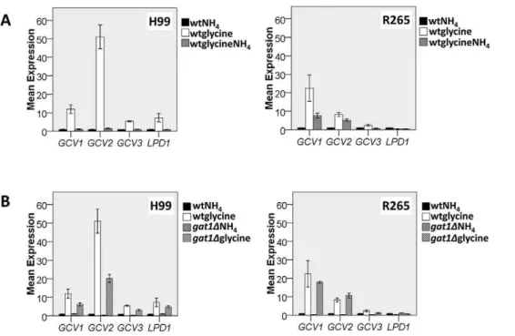
Since utilization of glycine as both the carbon and the nitrogen source has been used to differentiate between the two species [27], we compared the regulation of glycine utilization between H99 and R265 as representatives of the two species. In Saccharomyces cerevisiae, glycine is utilized as a nitrogen source via cleavage by the glycine decarboxylase complex which is controlled by NCR [48]. C. neoformans and C. gattii both contain theS. cerevisiae orthologs encoding 4 subunits of the glycine decarboxylase complex: the T-protein (encoded by GCV1 [49]), P-protein (encoded by GCV2 [50]), H-protein (encoded byGCV3[51]) and L-protein (encoded byLPD1[52]). Though the function of these proteins has not been confirmed experimentally, a similar function of the enzymes reported in S. cerevisiae indicates that these enzymes are highly conserved across fungal species. Thus, we analyzed the GAT1 regulation of these genes by comparing transcriptional profiles of these genes in the two species using quantitative PCR (qPCR). In H99, expression of all four genes was significantly up-regulated when glycine was used as the sole nitrogen source compared to
ammonium sulfate (Fig. 3A). In R265, however, such robust increases were not observed for the expressions ofGCV3andLPD1 (Fig. 3A). In H99, the addition of ammonium sulfate to the glycine media completely suppressed the expression of all 4 genes down to the levels observed with ammonium sulfate alone. However, in R265, the expression of GCV1 and GCV2 was only partially suppressed upon addition of ammonium sulfate (Fig. 3A). These results suggest that expression of two of the genes encoding glycine decarboxylase complex in R265 is not subject to the tight regulation by nitrogen catabolite repression as in H99.
We also examined the roles of GAT1 in regulating the expression of glycine decarboxylase complex genes. Although expression of the four glycine decarboxylase genes in H99gat1D
were induced when glycine was used as the sole nitrogen source, the induction levels ofGCV1, GCV2andGCV3were not as high as in the wild type (GCV1: wt/gat1D= 1.91, p = 0.05; GCV2: wt/ gat1D= 2.55, p = 0.01; GCV3: wt/gat1D= 1.8, p = 0.04, Fig. 3B). These data suggest thatGAT1in H99 plays a role in derepressing the expression of these four genes and some factor(s) yet to be identified also regulates their induction when glycine is used as the sole nitrogen source. In R265gat1D, on the other hand, the expression levels of these four genes were close to wild type levels. These data suggest that the role ofGAT1is less prominent in R265 than in H99 with respect to expressison of the four glycine decarboxylase genes when glycine is the sole nitrogen source.
GAT1regulates virulence differently between C. neoformansandC. gattii
Two previous studies have shown that disruption of GAT1in H99 altered its virulence [35,36]. We compared the impact of GAT1deletion in H99 and R265 on the expression of virulence factors in vitro including melanin and capsule production, ability to grow at 37uC and cell wall integrity in the presence of caffeine. Expression of theLAC1gene which encodes laccase responsible for Figure 1. Utilization of nitrogen sources by H99 and R265.5ml of cell suspensions at OD600nm of 10, 0.1 and 0.001 were spotted on 2% glucose YNB agar with 10 mM of the indicated nitrogen source and incubated for 5–7 days at 30uC. Amino acids differentially utilized by the two species are italicized and underlined.
Table 1.List of strains and their ability to utilize different nitrogen sources.
Name Country Species
Molecular
type D-Proline D-Alanine L-Phenylalanine L-Tryptophan CGB
B5763 unknown C. gattii VGI 2 2 2 2 +
B5765 unknown C. gattii VGI + + 2 2 +
B5788 unknown C. gattii VGI + + 2 2 +
NIH254 unknown C. gattii VGI 2 2 2 2 +
WM179 Australia C. gattii VGI 2 + 2 2 +
WM276 Australia C. gattii VGI + + + 2 +
B4506 Australia C. gattii VGI + + 2 2 +
NIH76 unknown C. gattii VGI + + 2 2 +
475061 Thailand C. gattii VGII + + 2 2 +
B4534 unknown C. gattii VGII + + 2 2 +
H001875 Colombia C. gattii VGII + + 2 2 +
H001941 Colombia C. gattii VGII 2 + 2 2 +
McBride Australia C. gattii VGII + + 2 2 +
NIH444 USA C. gattii VGII + + 2 2 +
R265 Canada C. gattii VGII + + 2 2 +
R272 Canada C. gattii VGII + + 2 2 +
Ram002 Australia C. gattii VGII + + 2 2 +
RB1 Canada C. gattii VGII + + 2 2 +
VPB058 Australia C. gattii VGII + + 2 2 +
WM178 Australia C. gattii VGII + + 2 2 +
NIH18 unknown C. gattii VGIII 2 + 2 2 +
NIH113 unknown C. gattii VGIII + + 2 2 +
NIH409 unknown C. gattii VGIII + + 2 2 +
CN043S New Zealand C. gattii VGIII + + 2 2 +
CN043W New Zealand C. gattii VGIII + + 2 2 +
H0078 Colombia C. gattii VGIII + + 2 2 +
H00818 Colombia C. gattii VGIII + + 2 2 +
NIH191 USA C. gattii VGIII + + 2 2 +
NIH312 unknown C. gattii VGIII + + 2 2 +
NIH744 unknown C. gattii VGIII 2 + 2 2 +
WM175 USA C. gattii VGIII + + 2 2 +
105 unknown C. gattii VGIV + + 2 2 +
LA390 Mexico C. gattii VGIV + + 2 2 +
H001686 Colombia C. gattii VGIV + + 2 2 +
NIH535 unknown C. gattii VGIV + + 2 2 +
WM779 South Africa C. gattii VGIV + + 2 2 +
Bt85 Botswana C. neoformans VNB 2 2 + + 2
Bt88 Botswana C. neoformans VNB 2 2 + + 2
Bt131 Botswana C. neoformans VNB 2 2 + + 2
NIH289 unknown C. neoformans VNI 2 2 + + 2
C48 USA C. neoformans VNI 2 2 + + 2
H99 USA C. neoformans VNI 2 2 + + 2
It743 Italy C. neoformans VNI 2 2 + + 2
JP1088 Japan C. neoformans VNI 2 2 + + 2
Mal212 Malawi C. neoformans VNI 2 2 + + 2
WM148 Australia C. neoformans VNI 2 2 + + 2
NIH394 unknown C. neoformans VNI 2 2 2 2 2
NIH281 unknown C. neoformans VNII 2 2 + 2 2
melanin production [53] and the amount of melanin produced are important for the virulence in both species [43,54]. The gat1D
strains of both H99 and R265 exhibited an increase in melanin production (gat1D/wt = 10.26, p= 0.001 and 2.9, p= 0.023 for H99 and R265, respectively) and the expression of LAC1 was found to be upregulated (gat1D/wt = 14.5, p= 0.001 and 3.2, p= 0.007 for H99 and R265, respectively) (Fig. 4A and 4B). When capsule production was compared in RPMI media with 5% CO2 at 37uC, only thegat1Dstrain of R265 but not of H99 produced a larger capsule than the wild type (gat1D/wt = 1.00,p= 0.915 and 1.17,p= 0.001 for H99 and R265, respectively) (Fig. 4C and 4D). However, neither R265gat1Dnor H99gat1Dshowed any difference in growth rate at 37uC compared to wild type (Fig. 5A). However,
a slight increase was observed in the tolerance to 1 mg/ml caffeine (Fig. 5B), a cell wall perturbing agent which has been used to test cell wall integrity of both species [43,55]. Next, we compared the virulence between gat1D and wild type strains using a murine inhalation model. As reported previously, virulence of H99gat1D
was slightly enhanced compared to H99 (p= 0.141, Fig. 6) [35,36]. Surprisingly, however, virulence of R265gat1D was significantly more reduced than the wild type (p= 0.024, Fig. 6).
Discussion
The ability to utilize specific nitrogen and carbon sources is among the standard methods used for the identification of yeast
Table 1.Cont.
Name Country Species
Molecular
type D-Proline D-Alanine L-Phenylalanine L-Tryptophan CGB
WM626 Australia C. neoformans VNII 2 2 + 2 2
CBS132 Japan C. neoformans VNIII 2 2 + + 2
KW5a Kuwait C. neoformans VNIII 2 2 + + 2
NIH304 unknown C. neoformans VNIII 2 2 + + 2
RKIM364 Germany C. neoformans VNIII 2 2 + + 2
SpaE13 Spain C. neoformans VNIII 2 2 + + 2
TBS54 India C. neoformans VNIII 2 2 + + 2
NIH487 unknown C. neoformans VNIV 2 2 2 2 2
NIH310 unknown C. neoformans VNIV 2 2 + + 2
NIH531 unknown C. neoformans VNIV 2 2 + + 2
B3501 USA C. neoformans VNIV 2 2 2 2 2
B3502 USA C. neoformans VNIV 2 2 2 2 2
JEC20 USA C. neoformans VNIV 2 2 2 2 2
JEC21 USA C. neoformans VNIV 2 2 2 2 2
NIH12 unknown C. neoformans VNIV 2 2 2 2 2
NIH430 unknown C. neoformans VNIV 2 2 2 2 2
NIH433 unknown C. neoformans VNIV 2 2 2 2 2
WM629 Australia C. neoformans VNIV 2 2 2 2 2
doi:10.1371/journal.pone.0034258.t001
Table 2.Utilization of different nitrogen sources according to molecular type.
Species Molecular type
C.n. C.g. VNI VNB VNII VNIII VNIV VGI VGII VGIII VGIV
D-proline negative 31 6 8 3 3 6 11 3 1 2 0
positive 0 30 0 0 0 0 0 5 11 9 5
D-alanine negative 31 2 8 3 3 6 11 2 0 0 0
positive 0 34 0 0 0 0 0 6 12 11 5
L-Phenylalanine negative 10 35 1 0 0 0 9 7 12 11 5
positive 21 1 7 3 3 6 2 1 0 0 0
L-Tryptophan negative 12 36 1 0 2 0 9 8 12 11 5
positive 19 0 7 3 1 6 2 0 0 0 0
CGB negative 31 0 8 3 3 6 11 0 0 0 0
positive 0 36 0 0 0 0 0 8 12 11 5
species [56,57] as well as some filamentous fungi [58]. Differen-tiation of the two species inCscalso has been effectively carried out by both nitrogen and carbon utilization tests [24,25,26,27,47]. However, the efficacies of these biochemical tests were established long before molecular strain typing methods became available and the validity of these tests could have been compromised since the populations tested could contain strains of the same genotype. For example, previous studies have reported 100% [24] and 95% [25] specificity for D-proline utilization byC. gattii while we found a much lower specificity; only 80% ofC. gattiistrains yielded positive results in contrast to the 100% that were negative forC. neoformans. The CGB media, on the other hand, showed 100% specificity between the two species regardless of molecular type. Remarkably, the false negatives observed for D-proline utilization were found mainly among strains of the VGI and VGIII (5 negatives among 6 strains) molecular types. According to the phylogenetic analysis of globalCscstrains based on multi gene sequence analyses, the VGI and VGIII molecular types formed a separate cluster with high bootstrap supports [6,7]. Our results underscore the biological variation between different molecular types withinC. gattii.
Upon screening for utilization of various amino acids as sole nitrogen sources, we discovered a new set of amino acids, phenylalanine, tryptophan and D-alanine, that could discriminate C. neoformansfromC. gattiistrains with reasonably high specificity. In fact, a previous study with much fewer numbers of strains also showed the discriminating power of phenylalanine and tryptophan utilization between the two species [35]. D-alanine has not been
studied as the sole nitrogen source for differentiation of the two species. Our study showed that utilization of D-alanine has a higher discrimination power than that of D-proline, the second most commonly used diagnostic reagent next to CGB medium. Although rare, false negatives or questionable results on CGB media have been reported [25,59,60,61] and these new sets of amino acid utilization panels can be applied to such rare strains. However, the strains with questionable CGB results might also yield ambiguous results with other nitrogen sources if the ambiguity is caused by a mutation in the genes associated with nitrogen metabolism. C. gattii strains that produce questionable CGB results can be tested for growth on tryptophan media in order to correct identify the questionable strains. Although molecular techniques enable confirmation of species identity, use of the one-step diagnostic media employing a species specific nitrogen source could be a cost effective option for developing countries with limited resources.
The nitrogen regulation by NCR inC. neoformanswas studied based on the role of the GATA-type transcriptional activator Gat1 [35,36]. As expected, Gat1 control of nitrogen regulation in the two species showed only minor differences. For example, Gat1 control of glycine utilizations inC. gattiiwas not as stringent as in C. neoformanswhile it was opposite in the utilization of aspartate. These results were supported by transcription analysis of the four genes encoding the glycine cleavage enzyme complex where the induction of these genes was positively controlled by Gat1 inC. neoformansbut to a less extent inC. gattii. In addition, the NCR of Figure 2. Disruption ofGAT1reduced ability to utilize all nitrogen sources.Wild type andgat1Dstrains were grown for 5–7 days at 30uC on 2% glucose YNB with 10 mM of different nitrogen sources. 5ml cell suspensions at OD600nm of 10, 0.1 and 0.001 were spotted on the media. YPD
Figure 3. Comparison of expression levels of the genes encoding glycine cleavage enzymes in H99 and R265.RNA levels of genes encoded enzymes in the glycine cleavage decarboxylase complex were determined in cells growing in ammonium sulfate (NH4), glycine or glycine plus ammonium sulfate (glycineNH4) as the sole nitrogen source. Expression is presented in folds of wild type levels growing in ammonium sulfate. A) Expression levels of the four genes in the wild type strains. B) Effect ofGAT1deletion on the expression of glycine decarboxylase complex genes. The experiment was carried out in triplicates. Bars = standard error, wt = wild type.
doi:10.1371/journal.pone.0034258.g003
Figure 4. Disruption ofGAT1enhanced melanin synthesis andLAC1expression in both species but capsule size increased only in R265.A) Cells were grown in melanin induction media using 10 mM proline as the nitrogen source. Melanin production was assayed by measuring the absorbance at 475 nm and B)LAC1expressions were quantified by real time PCR. C) Micrograph of cell grown in capsule induction media. D) The relative capsule size was measured in.100 cells for each strain grown in capsule induction media. Experiments were carried out in triplicates. *p#0.05 by the student t-test. Error bars = 2 standard deviation.
glycine utilization in the presence of ammonium was clear-cut in C. neoformansbut not inC. gattii. The difference in NCR of glycine utilization between the two species is reminiscent of creatinine metabolism where creatinine deiminase was inhibited by the accumulation of ammonium only inC. neoformans and not inC. gattii. As a result, growth of C. neoformanson creatinine media is considerably slower compared to C. gattii and creatinine bromthymol blue agar was the first one-step diagnostic media formulated to differentiate between the two species [47]. Moreover, it has been known that glycine metabolism is controlled by a complex process involving several genes [48,62,63]. For example, in S. cerevisiae, glycine can be synthesized from or converted to serine and threonine by serine hydroxymethyltrans-ferase (Shm1 and Shm2) and threonine aldolase (Gly1). However, both species ofCscdo not utilize threonine as effectively as other amino acids (Fig. 1) suggesting that the Gly1 role in glycine metabolism is minimal in Csc as has been reported in Candida albicans [64]. However, Shm1 may play a role in glycine metabolism in Csc since expression of SHM1 was up-regulated 10 fold in both species when glycine was used as the nitrogen source instead of ammonium sulfate (data not shown). The nitrogen catabolic pathway, therefore, appears to be complex and further studies are needed to elucidate the pathways inCsc.
The ability to survive in a limited nutrient environment in the host is crucial for the growth of pathogenic organisms. For example, Gat1 influences virulence of several pathogenic fungi including, Aspergillus fumigatus and C. albicans [32,33,35,36]. Our results on the virulence of H99gat1Dis similar to the findings of Lee et al. [35] but different from that of Kmetzsch et al. [36]. Interestingly, we found thatgat1Dbehaved differently between the
two species in controlling the expression of some virulence factors in vitroand virulence in animals. It was surprising to observe that several virulence factors including capsule production, melanin synthesis and cell wall integrity, were negatively controlled by Gat1 inC. gattiiwhile the virulence of R265gat1Dwas decreased in mice. In the host environment, the impact of impaired nitrogen utilization due to the lack of Gat1 function may be compensated by the increase in the virulence factors. The different impact of Gat1 in virulence between the two species might stem from the differences in their natural habitat and hence their pathobiology. It has been speculated that the virulence factors of the Cryptococcal species have evolved to combat their environmental predators such as soil amoeba and nematodes [65]. Pigeon droppings, the primary environmental niche of C. neoformans, is high in ammonium concentration and Gat1 may be constantly down-regulated while the primary natural habitat ofC. gattiiis detritus of trees which should be much lower in ammonium content than pigeon guano. This would have had a different impact on evolution of the role of Gat1 in regulation of virulence factors. However, whether the slight impact of Gat1 on the regulation of virulence factors observed in this study benefits survival ofCscin nature is unclear.
In conclusion, our study points out the different aspects of nitrogen metabolism betweenC. neoformansandC. gattii, two closely related etiologic agents of cryptococcosis. The range of nitrogen sources that can be used to differentiate between the two species has been expanded from previous studies to include a few more amino acids. The difference in nitrogen metabolism was confirmed by differences in nitrogen regulation by Gat1 and ammonium NCR which has a distinct effect on virulence between the two species. The difference in nitrogen utilization and its regulation by Figure 5. The role ofGAT1in cell growth.A)GAT1is not associated
with the growth rate of both species. Cell suspensions of 2 OD600were spotted on YPD agar in three 10 fold dilutions and incubated for three days at the designated temperatures B) Similar spot assays were carried out on YPD with or without caffeine and plates were incubated at 30uC for 3 days. Experiments were carried out in duplicates.
doi:10.1371/journal.pone.0034258.g005
Figure 6. Virulence ofH99gat1Dwas minimally enhanced while ofR265gat1Dwas significantly reduced in murine inhalation model.50,000 cells from each strain were inoculated intrapharyngeally to 10 Balb/c mice per group. Mice were fed with food and water ad libitum.
Gat1 shown in this study provides a compelling reason for characterization of the genes and metabolic pathways responsible for the differences. Global screening techniques namely transcrip-tome comparisons via cDNA microarrays and construction of Agrobacterium tumefaciens mediated insertional libraries [66] are currently being undertaken to elucidate the complex regulatory network of nitrogen metabolism inCsc.
Materials and Methods
Ethics Statement
The animal experiments were carried out with the approval and oversight of the Animal Care and Use Committee of the National Institute of Allergy and Infectious Diseases, United States National Institutes of Health, Bethesda MD.
Strains and media
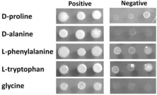
A global collection of 8 molecular types consisting of 31 C. neoformansand 36C. gattiistrains were revived from280uC freezers of Molecular Microbiology Section, Laboratory of Clinical Infectious Diseases, NIAID, NIH, Bethesda, MD, USA and Molecular Mycology Research Laboratory, Westmead Hospital, Westmead, NSW, Australia. Molecular types of each strain were confirmed by URA5-RFLP as previously described (data not shown) [3] (Table 1). Cells were maintained on YPD agar (2% glucose, 1% yeast extract, 2% peptone and 2% Bacto agar) until use. For nitrogen utilization tests, cell suspensions were diluted to 0.02 OD600 nm for a single spot test or 10, 0.1 and 0.001 for serial dilution spot tests. The cell suspensions were spotted on 2% glucose YNB media (Becton, Dickinson and company, Sparks, MD, USA) with or without 10 mM of each indicated nitrogen source (Sigma-Aldrich, St. Louis, MO, USA) and incubated for 5– 7 days at 30uC. For D-proline and D-alanine utilization tests, the results were recorded as negative when the growth was lower or equal to the growth on media without nitrogen source (Blank). For L-phenylalanine and L-tryptophan, results were recorded as negative when the strains grew poorly (see Fig. 7). CGB tests were carried out as described before and recorded as positive when the medium turns from yellow green to blue [27]. The nitrogen utilization tests were performed in duplicate or triplicate when differences were either evident or the experimental results were expected to fluctuate.
Identification of gene orthologs
Protein sequences of each gene fromS. cerevisiae genome project (http://www.yeastgenome.org/) were used to perform the BlastP inC. neoformans H99 genome project (http://www.broadinstitute.org/ annotation/genome/cryptococcus_neoformans/MultiHome.html) and C. gattii R265 genome project (http://www.broadinstitute. org/annotation/genome/cryptococcus_neoformans_b/MultiHome. html). Genes were identified as follows (C. neoformans/C. gattii):GAT1– CNAG_00193/CNBG_0368, GCV1 – CNAG_02818/CNBG_ 3472, GCV2 – CNAG_01594/CNBG_3846, GCV3 – CNAG_ 06316/CNBG_5135,LPD1– CNAG_02851/CNBG_5871.
Gene disruptions and complementations
Disruption constructs ofGAT1were created using overlapping PCR to link the 59 and 39 flanking region to nourseothricin antibiotic cassette [67]. The disruption constructs were trans-formed by biolistic transformation as described previously [68]. Transformants were selected on YPD containing nourseothricin. Homologous integrations were confirmed by PCR and southern hybridization. Disruptants were complemented by homologous integration ofGAT1gene intogat1Dusing biolistic transformation.
As none ofgat1Dwas able to utilize L-serine as the sole nitrogen source, transformants grown on serine media (2% glucose YNB with 10 mM L-serine) were analyzed for the homologous complementations by PCR and confirmed by southern hybrid-ization. (See table S1 for primers details)
Quantitative real-time PCR
Strains were grown in YPD broth overnight to mid log phase. Cells were washed 3 times with 16phosphate buffered saline (PBS: 137 mM NaCl, 2.7 mM KCl, 10 mM Na2HPO4, and 2 mM KH2PO4, pH 7.4) and re-suspended (1 OD600) in 2% glucose YNB supplemented with 10 mM of the specified nitrogen source. Cells were then grown at 30uC for 2 hrs and harvested for RNA extraction. Cell pellets were frozen and lyophilized. RNAs were extracted using RNA by TrizolH reagent (Invitrogen, Carlsbad, CA, USA) and cDNAs were generated by the Super-cript III first-strand synthesis system (Invitrogen, Carlsbad, CA, USA) according to the manufacturer’s protocol. Quantitative real-time PCR were performed using SYBR greener supermix (Invitrogen, Carlsbad, CA, USA) in Applied Biosystems 7500 Real-Time PCR System. Experiments were performed in triplicates using a relative standard curve method. (see Table S1 for primers details).
Melanin production
Quantification of laccase activity for melanin synthesis was performed in triplicate according to the described method [69] with modification, using 0.1% glucose YNB without amino acid and ammonium chloride (Becton, Dickinson and Company, MD, USA) with 10 mM proline and 1 mM epinephrine (Sigma-Aldrich, MD, USA) at 30uC. L-proline was used as a nitrogen source due to its minimal growth reduction uponGAT1disruption [35]. Culture supernatant was obtained after 24 hours and assayed by measuring the absorbance at OD475nm using DU-64 spectophotometer (Beckman coulter, Fullerton, CA).
Capsule formation
Amount of capsule produced was quantified by comparing the size ratio between cell with the capsules vs naked yeast (cell wall to cell wall diameter) as has been reported previously [43]. Briefly, each strain was grown overnight in YPD broth at 30uC. Yeast cells were harvested, washed and inoculated into capsule inducing RPMI with MOPS, HCO3, pH 7.3 and grown to a cell density of 106–107cells/ml at 37uC with 5% CO2for 72 hr [35,70]. Capsule size of the cells stained with India ink was quantified by light microscopy.
Figure 7. Examples of positive and negative growth on each nitrogen source.Cells were grown on 2% glucose YNB with 10 mM of each nitrogen source for 5–7 days at 30uC. 2ml cell suspensions at an
Ability to tolerate high temperature and cell wall perturbing agent
Strains were grown on YPD with or without 1 mg/ml caffeine, a known cell wall perturbing agent [43,55] at 30uC or 37uC. Serial dilution spot test were carried out as described in the figure legends.
Virulence in mice
BALB/c mice were inoculated with 50,000 yeast cells via intrapharyngeal inhalation as described [71,72]. Briefly, cells were diluted in phosphate buffered saline (PBS) to 2.56106 cells/ml. Mice were anesthetized by isoflurane and partially hung by placing their incisors on a string while their lower back lie to a support patch. Tongues of the mice were gently held in full extension with padded forceps while a 20ml suspension was pipetted onto the base of the tongue. Mice were allowed to breathe in the solution for 10 seconds. Tongues were then released and mice were placed in their cages to recover. Survival was monitored for 50 days.
Statistical analysis
All statistical analyses specified were performed in SPSS 17.0 (IBM Inc., Armonk, New York).
Supporting Information
Figure S1 Examples of GAT1/AREA complementation which restored nitrogen utilization to the wild type level. Wild type,gat1Dand complemented strains were grown on 2% glucose YNB with 10 mM of each nitrogen source for 5–7 days at 30uC. . 5ml of cells at OD600nm of 10, 0.1 and 0.001 were spotted on the media.
(TIF)
Table S1 Primers details. (DOCX)
Acknowledgments
We thank Wieland Meyer, Sydney University at Westmead Hospital, Westmead, NSW, Australia and Anastasia Litvintserva, Duke University, Durham, NC, USA for providing strains in this study.
Author Contributions
Conceived and designed the experiments: PN YC KJK. Performed the experiments: PN JR. Analyzed the data: PN YC KJK. Contributed reagents/materials/analysis tools: KJK. Wrote the paper: PN YC KJK.
References
1. Heitman J, Kozel TR, Kwon-Chung J, Perfect JR, Casadevall A (2011) Cryptococcus: from human pathogen to model yeast. Washington, DC: ASM press. 620 p.
2. Kwon-Chung J, Boekhout T, Wickes B, Fell J (2011) Systematics of the Genus Cryptococcusand its type speciesC. neoformans. In: Heitman J, Kozel TR, Kwon-Chung J, Perfect JR, Casadevall A, eds.Cryptococcus: From human pathogen to model yeast. Washington, DC: ASM press. pp 3–16.
3. Meyer W, Castaneda A, Jackson S, Huynh M, Castaneda E (2003) Molecular typing of IberoAmericanCryptococcus neoformansisolates. Emerg Infect Dis 9: 189–195. 4. Boekhout T, Theelen B, Diaz M, Fell JW, Hop WC, et al. (2001) Hybrid
genotypes in the pathogenic yeastCryptococcus neoformans. Microbiology 147: 891–907.
5. Meyer W, Aanensen DM, Boekhout T, Cogliati M, Diaz MR, et al. (2009) Consensus multi-locus sequence typing scheme forCryptococcus neoformansand Cryptococcus gattii. Med Mycol. pp 1–14.
6. Bovers M, Hagen F, Boekhout T (2008) Diversity of theCryptococcus neoformans-Cryptococcus gattiispecies complex. Rev Iberoam Micol 25: S4–12.
7. Ngamskulrungroj P, Gilgado F, Faganello J, Litvintseva AP, Leal AL, et al. (2009) Genetic diversity of the Cryptococcus species complex suggests that Cryptococcus gattiideserves to have varieties. PLoS One 4: e5862.
8. Bennett JE, Kwon-Chung KJ, Howard DH (1977) Epidemiologic differences among serotypes ofCryptococcus neoformans. Am J Epidemiol 105: 582–586. 9. Franzot SP, Salkin IF, Casadevall A (1999)Cryptococcus neoformansvar.grubii:
separate varietal status forCryptococcus neoformansserotype A isolates. J Clin Microbiol 37: 838–840.
10. Levitz SM (1991) The ecology ofCryptococcus neoformansand the epidemiology of cryptococcosis. Rev Infect Dis 13: 1163–1169.
11. Kwon-Chung KJ, Boekhout T, Fell JW, Diaz M (2002) (1557) Proposal to conserve the name Cryptococcus gattii against C.hondurianusand C.basillisporus (Basidiomycota, Hymenomycetes, Tremellomycetidae). Taxon 51: 804–806. 12. Litvintseva AP, Thakur R, Vilgalys R, Mitchell TG (2006) Multilocus sequence
typing reveals three genetic subpopulations ofCryptococcus neoformansvar.grubii (serotype A), including a unique population in Botswana. Genetics 172: 2223–2238.
13. Choi YH, Ngamskulrungroj P, Varma A, Sionov E, Hwang SM, et al. (2010) Prevalence of the VNIc genotype ofCryptococcus neoformansin non-HIV-associated cryptococcosis in the Republic of Korea. FEMS Yeast Res 10: 769–778. 14. Chen J, Varma A, Diaz MR, Litvintseva AP, Wollenberg KK, et al. (2008)
Cryptococcus neoformans strains and infection in apparently immunocompetent patients, China. Emerg Infect Dis 14: 755–762.
15. Kidd SE, Hagen F, Tscharke RL, Huynh M, Bartlett KH, et al. (2004) A rare genotype ofCryptococcus gattiicaused the cryptococcosis outbreak on Vancouver Island (British Columbia, Canada). Proc Natl Acad Sci U S A 101: 17258–17263.
16. Fraser JA, Giles SS, Wenink EC, Geunes-Boyer SG, Wright JR, et al. (2005) Same-sex mating and the origin of the Vancouver Island Cryptococcus gattii outbreak. Nature 437: 1360–1364.
17. Byrnes EJ, 3rd, Li W, Lewit Y, Ma H, Voelz K, et al. (2010) Emergence and pathogenicity of highly virulentCryptococcus gattiigenotypes in the northwest United States. PLoS Pathog 6: e1000850.
18. MacDougall L, Kidd SE, Galanis E, Mak S, Leslie MJ, et al. (2007) Spread of Cryptococcus gattiiin British Columbia, Canada, and detection in the Pacific Northwest, USA. Emerg Infect Dis 13: 42–50.
19. Ellis DH, Pfeiffer TJ (1990) Natural habitat ofCryptococcus neoformansvar.gattii. J Clin Microbiol 28: 1642–1644.
20. Kwon-Chung KJ, Bennett JE (1984) Epidemiologic differences between the two varieties ofCryptococcus neoformans. Am J Epidemiol 120: 123–130.
21. Granados DP, Castaneda E (2006) Influence of climatic conditions on the isolation of members of theCryptococcus neoformansspecies complex from trees in Colombia from 1992-2004. FEMS Yeast Res 6: 636–644.
22. Bennett JE, Kwon-Chung KJ, Theodore TS (1978) Biochemical differences between serotypes ofCryptococcus neoformans. Sabouraudia 16: 167–174. 23. Kwon-Chung KJ, Bennett JE, Theodore TS (1978)Cryptococcus bacillisporussp.
nov.: serotype B–C ofCryptococcus neoformans. International Journal of Systematic Bacteriology 28: 616–620.
24. Dufait R, Velho R, De Vroey C (1987) Rapid identification of the two varieties of Cryptococcus neoformans by D-proline assimilation. Mykosen 30: 483. 25. Nishikawa MM, Sant’Anna OD, Lazera MS, Wanke B (1996) Use of D-proline
assimilation and CGB medium for screening BrazilianCryptococcus neoformans isolates. J Med Vet Mycol 34: 365–366.
26. Salkin IF, Hurd NJ (1982) New medium for differentiation of Cryptococcus neoformans serotype pairs. J Clin Microbiol 15: 169–171.
27. Kwon-Chung KJ, Polacheck I, Bennett JE (1982) Improved diagnostic medium for separation ofCryptococcus neoformansvar.neoformans(serotypes A and D) and Cryptococcus neoformans var. gattii (serotypes B and C). J Clin Microbiol 15: 535–537.
28. Magasanik B, Kaiser CA (2002) Nitrogen regulation inSaccharomyces cerevisiae. Gene 290: 1–18.
29. Wong KH, Hynes MJ, Davis MA (2008) Recent advances in nitrogen regulation: a comparison betweenSaccharomyces cerevisiaeand filamentous fungi. Eukaryot Cell 7: 917–925.
30. Fu YH, Marzluf GA (1987) Characterization of nit-2, the major nitrogen regulatory gene ofNeurospora crassa. Mol Cell Biol 7: 1691–1696.
31. Mitchell AP, Magasanik B (1984) Regulation of glutamine-repressible gene products by the GLN3 function inSaccharomyces cerevisiae. Mol Cell Biol 4: 2758–2766.
32. Limjindaporn T, Khalaf RA, Fonzi WA (2003) Nitrogen metabolism and virulence ofCandida albicansrequire the GATA-type transcriptional activator encoded by GAT1. Mol Microbiol 50: 993–1004.
33. Hensel M, Arst HN, Jr., Aufauvre-Brown A, Holden DW (1998) The role of the Aspergillus fumigatusareA gene in invasive pulmonary aspergillosis. Mol Gen Genet 258: 553–557.
34. Snoeijers SS, Vossen P, Goosen T, Van den Broek HW, De Wit PJ (1999) Transcription of the avirulence gene Avr9 of the fungal tomato pathogen Cladosporium fulvum is regulated by a GATA-type transcription factor in Aspergillus nidulans. Mol Gen Genet 261: 653–659.
36. Kmetzsch L, Staats CC, Simon E, Fonseca FL, Oliveira DL, et al. (2010) The GATA–type transcriptional activator Gat1 regulates nitrogen uptake and metabolism in the human pathogenCryptococcus neoformans. Fungal Genet Biol 48: 192–199.
37. Casadevall A, Perfect JR (1998) Cryptococcus neoformans. Washington, DC: ASM press.
38. Kwon-Chung KJ (1975) A new genus, filobasidiella, the perfect state of Cryptococcus neoformans. Mycologia 67: 1197–1200.
39. Kwon-Chung KJ (1976) A new species ofFilobasidiella, the sexual state of Cryptococcus neoformansB and C serotypes. Mycologia 68: 943–946.
40. Kwon-Chung KJ, Wickes BL, Stockman L, Roberts GD, Ellis D, et al. (1992) Virulence, serotype, and molecular characteristics of environmental strains of Cryptococcus neoformansvar.gattii. Infect Immun 60: 1869–1874.
41. Ma H, Hagen F, Stekel DJ, Johnston SA, Sionov E, et al. (2009) The fatal fungal outbreak on Vancouver Island is characterized by enhanced intracellular parasitism driven by mitochondrial regulation. Proc Natl Acad Sci U S A 106: 12980–12985.
42. Ngamskulrungroj P, Himmelreich U, Breger JA, Wilson C, Chayakulkeeree M, et al. (2009) The trehalose synthesis pathway is an integral part of the virulence composite forCryptococcus gattii. Infect Immun 77: 4584–4596.
43. Ngamskulrungroj P, Price J, Sorrell T, Perfect JR, Meyer W (2011)Cryptococcus gattiivirulence composite: candidate genes revealed by microarray analysis of high and less virulent Vancouver island outbreak strains. PLoS One 6: e16076. 44. Narasipura SD, Chaturvedi V, Chaturvedi S (2005) Characterization of Cryptococcus neoformans variety gattii SOD2 reveals distinct roles of the two superoxide dismutases in fungal biology and virulence. Mol Microbiol 55: 1782–1800.
45. Ngamskulrungroj P, Serena C, Gilgado F, Malik R, Meyer W (2011) Global VGIIa isolates are of comparable virulence to the major fatalCryptococcus gattii Vancouver Island outbreak genotype. Clin Microbiol Infect 17: 251–258. 46. Perfect JR, Ketabchi N, Cox GM, Ingram CW, Beiser CL (1993) Karyotyping
of Cryptococcus neoformans as an epidemiological tool. J Clin Microbiol 31: 3305–3309.
47. Polacheck I, Kwon-Chung KJ (1980) Creatinine metabolism in Cryptococcus neoformansandCryptococcus bacillisporus. J Bacteriol 142: 15–20.
48. Piper MD, Hong SP, Eissing T, Sealey P, Dawes IW (2002) Regulation of the yeast glycine cleavage genes is responsive to the availability of multiple nutrients. FEMS Yeast Res 2: 59–71.
49. McNeil JB, Zhang F, Taylor BV, Sinclair DA, Pearlman RE, et al. (1997) Cloning, and molecular characterization of theGCV1gene encoding the glycine cleavage T-protein fromSaccharomyces cerevisiae. Gene 186: 13–20.
50. Sinclair DA, Hong SP, Dawes IW (1996) Specific induction by glycine of the gene for the P-subunit of glycine decarboxylase fromSaccharomyces cerevisiae. Mol Microbiol 19: 611–623.
51. Nagarajan L, Storms RK (1997) Molecular characterization of GCV3, the Saccharomyces cerevisiaegene coding for the glycine cleavage system hydrogen carrier protein. J Biol Chem 272: 4444–4450.
52. Ross J, Reid GA, Dawes IW (1988) The nucleotide sequence of theLPD1gene encoding lipoamide dehydrogenase in Saccharomyces cerevisiae: comparison between eukaryotic and prokaryotic sequences for related enzymes and identification of potential upstream control sites. J Gen Microbiol 134: 1131–1139.
53. Williamson PR (1994) Biochemical and molecular characterization of the diphenol oxidase ofCryptococcus neoformans: identification as a laccase. J Bacteriol 176: 656–664.
54. Williamson PR (1997) Laccase and melanin in the pathogenesis ofCryptococcus neoformans. Front Biosci 2: e99–107.
55. Gerik KJ, Donlin MJ, Soto CE, Banks AM, Banks IR, et al. (2005) Cell wall integrity is dependent on thePKC1signal transduction pathway inCryptococcus neoformans. Mol Microbiol 58: 393–408.
56. Kurtzman CP, Fell J, Boekhout T (2011) The yeasts: A taxonomic study. Amsterdam: Elsevier.
57. Segal E, Ajello L (1976) Evaluation of a new system for the rapid identification of clinically important yeasts. J Clin Microbiol 4: 157–159.
58. Philpot CM (1977) The use of nutritional tests for the differentiation of dermatophytes. Sabouraudia 15: 141–150.
59. Klein KR, Hall L, Deml SM, Rysavy JM, Wohlfiel SL, et al. (2009) Identification ofCryptococcus gattiiby use of L-canavanine glycine bromothymol blue medium and DNA sequencing. J Clin Microbiol 47: 3669–3672. 60. Leal AL, Faganello J, Bassanesi MC, Vainstein MH (2008)Cryptococcusspecies
identification by multiplex PCR. Med Mycol 46: 377–383.
61. McTaggart L, Richardson SE, Seah C, Hoang L, Fothergill A, et al. (2011) Rapid identification of Cryptococcus neoformans var. grubii, C. neoformans var. neoformans, andC. gattiiby use of rapid biochemical tests, differential media, and DNA sequencing. J Clin Microbiol 49: 2522–2527.
62. Monschau N, Stahmann KP, Sahm H, McNeil JB, Bognar AL (1997) Identification ofSaccharomyces cerevisiaeGLY1 as a threonine aldolase: a key enzyme in glycine biosynthesis. FEMS Microbiol Lett 150: 55–60.
63. Kastanos EK, Woldman YY, Appling DR (1997) Role of mitochondrial and cytoplasmic serine hydroxymethyltransferase isozymes in de novo purine synthesis inSaccharomyces cerevisiae. Biochemistry 36: 14956–14964.
64. McNeil JB, Flynn J, Tsao N, Monschau N, Stahmann K, et al. (2000) Glycine metabolism inCandida albicans: characterization of the serine hydroxymethyl-transferase (SHM1,SHM2) and threonine aldolase (GLY1) genes. Yeast 16: 167–175.
65. Casadevall A, Steenbergen JN, Nosanchuk JD (2003) ‘Ready made’ virulence and ‘dual use’ virulence factors in pathogenic environmental fungi–the Cryptococcus neoformansparadigm. Curr Opin Microbiol 6: 332–337.
66. McClelland CM, Chang YC, Kwon-Chung KJ (2005) High frequency transformation ofCryptococcus neoformansandCryptococcus gattiiby Agrobacterium tumefaciens. Fungal Genet Biol 42: 904–913.
67. Davidson RC, Blankenship JR, Kraus PR, de Jesus Berrios M, Hull CM, et al. (2002) A PCR-based strategy to generate integrative targeting alleles with large regions of homology. Microbiology 148: 2607–2615.
68. Toffaletti DL, Rude TH, Johnston SA, Durack DT, Perfect JR (1993) Gene transfer inCryptococcus neoformansby use of biolistic delivery of DNA. J Bacteriol 175: 1405–1411.
69. Pukkila-Worley R, Gerrald QD, Kraus PR, Boily MJ, Davis MJ, et al. (2005) Transcriptional network of multiple capsule and melanin genes governed by the Cryptococcus neoformanscyclic AMP cascade. Eukaryot Cell 4: 190–201. 70. Zaragoza O, Fries BC, Casadevall A (2003) Induction of capsule growth in
Cryptococcus neoformans by mammalian serum and CO2. Infect Immun 71:
6155–6164.
71. Sugui JA, Vinh DC, Nardone G, Shea YR, Chang YC, et al. (2010)Neosartorya udagawae(Aspergillus udagawae), an emerging agent of aspergillosis: how different is it from Aspergillus fumigatus? J Clin Microbiol 48: 220–228.