LEILA MARIA GUISSONI CAMPOS
Projeções hipotalâmicas do núcleo supraquiasmático com base na distribuição de fibras imunorreativas para VIP e AVP no Cebus apella.
São Paulo 2013
Tese apresentada ao Programa de Pós Graduação em Ciências Morfofuncionais do Instituto Ciências Biomédicas da Universidade de São Paulo, para obtenção do título de Doutor em Ciências.
Área de concentração: Ciências Morfofuncionais
RESUMO
Campos LMG. Projeções hipotalâmicas do núcleo supraquiasmático com base na distribuição de fibras imunorreativas para VIP e AVP no Cebus apella. [tese
(Doutorado em Ciências Morfofuncionais)]. São Paulo: Instituto de Ciências Biomédicas, Universidade de São Paulo; 2013.
O núcleo hipotalâmico supraquiasmático (SCh), apresenta caracterização neuroquímica com duas subpopulações principais de células produtoras de polipeptídeo intestinal vasoativo (VIP) e argenina vasopressina (AVP). As fibras imunorreativas (IR) a AVP e VIP eferentes do SCh apresentam características morfológicas que possibilitam seu rastreamento a longas distâncias dentro do hipotálamo. No presente estudo mapeamos as fibras e terminais VIP e AVP IR nas áreas hipotalâmicas do primata Cebus apella utilizando a técnica de
imuno-histoquímica. Assim, foi realizada a identificação das áreas hipotalâmicas recipientes do SCh, utilizando o mapeamento associado à análise morfológica das fibras destas duas substâncias neuroativas. As fibras VIP e AVP IR com características de fibras eferentes do SCh foram identificadas em porções anteriores: hipotálamo anterior, área pré-óptica, área hipotalâmica lateral, SPZV; e porções mais caudais: porção retroquiasmática, área tuberal. Os resultados indicam um padrão similar de distribuição de fibras VIP e AVP IR em áreas hipotalâmicas descritas como recipientes das projeções do SCh, quando comparado com outras espécies descritas na literatura.
Palavras-chave: Núcleo supraquiasmático. Cebus apella. Hipotálamo. VIP. AVP.
ABSTRACT
Campos LMG. Suprachiasmatic nucleus projections for hypothalamic areas according to VIP and AVP immunoreactivity in the Cebus apella monkey. [Ph. D.
thesis (Sciences Morphofunctional)]. São Paulo: Instituto de Ciências Biomédicas, Universidade de São Paulo; 2013.
The suprachiasmatic nucleus (SCN) of the hypothalamus, contains a variety of different neurons that tend to form two major subpopulations within the nucleus, the vasoactive intestinal peptide (VIP) and vasopressin (AVP). The immunoreactive (IR) fibers derived from the VIP and AVP IR cells of the SCN present morphological characteristics that allow their specific tracking in long distances within the hypothalamus. In the present investigation we mapped VIP and AVP IR terminals in hypothalamic areas of the primate Cebus apella using immunohistochemistry, and
identified SCN recipient areas in the hypothalamus using the mapping distribution of IR fibers associated with morphological analysis of these two neuroactive substances fibers. VIP and AVP IR fibers with characteristics from SCN were identified in the rostral anterior hypothalamic, medial preoptic area, lateral hypothalamic area, and more caudally in SPZV and retrochiasmatic tuberal area. The results indicate that there is a similarity in the pattern of VIP and AVP IR fibers in hypothalamic recipients areas from SCN projections when compared with species described in the literature.
1 INTRODUÇÃO
A maioria das espécies apresenta ritmos biológicos fisiológicos, endócrinos e comportamentais marcados por variações diárias ou sazonais de temperatura, umidade e períodos fóticos. Esta capacidade adaptativa que aperfeiçoa a eficiência dos sistemas biológicos está baseada em mecanismos que promovem a organização temporal dos fenômenos fisiológicos do organismo com o meio ambiente em que ele vive (Moore-Ede et al., 1982; Reppert, Weaver, 2002).
Os chamados ritmos biológicos circadianos oscilam em períodos ao redor de 24 horas e persistem em condições ambientais constantes o que indica a existência de um sistema de oscilação endógeno composto por um marca-passo circadiano central dominante, o núcleo supraquiasmático (SCh), e por vias aferentes e eferentes deste núcleo (Moore-Ede et al., 1982). Este sistema de oscilação ou sistema de temporização circadiano (STC) permite ao organismo antecipar-se e preparar-se para as alterações que ocorrem no ambiente natural, podendo assim otimizar os seus comportamentos de acordo com os padrões rítmicos ambientais.
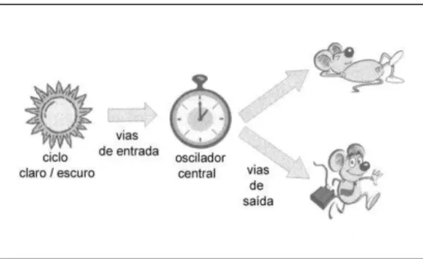
Figura 1 - Esquema de modelo simplificado do sistema de temporização circadiano (STC).
Esquema simplificado do STC mostrando o oscilador central, que representa o núcleo supraquiasmático (SCh), sob influência de fatores de entrada como o ciclo claro-escuro e a expressão de ritmos em vários aspectos fisiológicos como a atividade e repouso por meio de estruturas consideradas vias de saída que podem representar conexões neurais diretas e/ou fatores secretados. Fonte: Adaptado de Sehgal (2004).
(Gooley et al., 2006) e pode ser influenciado por vias neurais e por secreções endócrinas (Cipolla-Neto et al., 1988).
A oscilação circadiana do SCh deve-se à capacidade oscilatória autônoma de suas células que por sua vez decorre de alças de retro-alimentação de transcrição e translação resultando na expressão rítmica dos genes do relógio ou “clock genes” (Reppert, Weaver, 2001). Estes componentes “clock” incluem, entre outros, três genes da família Per: Per1, Per2 e Per3 (Zylka et al., 1998), o gene Clock (King et al., 1997), genes “cryptochrome” Cry1 e Cry2 (Kume et al., 1999) e o BMAL1 (Gekakis et al., 1998).
A determinação do SCh como oscilador central veio por meio de experimentos clássicos que mostraram que: 1) lesões no SCh levam a perda dos ritmos circadianos para uma série de parâmetros comportamentais e fisiológicos (Moore, Eichler, 1972), 2) Existem ritmos de atividade elétrica dentro do SCh após isolamento do resto do cérebro, numa preparação conhecida como ‘ilha hipotalâmica’ (Inouye, Kawamura, 1979), 3) partes do cérebro que normalmente exibem uma variação circadiana na atividade neuronal deixam de fazê-lo após isolamento cirúrgico do SCh, 4) O transplante de SCh fetal recupera a ritmicidade circadiana em roedores arrítmicos por lesão bilateral prévia do SCh (Lehman et al., 1987) e 5) Quando é feito esse tipo de transplante, se forem utilizados animais mutantes com período circadiano mais curto como doadores, o hospedeiro recupera a ritmicidade circadiana com período similar ao do doador (Ralph et al., 1990).
1.2 Aspectos anatômicos do SCh e o Hipotálamo
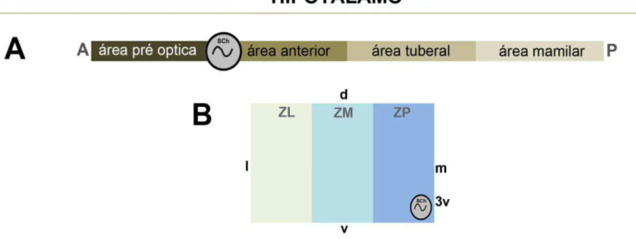
O hipotálamo tem sido didaticamente dividido em 4 regiões antero-posteriores identificadas como área pré óptica, área anterior, área tuberal e área mamilar (Le Gros Clark, 1938 apud Simerly, 1995) nas quais estão distribuídas no sentido médio lateral 3 zonas longitudinais distintas denominadas zonas periventricular, medial e lateral (Crosby, Woodsburne, 19401 apud Simerly, 1995).
A zona periventricular contém a maioria dos neurônios hipofisários, que projetam para eminência mediana e modulam a hipófise anterior (adeno hipófise), e
1 Crosby EC, Woodburne RT. The comparative anatomy of the preoptic area and the hypothalamus.
também grande parte dos neurônios magnocelares que projetam para hipófise posterior (neuro hipófise).
Na zona medial, núcleos bem distintos constituem áreas com amplas conexões com o telencéfalo, regiões límbicas e intra-hipotalâmicas além de algumas conexões com tálamo e tronco encefálico. Este conjunto de núcleos é responsável pela iniciação de diversos comportamentos e pela integração entre as zonas periventricular e lateral.
Na zona lateral, os neurônios estão distribuídos entre as fibras do feixe prosencefálico medial, formando um núcleo intersticial. Esta zona é reconhecida como área integradora, com núcleos envolvidos em fenômenos como alerta e comportamentos motivados (Saper et al., 1979; Simerly, 1995; Watts, Swanson, 1987).
O SCh está localizado na zona periventricular e embora apresente variações interespecíficas quanto à forma tridimensional, volume, densidade e tamanho das células, apresenta organização conservada em mamíferos, como um par de aglomerados de pequenos neurônios situados no hipotálamo anterior, adjacentes ao terceiro ventrículo (3V) e imediatamente dorsal ao quiasma óptico (ox) (Cassone et al., 1988; Lydic et al., 1982; Pinato et al., 2007).
Figura 2 - Esquema de divisão do hipotálamo.
1.3 Neuroquímica do SCh
A grande complexidade funcional deste núcleo tem sido atribuída em parte, à sua composição neuroquímica, caracterizada por diferentes populações neuronais e terminais que produzem e/ou liberam diferentes substâncias neuroativas. Esta composição apresenta pequenas variações interespecíficas entre roedores, e importantes diferenças entre roedores e primatas.
No SCh de roedores foram descritos neurônios imunorreativos (IR) a óxido nítrico sintase (NOS) (Decker, Reuss, 1994; Wang, Morris, 1996), proteínas ligantes de cálcio calbindina (CB), parvalbumina (PV) e calretinina (CR) (Marshall et al., 2000; Résibois, Rogers, 1992), ácido gama-amino-butírico (GABA), descarboxilase do ácido glutâmico (GAD) (Abrahamson, Moore, 2001; Moore, Speh, 1993), encefalina (ENK) (Smale et al., 1991), colecistoquinina e somatostatina (Moore et al., 2002), proteína nuclear específica de neurônios (NeuN) (Geoghegan, Carter, 2008), argenina vasopressina (AVP) e polipetídeo intestinal vasoativo (VIP) (para revisão ver Moore et al., 2002; Morin, Alen, 2006).
Ainda em roedores, o neuropeptídeo Y (NPY) foi descrito apenas em terminais e fibras, predominando na porção ventral do SCh de hamster (Card, Moore, 1984; Morin et al., 1992; Ueda et al., 1986), e rato (Morin et al., 2006; Ueda et al., 1986; Van den Pol, Tsujimoto, 1985). Fibras e terminais imunorreativos ao polipeptídeo liberador de gastrina (GRP) foram encontrados em ratos. O aminoácido excitatório glutamato (GLU), e a colecistoquinina (CCK) em ratos e camundongos respectivamente (Meziane et al., 1997; Van den Pol, 1991), enquanto que os terminais imunorreativos a serotonina (5-HT) foram observados em todas as espécies estudadas (Abrahamson, Moore, 2001; Card, Moore, 1984; Cassone et al., 1988; Moore, Speh, 2004; Negroni et al., 2003; Ueda et al., 1983). A substância P (SP) apesar de apresentar denso plexo de terminais IR no SCh do rato, apresenta escassez de terminais no camundongo e no Octodon degus (Abranhamson, Moore,
Em primatas, foram encontrados, até o momento, neurônios NPY IR no SCh em duas espécies, lêmures (Bons et al., 1990) e humanos (Mai et al., 1991). Células imunorreativas a CB também foram descritas nesta classe (Costa et al., 1998; Mai et al., 1991) Quanto aos neurônios imunorreativos à AVP (AVP IR) e VIP (VIP IR), em humanos, assim como em roedores, apresentam-se em porções topograficamente distintas do SCh, sendo a distribuição dos neurônios AVP IR médio dorsal e VIP IR ventral (Dai et al., 1997). Contrariamente, em outros primatas os neurônios VIP IR foram observados na porção dorsal (Costa et al., 1998), ou central do SCh (Moore, Speh, 2004).
Em relação às fibras e terminais foram observados no SCh de primatas 5-HT IR (Cavalvante et al., 2002; Moore, Seph, 2004, Pinato et al., 2007; Ueda et al., 1983), NPY IR (Cavalcante et al., 2002; Pinato et al., 2007; Pinato et al., 2009; Ueda et al., 1986), SP IR (Costa et al., 1998), peptídeo gastrina (GRP IR) e GLU IR (Abrahamson, Moore, 2001; Card, Moore, 1984; Morin et al., 2006; Ueda et al., 1983; Van den Pol, 1991; Van den Pol, Tsujimoto, 1985). A proteína GFAP também foi descrita em astrócitos do SCh de primatas (Morin et al., 1989; Stopa et al., 1999).
1.4 VIP e AVP do SCh
Dentre todos os constituintes neuroquímicos citados, VIP e AVP são as principais substâncias neuroativas do SCh. Além de estar relacionado diretamente à complexidade funcional, o arranjo diferencial destas duas populações de neurônios faz parte dos critérios funcionais e neuroquímicos que determinam a divisão em porções ventrolateral e dorsomedial desse núcleo (Abrahamson, Moore, 2001; Ueda et al., 1983; Watts, Swanson, 1987).
A região ventrolateral "core" neuroquimicamente caracterizada como produtora de VIP, possui fibras eferentes VIP IR para a porção shell e para fora do núcleo (Dai et al., 1997; Jacomy et al, 1999, para revisão Moore, 2013), porém está principalmente relacionada ao mecanismo de sincronização dos ritmos circadianos aos eventos externos sendo assim o local de entrada para as três principais aferências do SCh:
O trato retino hipotalâmico (TRH) formado por projeções retinianas (Hendrickson et al., 1972; Moore, Lenn, 1972) cujos principais neurotransmissores, identificados em roedores, são o aminoácido excitatório GLU (Moffet et al., 1990), e o Polipeptídeo Hipofisário Ativador de Adenilato Ciclase (PACAP) (Antle, Silver, 2005).
O trato geniculo hipotalâmico (TGH), que em roedores, tem origem no folheto intergeniculado do tálamo (FIG) (Hickey, Spear, 1976), cujo núcleo homólogo em primatas é o pré-geniculado PGN (Pinato et al., 2009) tem como principal neurotransmissor o NPY (Moore et al., 2002; Morin et al., 2006; Ueda et al., 1986).
Projeções serotonérgicas que caracterizam a terceira maior fonte de aferências do SCh proveniente do núcleo mediano da rafe (Hay-SChimidt et al., 2003; Moga, Moore, 1997). Fibras e terminais IR a 5-HT ocupam toda região ventral do SCh de ratos (Van den Pol, Tsujimoto, 1985) e outros roedores (Negroni et al., 1997), já no primata
Callithrix jacchus apresentam maior concentração nas áreas central e
dorsal (Cavalcante et al., 2002) enquanto que no primata Cebus apella
estão esparsamente distribuídos pela periferia do núcleo (Pinato et al., 2007).
A região dorsomedial "shell" do SCh relacionada com geração de ritmos circadianos é constituída por uma população de células neuronais produtoras de AVP (Cassone et al., 1988; Ramanathan et al., 2006), que se projetam em menor número para a porção core e em maior número do SCh para outras regiões cerebrais caracterizadas como áreas recipientes, representa a principal área de saída do SCh (Abranhamson, Moore, 2001; Cassone et al., 1988).
apresenta projeções para a chamada zona subparaventricular (SPVZ), núcleo paraventricular do hipotálamo (PaV) e região septal (Watts, Swanson, 1987). Algumas destas áreas projetam de volta para o SCh, sendo que a SPVZ parece atuar como centro redistribuidor e amplificador, das funções do SCh (Morin et al. 1994). As conexões com o sistema límbico estão estabelecidas principalmente por meio de conexões com os núcleos adjacentes, como por exemplo, o paraventricular do hipotálamo (PaV), que representa importante elo neuroanatômico entre os efeitos fóticos e o mecanismo neuroendócrino, com implicações na coordenação do sistema nervoso autônomo (SNA) e controle do comportamento emocional (Watts, 1991; Watts e Swanson, 1987). Em humanos foi demonstrado que o SCh projeta-se principalmente para porções centrais e mediais da AHA, PaV e DMH (Daí et al., 1997).
A comprovada variação interespecífica dos diferentes componentes do STC, aliada à possibilidade de investigação desse sistema em modelo primata, ressaltam a importância de mapear a distribuição de fibras imunorreativas (IR) às substâncias neuroativas AVP e VIP no primata Cebus apella, a fim de identificar as áreas
recipientes destas fibras. A significância funcional dessas fibras tem relação direta com as funções do SCh. Fibras IR a AVP e VIP originadas do SCh marcam as principais eferências do SCh em mamíferos (Abrahamson, Moore, 2001; Buijs, 1997; Watts, Swanson, 1987). Elas apresentam características morfológicas tais como: fibras finas e varicosas, botões irregularmente espaçados ao longo de suas trajetórias com presença de botões terminais dilatados, que permitem diferenciá-las de fibras oriundas de outras populações neuronais, por exemplo, com as fibras AVP e VIP oriundas de neurônios magnocelulares do PaV, núcleo supra ótico (SO), córtex e amigdala respectivamente, que apresentam calibre maior, e poucas varicosidades ao longo do trajeto (Abrahamson, Moore, 2001). Os estudos que associaram a técnica imuno-histoquímica ao uso de traçadores obtiveram resultados similares, confirmando a eficácia dessa metodologia (Abrahamson, Moore, 2001; Dai et al., 1997; Morin et al., 1994; Watts, Swanson, 1987). Portanto, este trabalho teve como intuito mapear a distribuição das fibras neuroativas AVP e VIP no hipotálamo do primata Cebus apella utilizando-se a técnica de imuno-histoquímica associada ao
7 CONCLUSÕES
– A população neuronal AVP IR intrínseca ao SCh é composta por dois subgrupos de tamanhos diferentes na porção shell do núcleo.
– Diferenças morfológicas nos axônios AVP e VIP IR permitiram distinguir as fibras com origem no SCh de fibras com origem em outros locais produtores de AVP e VIP.
– Fibras AVP e VIP IR com morfologia similar as fibras com origem no SCh coexistem nas seguintes áreas hipotalâmicas do primata Cebus apella:
VMH, DHM, MPA, AHA, PaV, SPVZ e SV.
– O padrão de distribuição de fibras AVP e VIP IR encontrada nas áreas hipotalâmicas do Cebus apella corrobora com os dados descritos em
outras espécies.
– As fibras AVP IR aparecem em porções mais mediais da SPVZ, e PaV em relação as fibras VIP IR. Essa característica pode representar populações neuronais distintas recipientes para estas duas substâncias neuroativas dentro de uma mesma área, o que deve ser explorado em futuro estudo.
REFERÊNCIAS2
Abrahamson EE, Moore RY. Suprachiasmatic nucleus in the mouse: retinal innervation intrinsic organization and efferent projections. Brain Res. 2001;916:172-91.
Acosta-Galvan G, Yi CX, van der Vliet J, Jhamandas JH, Panula P, Angeles-Castellanos M, Del Carmen Basualdo M, Escobar C, Buijs RM. Interaction between hypothalamic dorsomedial nucleus and the suprachiasmatic nucleus determines intensity of food anticipatory behavior. Proc Natl Acad Sci U S A. 2011;5;108(14):5813-8.
Antle MC, Silver R. Orchestrating time: arrangements of the brain circadian clock. Trends Neurosci. 2005;28:145–51.
Bons N, Mestre N, Netter A, Danger JM, Plletier G, Vaudry H. Localization and characterization of neuropeptide Y in the brain of Microcebus murinus (Primate, Lemurian). J Comp Neurol. 1990;298:343-61.
Bourne JA, Warner CE, Upton DJ, Rosa MGP. Chemoarchitecture of the middle temporal visual area in the marmoset monkey (Callithrix jacchus): laminar distribution of calcium-binding proteins (calbindin, parvalbumin) and nonphosphorylated neurofilament. J Comp Neurol. 2007;500:832–49.
Buijs RM, Markman MM, Hou YX and Shinn S. Projections of the suprachiasmatic nucleus to stress-related areas in the rat hypothalamus: A light and electron microscopical study. J. Comp. Neurol. 1993;335:42-54.
Buijs RM. Anatomical basis for the expression of circadian rhythms: efferent projetions of the suprachiasmatic nucleus. J. Comp. Neurol. 1997;111:229-40.
Card JP, Moore RY. The organization of visual circuits influencing the circadian activity of the suprachiasmatic nucleus. In: Klein DC, Moore RY, Reppert SM, editors. Suprachiasmatic nucleus: The mind`s clock. New York: Oxford University Press. 1991;p51-71.
Card JP, Moore RY. The siprachiasmatic nucleus of the golden hamster: immunohistochemical analysis of the cell and fiber distribution. Neuroscience. 1984;13:415-31.
Cassone VM, Speh JC, Card JP, Moore RY. Comparative anatomy of the mammalian hypothalamic suprachiasmatic nucleus. J Biol Rhythms. 1988;3:71-91.
2De acordo com:
Cavalcante JS, Alves AS, Costa MSMO, Britto LRG. Differential distribution of afferents containing serotonin and neuropeptide Y within the marmoset suprachiasmatic nucleus. Brain Res. 2002;927:200-3.
Chou TC, Scammell TE, Gooley JJ, Gaus SE, Saper CB, Lu J: Critical role of dorsomedial hypothalamic nucleus in a wide range of behavioral circadian rhythms. J Neurosci 2003,23:10691-702.
Cipolla-Neto J, Marques N, Menna-Barreto, L, editors. Introdução ao estudo da cronobiologia. São Paulo: Ícone/EDUSP; 1988.
Costa MSMO, Moreira LF, Alones V, Lu J, Santee UR, Cavalcante JS, Moraes PRA, Britto LRG, Menaker M. Characterization of the circadian system in a brazilian species of monkey (Callithrix jacchus): Immunohistochemical analysis and retinal
projections. Biol Rhythm Res. 1998;29:510-20.
Dai J, Swaab DF, Van der Vliet J, Buijs RM. Postmortem tracing reveals the organization of hypothalamic projections of the suprachiasmatic nucleus in the human brain. J Comp Neurol. 1998 Oct;400(1):87-102.
Dai J, Swaab DF, Buijs RM. Distribution of vasopressin and vasoactive intestinal polypeptide (VIP) fibers in the human hypothalamus with special emphasis on suprachiasmatic nucleus efferent projections. J Comp Neurol. 1997 Jul 14;383(4):397-414.
Dalton LD, Carpenter RG, Grossman SP. Ingestive behavior in adult rats with dorsomedial hypothalamic lesions. Physiol Behav. 1981Jan;26(1):117-23.
Decker K, Reuss S. Nitric oxide-sinthesizing neurons in the hamster suprachiasmatic nucleus: A combined NOS- and NADPH- staining and retinohipothalamic tract tracing study. Brain Res. 1994;666:284-288.
Evans JA, Leise TL, Castanon-Cervantes O, Davidson AJ. Intrinsic regulation of spatiotemporal organization within the suprachiasmatic nucleus. PLoS One. 2011Jan 7;6(1):e15869.
Fiorani JR M, Gattass R, Rosa MGP, Sousa APB. Visual area MT in the Cebus monkey: location, visuotopic organization, and variability. J Comp Neurol. 1989;287:98–118.
Fragaszy DM, Visalberghi E, Robinson JG. Variability and Adaptability in the Genus Cebus. Folia Primatologica. 1990;54:114-8.
Frazão R, Pinato L, da Silva AV, Britto LR, Oliveira JA, Nogueira MI. Evidence of reciprocal connections between the dorsal raphe nucleus and the retina in the monkey Cebus apella. Neurosci Lett. 2008 Jan 10;430(2):119-23.
Gallo RV. Effect of electrical stimulation of the dorsomedial hypothalamic nucleus on pulsatile LH release in ovariectomized rats. Neuroendocrinology. 1981 Mar;32(3):134-8.
Gattass R, Gross CG. Visual topography of the striate projection zone in the posterior superior temporal sulcus (MT) of the macaque. J Neurophysiol. 1981;46:621–38. Gattass R, Gross CG, Sandell JH. Visual topography of V2 in the macaque. J Comp Neurol. 1981;201:519–39.
Gattass R, Sousa APB, Rosa MGP. Visual topography of V1 in the Cebus monkey. J Comp Neurol. 1987;259:529–48.
Gekakis N, Staknis D, Nguyen HB, Davis FC, Wilsbacher LD, King DP, Takahashi JS, Weitz CJ. Role of the CLOCK protein in the mammalian circadian mechanism. Science. 1998 Jun;280(5369):1564-9.
Geoghegan D, Carter DA. A novel site of adult doublecortin expression: neuropeptide neurons within the suprachiasmatic nucleus circadian clock. BMC Neurosci. 2008; Jan4;9:2.
Goel N, Lee TM, Smale L. Suprachiasmatic nucleus and intergeniculate leaflet in the diurnal rodent Octodon degus: retinal projections and immunocytochemical characterization. Neuroscience. 1999;92(4):1491-509.
Gooley JJ, Schomer A, Saper CB. The dorsomedial hypothalamic nucleus is critical for the expression of food-entrainable circadian rhythms. Nat Neuroscience. 2006,9:398-407.
Hay-Schmidt A, Vrang N, Larsen PJ, Mikkelsen JD. Projetions from the rafhe nuclei to the suprachiasmatic nuclei of the rat. J Chem Neuroanat. 2003;25:293-310.
Hendrickson AE, Wagoner N, Cowan WM. an autoradiographic and electron microscope study of retino-hypothalamic connections. Z Zellforsch. 1972;135:1-26. Hickey TL, Spear PD. Retinogeniculate projetions in hooded and albino rats. Exp Brain Res. 1976;24:523-529.
Honma S, Honma KI, Nagasaka T, Hiroshige T. The ventromedial hypothalamic nucleus is not essential for the prefeeding corticosterone peak in rats under restricted daily feeding. Physiol. Behav. 1987;39:211-5.
Hoorneman EMD and Buijs RM. Vasopressin ®bers pathways in the rat brain following suprachiasmatic nucleus lesioning. Brain Res. 1982;243:235-41.
Ibata Y, Okamura H, Tanaka M, Tamada Y, Hayashi S, Iijima N, Matsuda T, Munekawa K, Takamatsu T, Hisa Y, Shigeyoshi Y, Amaya F. Functional morphology of the suprachiasmatic nucleus. Front Neuroendocrinol. 1999Jul;20(3):241-68.
Inouye ST, Kawamura H. Persistence of circadian rhythmicity in a mammalian hypothalamic “island” containing the suprachiasmatic nucleus. Proc Natl Acad Sci USA. 1979;76:5962-5966.
Jacomy H, Burlet A, Bosler O. Vasoactive intestinal peptide neurons as synaptic targets for vasopressin neurons in the suprachiasmatic nucleus. Double-label immunocytochemical demonstration in the rat. Neuroscience. 1999;88(3):859-70. Kalsbeek A. SCN outputs and the hypothalamic balance of life. J Biol Rhythms. 2006;21(6):458-69.
Kalsbeek A, Teclemariam-Mesbah R, Pevet P. Efferent projections of the suprachiasmatic nucleus in the golden hamster (Mesocricetus auratus), J. Comp. Neurol. 1993;332:293–314.
Kalsbeek A, La Fleur S, Van Heijningen C, Buijs R M. Suprachiasmatic GABAergic inputs to the paraventricular nucleus control plasma glucose concentrations in the rat via sympathetic innervation of the liver. J Neurosci. 2004;24(35):7604-13.
Kiba T, Tanaka K, Endo O, Inoue S. Role of vagus nerve in increased DNA synthesis after hypothalamic ventromedial lesions in rat liver. Am J Physiol. 1992 Mar;262(3 Pt 1):G483-7.
King DP, Zhao Y, Sangoram AM, Wilsbacher LD, Tanaka M, Antoch MP, Steeves TD, Vitaterna MH, Kornhauser JM, Lowrey PL, Turek FW, Takahashi JS. Positional cloning of the mouse circadian clock gene. Cell. 1997;89(4):641-53.
Kume K, Zylka MJ, Sriram S, Shearman LP, Weaver DR, Jin X, Maywood ES, Hastings MH, Reppert SM. mCRY1 and mCRY2 are essential components of the negative limb of the circadian clock feedback loop. Cell. 1999;98(2):193-205.
Lavalle AC. Capuchin (Cebus paella) Tool Use in Captive Naturalistic Enviroment. International Journal os Primatology. 1999;20(3):399-413.
Leak RK, Moore RY. Topographic organization of suprachiasmatic nucleus projection neurons. J Comp Neurol. 2001;433:312-334.
Le Gros Clark WE. Morphological aspects of the hypothalamus. In Le Gros Clark WE, Beattie J, Riddoch G, Dott NM, editors. The hypothalamus. Oliver & Boyd; 1938. p. 1-68.
Lu J., Zhang YH, Chou TC, Gaus SE, Elmquist JK, Shiromani P, Saper CB. Contrasting effects of ibotenate lesions of the paraventricular nucleus and subparaventricular zone on sleep–wake cycle and temperature regulation. J. Neurosci. 2001;21:4864–74.
Lydic R, Albers HE, Tepper B, Moore-Ede MC. Three-dimensional structure of the mammalian suprachiasmatic nuclei: a comparative study of five species. J Comp Neurol. 1982;204:225-37.
Mai JK, Kedziora O, Teckhaus L, Sofroniew MV. Evidence for subdivisions in the human suprachiasmatic nucleus. J Comp Neurol. 1991;305:508-25.
Mahoney CE, Brewer JM, Bittman EL. Central control of circadian phase in arousal-promoting neurons. PLoS One. 2013;8(6):e67173.
Marshall ST, Faanunu AI, Bult A. Calretinin is not a marker for subdivisions within the suprachiasmatic nucleus. Brain Res 2000:854:216-9.
McGinty D, Szymusiak R. Keeping cool: a hypothesis about the mechanisms and functions of slow-wave sleep. Trends Neurosci. 1990 Dec;13(12):480-7.
Menaker M. Circadian photoreception. Science. 2003; 299:213-4.
Meziane H, Devigne C, Tramu G, Soumireu-Mourat B. Distribution of cholecystokinin imunnoreactivy in the BALB/c mouse forebrain: na immunocytochemical study. J chem. Neuroant. 1997;12:191-209.
Moffet JR, Williamson L., Palkovits M, Namboodiri MAA. N-acetylaspartyglutmate: A transmitter candidate for the retinohypothalamic tract. Proc Natl Acad Sci USA. 1990;87:8065-9.
Moga MM, Moore RY. Organization of neural inputs to the suprachiasmatic nucleus in the rat. J Comp Neurol. 1997;389:508-34.
Moore-Ede MC, Sulaman FM, Fuller CA. The clocks that time us: physiology of the circadian timing system. Cambridge: Harvard Univ Press; 1982. 448 p.
Moore RY, Eichler VB. Loss of circadian adrenal corticosterone rhythm following suprachiasmatic nucleus lesions in the rat. Brain Res. 1972;42:201-6.
Moore RY, Lenn NJ. A retinohypothalamic projetions in the rat. J Comp Neurol. 1972; 180:1-14.
Moore RY. Organization of the primate circadian system. J. Biol. Rhythms 1993;8:s3-s9.
Moore RY, Speh JC. GABA is the principal neurotransmitter of the circadian system. Neusoci Lett. 1993;150:112-6.
Moore RY, Speh JC. Serotonin innervations of the primate surpachiasmatic nucleus. Brain Res. 2004;1010:169-73.
Moore RY, Speh JC, Leak RK. Suprachiasmatic nucleus organization. Cell Tiss Res. 2002;309:89-98.
Moore RY. The suprachiasmatic nucleus and the circadian timing system. Prog Mol Biol Transl Sci. 2013;119:1-28.
Morin LP. The Circadian Visual System. Brain Reserch Reviews. 1994;67:102-27. Morin LP and Allen CN. The circadian visual system. Brain Res Rev. 2006; 51:1-60. Morin LP, Blanchard J, Moore RY. Intergeniculate leaflet and suprachiasmatic nucleus organization and connections in the golden hamster. Vis Neurosci. 1992;8:219-30.
Morin LP, Johnson RF, Moore RY. Two brain nuclei controlling circadian rhythms are identified by GFAP immunoreactivity in hamsters and rats. Neurosci Lett. 1989;99:55-60.
Mrosovsky N, Salmon P A. A behaviourai method for accelerating reentrainment of rhythms to new light-dark cycles. Nature. 1987;330:372-3.
Negroni J, Nevo E, Cooper HM. Neuropeptidergic organization of the suprachiasmatic nucleus in the blind mole rat (Spalax ehrenbergi). Brain Res Bull. 1997:44:633-9.
Padberg J, Disbrow E, Krubitzer L. The organization and connections of anterior and posterior parietal cortex in titi monkeys: do New World monkeys have an area 2? Cereb Cortex. 2005;15:1938-63.
Paxinos P, Petrides M, Toga AW. The rhesus monkey brain – in stereotaxic coordnates. San Diego: Academic Press; 2009.409 p.
Pinato L, Allemandi W, Abe LK, Frazão R, Cruz-Rizzolo RJ, Cavalcante JS, Costa MS, Nogueira MI. A comparative study of cytoarchitecture and serotonergic afferents in the suprachiasmatic nucleus of primates (Cebus apella and Callithrix jacchus) and rats (Wistar and Long Evans strains). Brain Res. 2007.May 29;1149:101-10.
Poulain DA, Wakerley JB. Electrophysiology of hypothalamic magnocellular neurones secreting oxytocin and vasopressin. Neuroscience. 1982;7(4):773-808. Provencio I, Rollag MD, Castrucci AM. Photoreceptive net in the mammalian retina. This mesh of cells may explain how some blind mice can still tell day from night. Nature. 2002;415-493.
Ralph MR, Foster RG, Davis FC, Menaker M. Transplanted suprachiasmatic nucleus determines circadian rhythms period. Science. 1990;247:975-8.
Ramanathan C, Nunez AA, Martinez GS, Schwartz MD, Smale L. Temporal and spatial distribution of immunoreactive PER1 and PER2 proteins in the suprachiasmatic nucleus and peri-suprachiasmatic region of the diurnal grass rat (Arvicanthis niloticus). Brain Res. 2006 Feb 16;1073-1074:348-58.
Résibois A, Rogers JH. Calretinin in the brain: an immunohistochemical study. Neuroscience. 1992;46:101-34.
Reuss S, Burger K. Substance P-like immunoreactivity in the hypothalamic suprachiasmatic nucleus of Phodopus sungorus – relation to daytime, photoperiod, sex and age. Brain res. 1994;638:189-95.
Reghunandanan V, Reghunandanan R. Neurotransmitters of the suprachiasmatic nuclei. J Circadian Rhythms. 2006;4:2.
Reppert SM, Weaver DR. Molecular analysis of mammalian circadian rhythms. Annu Rev Physiol. 2001;63:647-76.
Reppert SM, Weaver DR. Coordination of circadian timing in mammals. Nature 2002;418:935–41.
Romijn HJ, van Uum JF, Emmering J, Goncharuk V, Buijs RM. Colocalization of VIP with AVP in neurons of the human paraventricular, supraoptic and suprachiasmatic nucleus. Brain Res. 1999 Jun 19;832(1-2):47-53.
Rosa MGP, Sousa APB, Gattass R. Representation of the visual field in the second visual area in the Cebus monkey. J Comp Neurol. 1988;275:326–45.
Sage D., Watershed Segmentation; École Polytechnique Fédérale de Lausanne Disponível em: http://bigwww.epfl.csage/soft/watershed/index.html. Acesso em: 02 abr. 2011
Saper CB. Image is everything. J Comp Neurol.1999;412(3):381-2.
Saper CB; Swanson LW; Cowan WN. An autoradiographic study of the efferent connections of the hypothalamic area in the rat. J Comp Neurol. 1979;183(4):689-706.
Schlenker, E. H. Integration in the PVN: another piece of the puzzle. Am J Physiol Regul Integr Comp Physiol. 2005;289(3):R653-5.
Sehgal A. Molecular biology of circadian rhythms. Hoboken, NJ. Wiley-Liss; 2004. Shekhar A, Katner JS. Dorsomedial hypothalamic GABA regulates anxiety in the social interaction test. Pharmacol Biochem Behav. 1995 Feb;50(2):253-8.
Shepherd GM, Raastad M. Axonal varicosity distributions along parallel fibers: a new angle on a cerebellar circuit. Cerebellum. 2003;2(2):110-3.
Smale L, Blanchard J, Moore RY, Morin LP. Immunocytochemical characterization of the suprachiasmatic nucleus and the intergeniculate leaflet in the diurnal ground squirrel, Spermophilus lateralis. Brain res. 1991;563:77-86.
Simerly RB. Anatomical substrates of hypothalamic integration. In: Paxinos G, editor. The rat nervous system. 2nd ed. Academic Press; 1995. p. 353-76.
Simerly RB. Organization and regulation of sexually dimorphic neuroendocrine pathways. Behav Brain Res. 1998; v.92, p.195-203.
Stephan FK, Berkley KJ, Moss RL. Efferent connections of the rat suprachiasmatic nucleus. Neuroscience. 1981;6:2625-41.
Stopa EG, Volicer L, Kuo-Leblank V, Harper D, Lathi D, Tate B, Satlin A. Pathologic evaluation of the human suprachiasmatic nucleus in severe dementia. J Neuropath Exp Neurol. 1999;58:29-39.
Terborgh, J. Five New World pimates: a study in comparative ecology. Princeton: Princeton University Press. 1983:260.
Thompson RH, Canteras NS, Swanson LW. Organization of projections from the dorsomedial nucleus of the hypothalamus: a PHA-L study in the rat. J Comp Neurol. 1996;376 (1):143-73.
Trudel E, Bourque CW. Central clock excites vasopressin neurons by waking osmosensory afferents during late sleep. Nat Neurosci. 2010;13(4):467-74.
Ueda S, Kawata M, Sano Y. Identification of serotonin- and vassopressin immunoreactivities in the suprachiasmatic nucleus of four mammalian species. Cell Tiss. 1983;Res 234:237-48.
Ueda S, Kawata M, Sano Y. Identification of neuropeptide Y immunoreactivity in the suprachiasmatic nucleus and the lateral geniculate nucleus of rat: intrinsic anatomy. J. comp Neurol. 1986;191:661-702.
Van den Pol AN, Tsujimoto KL. Neurotransmitters of the hypothalamic suprachiasmatic nucleus: immunocytochemical analysis of 25 neuronal antigens. Neuroscience. 1985;15:1049-86.
Vasalou C, Henson MA. A multicellular model for differential regulation of circadian signals in the core and shell regions of the suprachiasmatic nucleus J Theor Biol. 2011 Nov 7; 288:44-56. Aug 22.
Vida B, Deli L, Hrabovszky E, Kalamatianos T, Caraty A, Coen CW, Liposits Z, Kalló I. Evidence for suprachiasmatic vasopressin neurones innervating kisspeptin neurones in the rostral periventricular area of the mouse brain: regulation by oestrogen. J Neuroendocrinol. 2010 Sep;22(9):1032-9.
Wang H, Morris JF. Presence of neuronal nitric oxide synthase in the suprachiasmatic nuclei of mouse and rat. Neuroscience. 1996;74:1059-68.
Watts AG, Swanson LW. Efferent projection of the suprachiasmatic nucleus II Studies using retrograde transport of Phuorescente Dyes and Simultaneous Peptides Immunohistochemistry in the Rat. J. Comp Neurol. 1987;258:230-52.
Watts AG, Swanson LW. Efferent projection of the suprachiasmatic nucleus: I. Studies using anterograde transport of Phaseolus vulgares leucoagglutinin in the rat. J Comp Neurol. 1987;258:204-229.
Watts AG. The efferent projections of the suprachiasmatic nucleus: anatomical insights into the control of circadian rhythms. In Suprachiasmatic nucleus: the mind's clock Edited by: Klein DC, Moore RY Reppert SM. New York: Oxford Univ Press; 1991. p. 77-106.
Watson RE Jr, Langub MC Jr, Engle MG, Maley BE. Estrogen-receptive neurons in the anteroventral periventricular nucleus are synaptic targets of the suprachiasmatic nucleus and peri-suprachiasmatic region. Brain Res. 1995;689:254–64.
Vrang N, Larsen PJ, Moller M, Mikkelsen JD. Topographical organization of the rat suprachiasmatic-paraventricular projection.J Comp Neurol. 1995;353:585-603. Zaborsky L and Makard GB. Intrahypothalamic connections: An electron microscopic study in the rat brain. Exp. Brain Res. 1979;34:201-15.
Zhang ZW, Kang JI, Vaucher E. Axonal varicosity density as an index of local neuronal interactions. PLoS One. 2011;6(7):e225-43.