Copyright
© ABE&M todos os dir
eitos r
eser
vados.
Analysis of testosterone
pulsatility in women with
ovulatory menstrual cycles
Análise da pulsatilidade da testosterona em mulheres com ciclos menstruais ovulatórios
Lucia H. C. Nóbrega1, George D. Azevedo2, Josivan G. Lima1,
Rui A. Ferriani3, Poli Mara Spritzer4, Marcos F. S. Sá3, Técia M. O. Maranhão2
ABSTRACT
Objective: To evaluate the pattern of the pulsatile secretion of testosterone in normal menstrual cycle. Methods: Eight healthy women with ovulatory menstrual cycles were enrolled. Blood samples were collected at ten-minute intervals for six hours, starting between 7 and 8 am, after a ten-hour fasting, in three phases: mid-follicular (Day 7), late follicular (Day 12) and mid-luteal phase (Day 21). Samples were assayed for testosterone, LH and the baseline also for SHBG.
Results: Testosterone pulse frequency, mean amplitude pulse, percentage of increment in pulse amplitude, mean duration of pulses and pulse interval were similar in the three phases. LH pulsa-tility was statistically different among the three phases (p < 0.001) representing normal ovulatory cycles. Conclusions: These data increase the knowledge about the testosterone secretion profile in the human menstrual cycle and can be used as a contribution to clinical investigation in both hyperandrogenism and androgen insufficiency syndrome. Arq Bras Endocrinol Metab. 2009;53(8):1040-6
Keywords
Testosterone; pulse; menstrual cycle
RESUMO
Objetivo: Avaliar o padrão pulsátil da secreção da testosterona em mulheres normais. Méto-dos: Oito mulheres saudáveis com ciclos ovulatórios foram selecionadas. Amostras sanguí-neas foram coletadas a cada dez minutos durante seis horas, começando entre 7 e 8 h da manhã, após dez horas de jejum, nas três fases do ciclo menstrual: folicular média (Dia 7), foli-cular tardia (Dia 12) e lútea (Dia 21). Foram mensurados: testosterona, LH e, no basal, também SHBG. Resultados: A frequência dos pulsos de testosterona, média da amplitude do pulso, porcentagem do incremento da amplitude, duração e intervalos dos pulsos foram similares nas três fases (p > 0,05). A pulsatilidade do LH foi estatisticamente diferente entre as três fases (p < 0,001), caracterizando padrão característico do ciclo ovulatório normal. Conclusões: Esses dados aumentam o conhecimento sobre o padrão de secreção da testosterona no ciclo mens-trual humano e representam uma contribuição para a investigação clínica, tanto no hiperandro-genismo como na síndrome de insuficiência androgênica. Arq Bras Endocrinol Metab. 2009;53(8):1040-6
Descritores
Testosterona; pulsatilidade; ciclo menstrual 1 Programa de Pós-graduação em
Ciências da Saúde, Universidade Federal do Rio Grande do Norte (UFRN), Natal, RN, Brasil
2 Programa de Pós-graduação em
Ciências da Saúde, Departamentos de Morfologia e Tocoginecologia, UFRN, Natal, RN, Brasil
3 Departamento de Ginecologia
e Obstetrícia, Faculdade de Medicina de Ribeirão Preto (FMRP), Universidade de São Paulo (USP), Ribeirão Preto, SP, Brasil
4 Departamento de Fisiologia,
Universidade Federal do Rio Grande do Sul (UFRGS), Porto Alegre, RS, Brasil
Correspondence to:
Lucia H. C. Nóbrega Av. Afonso Pena, 757 59020-100 – Natal, RN, Brasil [email protected]
Received on Mar/9/2009 Accepted on Oct/16/2009
INTRODUCTION
T
he women menstrual cycle involves a complex andregular change in female anatomy and physiology, that occurs every month, between puberty and
Copyright
© ABE&M todos os dir
eitos r
eser
vados.
menstrual cycle. The hormone synthesis depends on several factors, such as gonadotrophin levels and its re-ceptors, level of precursor substrate for steroidogenesis and presence of specific enzymes (1,2). An essential fac-tor for this regulation is the pulsatile pattern of gona-dotrophin secretion by pituitary (3,4).
Androgen synthesis by the ovaries occurs at the theca cells, and is mainly regulated by LH (5,6), but also by insulin (7,8). These androgens are transferred to the pre-ovulatory granulosa cells, where the androstenedione and testosterone are aromatized to estrone and estradiol by 17-b-hydroxysteroid dehydrogenase type I, stimula-ted by FSH (9). The ovarian androgen production and its conversion to estradiol are essential to the physiologic ovulatory process, and an insufficient androgen produc-tion at the follicular phase can lead to anovulaproduc-tion (10). Some studies have shown fasting testosterone and androstenedione present some fluctuation in menstrual cycle with higher levels in follicular phase than in lute-al phase, especilute-ally if cycles are ovulatory (11,12). The majority of the hormones is synthesized and secreted in a pulsatile pattern. A pulse is identified by an abrupt increase with a subsequent decrease in the hormone output, which reflects in its serum concentration (13). Although normal pulsatility of androgen secretion was demonstrated in men (14,15) as well as in some disea-ses (16), the normal pulsatile pattern of secretion and its variability among the different phases of the mens-trual cycle have not been established yet.
An adequate androgen secretion is important to the follicle recruitment and selection but high androgenic levels can lead to anovulation (17) with changes in the pattern of secretion of gonadotrophins (18). Barontini and cols. (19) and Veldhuis and cols. (20) have suggested mechanism for pathogenesis of chronic anovulation and have shown disruption of the synchronous secretion of LH and testosterone in adolescents with polycystic ovary syndrome. Considering the related data and the actual fa-cilities in prescribing testosterone for women by gyneco-logists, it is important to know if there is any variation of this hormone secretion on distinct phases of the normal menstrual cycle, enhancing the knowledge of the woman physiology. Thus, the aim of this study was to analyze the pulsatile secretion profile of testosterone in different phases of ovulatory menstrual cycle in healthy women.
METHODS
Eight young, healthy voluntary women were enrolled in this study. They were required to be between 18 and 30
years-old, non-smokers and with menstrual cycles oc-curring at regular intervals of 26 to 32 days. Menstrual cycles were considered as regular by menstrual recordings. The first day of menstrual bleeding was considered as the first day of the cycle. Ovulation was confirmed by serum progesterone level performed in the luteal phase
(21st day of the cycle) above 5.0 ng/mL (21) of each
studied cycle. In the 21st day of menstrual cycle, if
pro-gesterone levels were under 5.0 ng/mL, the cycle was considered as anovulatory and discharged. Volunteers with history or evidence of heart, liver, or kidney disea-se, diabetes, menstrual or thyroid disorders, pregnancy, lactation and hypothalamic, pituitary or ovarian disor-ders were excluded. All patients who were on any drugs which could influence menstrual cycle during last three months were also excluded. The protocol of this experi-ment was approved by the Institutional Ethics Commit-tee (UFRN), and all participants signed consent forms and received a full verbal and written description of the nature of the experiment, its risks and benefits and their ability to withdraw from the experiment at any time.
Volunteers were screened through interviews, and a clinical examination by a physician was performed be-fore inclusion in the study. Body mass index (BMI) was calculated as weight (kg) divided by the squared height
(m2) (22). All women were kept on dietary and exercise
previous habits as well as used a barrier contraception method (condom) during the period of the study.
Experimental data collection
Considering the first day of menstrual cycle as Day 1, blood samples were collected in three phases:
mid-folli-cular (7th day of the cycle or Day 7), late follicular (Day
12) and mid-luteal phase (Day 21). Blood samples were collected in three consecutive cycles for each subject and hematocrits above 35% were required.
Blood samples were collected via a venous catheter at ten-minute intervals for six hours, starting between 7 and 8 am, after a ten-hour of fasting (n = 37 points/subject). The subjects remained resting during the time of the ex-periment and were permitted to drink water or juice and to eat fruits at least every two hours. Blood sample were centrifuged and sera were stored at -20 °C until assayed.
Hormone assays
Copyright
© ABE&M todos os dir
eitos r
eser
vados.
with a “coat-a-count” kit (Diagnostic Products Cor-poration, Los Angeles, CA, USA), and LH and SHBG levels were performed with chemoluminescent kits at
Immullite® 2000 equipment (Diagnostic Products
Corporation, Los Angeles, CA, USA).
Method sensitivities for LH, testosterone and SHBG were 0.05 mUI/mL, 4 ng/dL, and 0.02 nmol/L, res-pectively. Samples for each subject were done at the same assay to minimize the interassay variation. The intra-as-say coefficients of variation for these asintra-as-says were 3.8% for the LH, 4.8% for testosterone, and 4.2% for SHBG.
Free androgen index (FAI) was calculated from to-tal testosterone and SHBG: FAI = (100 x testostero-ne)/SHBG with both expressed in nanomoles per li-ter. FAI results were analyzed in the different phases of menstrual cycle. LH pulsatility was studied to confirm normal pattern of the studied cycles.
Data analysis
The time series of testosterone and LH concentrations over six hours were analyzed for mean concentration, pulse frequency, pulse amplitude, mean % amplitude elevation and pulse duration by the computer program Cluster, developed by Veldhuis and Johnson in 1986 at the University of Virginia. A 2x1 pulse configuration was used with up and down T-ratios of 2 to give a false detection rate less than 10%.
Statistical analysis
Descriptive analysis was performed using GraphPad Prism, version 3.00 for Windows (GraphPad Softwa-re, San Diego, CA, USA). All data sets were tested for non-normality. One-way ANOVA and Kruskal-Wallis tests were performed to compare the three phases of menstrual cycle. P-value less than 0.05 was considered statistically significant.
RESULTS
Table 1 presents the characteristics of the subjects at the
initial evaluation. Mean BMI was 21.33 ± 0.97 kg/m2
and all volunteers had ovulatory menstrual cycles. Mean serum values of testosterone, LH, SHBG and FAI are shown in table 2. There was no statistically sig-nificant difference in these parameters among the three menstrual cycle phases evaluated.
LH pulsatility was statistically different among the three phases of the menstrual cycle (p < 0.001) with mean pulse frequency of 4.5 ± 1.51 in mid-follicular
phase, 5.125 ± 1.35 in late follicular phase and 1.0 ± 0.75 in luteal phase (Day 21) (Figure 1). Mean pul-se interval was 66.39 ± 15.19 in mid-follicular phapul-se, 60.00 ± 17.43 in late follicular phase and 100.00 ± 102.50 in the luteal phase (p = 0.02). There was no di-fference among the three phases in LH mean amplitude pulse, percentage of increment in pulse amplitude and mean duration of pulses (Figure 1).
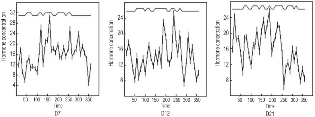
Figure 2 illustrates serum testosterone pulsatile se-cretion profile in one of the eight ovulatory women, each of whom underwent repetitive blood sampling at ten-minute intervals in the mid-follicular (Day 7), late follicular (Day 12) and mid-luteal (Day 21) phase of menstrual cycle. Testosterone pulse frequency was si-milar in the three phases, with a mean of 5.25 ± 0.70 in the mid-follicular phase, 5.0 ± 0.92 in late follicular phase and 6.12 ± 1.45 in the luteal phase (p > 0.05) (Figure 3). There was also no difference among the three phases in testosterone mean amplitude pulse, percentage of increment in pulse amplitude and mean duration of pulses (Figure 3). Mean frequency of tes-tosterone nadir was 5.25 in the mid-follicular phase, 6.0 in late follicular phase and 6.25 in the luteal phase (p > 0.05). There was no difference in pulse interval and in nadir duration among the three phases (p > 0.05).
DISCUSSION
Ovarian function involves hormonal secretion, but also gametogenesis and ovulation. For this, a complex hor-monal secretion and a coordinated relationship among
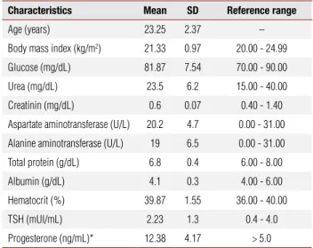
Table 1. Clinical, biochemical and hormonal characteristics of the subjects at the initial evaluation
Characteristics Mean SD Reference range
Age (years) 23.25 2.37 –
Body mass index (kg/m2) 21.33 0.97 20.00 - 24.99
Glucose (mg/dL) 81.87 7.54 70.00 - 90.00
Urea (mg/dL) 23.5 6.2 15.00 - 40.00
Creatinin (mg/dL) 0.6 0.07 0.40 - 1.40
Aspartate aminotransferase (U/L) 20.2 4.7 0.00 - 31.00
Alanine aminotransferase (U/L) 19 6.5 0.00 - 31.00
Total protein (g/dL) 6.8 0.4 6.00 - 8.00
Albumin (g/dL) 4.1 0.3 4.00 - 6.00
Hematocrit (%) 39.87 1.55 36.00 - 40.00
TSH (mUI/mL) 2.23 1.3 0.4 - 4.0
Progesterone (ng/mL)* 12.38 4.17 > 5.0
* Collected on the 21st day of the menstrual cycle.
Copyright
© ABE&M todos os dir
eitos r
eser
vados.
Table 2. Fasting LH, testosterone, SHBG and FAI of the subjects (S) in mid-follicular phase, late follicular phase and mid-luteal phase
Clinical characteristic Mid-follicular phase (mean ± SD)
Late follicular phase (mean ± SD)
Luteal phase
(mean ± SD) p-value
LH (mUI/mL) 5.45 ± 1.68 6.90 ± 1.62 5.61 ± 4.37 NS
Testosterone (nmol/L) 0.90 ± 0.54 1.34 ± 0.47 1.05 ± 0.20 NS
SHBG (nmol/L) 42.45 ± 16.23 47.27 ± 23.71 46.81 ± 15.61 NS
FAI (nmol/L) 2.34 ± 1.49 3.19 ± 1.40 2.41 ± 0.76 NS
SD: standard deviation; NS: non significant.
Frequency
D7 D12 D21
0 1 2 3 4 5 6 7
Day of the menstrual cycle
Pulses/6 hours
Mean amplitude
D7 D12 D21
0.0 2.5 5.0 7.5 10.0 12.5
Day of the menstrual cycle
mUI/mL
% Increase amplitude
D7 D12 D21
0 100 200 300 400
Day of the menstrual cycle
%
Pulse duration
D7 D12 D21
0 100 200
Day of the menstrual cycle
Minutes
Figure 1. LH pulsatility in the three phases of the menstrual cycle. Frequency (p < 0.01) and mean amplitude (p = 0.02). Increase amplitude and pulse duration was not significant.
hypothalamus, pituitary and ovary are necessary. The ste-roidal ovary production is also involved in this process.
Gonadotrophins have different pattern of secretion in men and women since the first day of life (23). Ne-vertheless, this difference remains in reproductive life with stable frequency and amplitude in men (24,25), but shows great variability in women (26,27), inclu-ding ethnic variations (28). This gonadotrophin pul-satile liberation reflects the influence of the ovarian steroidal secretion on the hypothalamus pituitary axis.
Androgens are crucial factors for normal develop-ment of female gonadal function (10). There are
suffi-cient data about gonadotrophin pulsatility (27,29,30), but much less is seen about testosterone in the litera-ture. In the present study, LH did show different pulse frequency from the studied phases as already registered by other authors (26,27).
testostero-Copyright
© ABE&M todos os dir
eitos r
eser
vados.
D7 D12 D21
Figure 2. Testosterone (ng/dL) pulsatility on Day 7, Day 12 and Day 21 of one of the eight subjects. Time expressed in minutes.
Frequency
D7 D12 D21
4 5 6 7 8
Day of the menstrual cycle
Pulses/6 hours
Mean amplitude
D7 D12 D21
0 10 20 30 40 50 60 70
Day of the menstrual cycle
ng/dL
% Increase amplitude
D7 D12 D21
120 130 140 150 160 170 180 190 200 210 220
Day of the menstrual cycle
%
Pulse duration
D7 D12 D21
0 10 20 30 40 50 60
Day of the menstrual cycle
Minutes
Figure 3. Testosterone pulsatility in the three phases of the menstrual cycle. No statistical significance in any parameter.
ne (31), but it also did not show significant difference among the three phases.
By using adequate methodology, it has been shown that testosterone, as other hormones involved with
re-productive physiology (27), is secreted in a pulsatile pat-tern. However, testosterone pulsatility pattern does not vary among the three different menstrual cycle phases studied: mid-follicular, late follicular and luteal phase.
32
24 24
20 20
16 16
12 12
8 8
28
24
20
16
12
Hormone concentration
Hormone concetration Hormone concetration
8 4
50 100 150 200 250 300 350 50 100 150 200 250 300 350 50 100 150 200 250 300 350
Copyright
© ABE&M todos os dir
eitos r
eser
vados.
It should be considered that, during the reproductive years, androgens are produced by two sources: ovaries and adrenal glands (32), and these are not under LH control. In addition, other hormones from ovary and adrenal, which production can vary during menstrual cycle, can influence in different ways the activated en-zymatic mechanisms of the follicle and luteolysed cells and so could influence testosterone production beyond the LH (2).
Pulsatility studies are usually related to considerable difficulty in volunteer’s recruitment. Multiple samples are required and total blood loss is a constant preoc-cupation (20,33). The assay limitation is also impor-tant as testosterone assays are designed for higher levels presented in men and could not be accurate enough to determine lower women levels (31,32). In addition, serum androgen levels do not necessarily reproduce intraovarian androgen concentrations. Further studies with assisted reproduction techniques could evaluate the correlation between intraovarian androgens and its serum pulsatility.
Variations in androgen levels or in its pulsatility could contribute to the understanding and manage-ment of some conditions related to androgen action, such as women mood and sexual behavior in the con-troversial androgen insufficiency syndrome which was characterized by a team of investigators in 2002 (34). It is well known that testosterone influence behavior and both cognitive and sexual function in men (35). However, evidence in women is limited by lack of nor-mative data for androgen levels and much controversy relies about types of treatment. As sexual nor mood questionnaires were not used, comparison between an-drogen secretion pattern and possible behavior changes in menstrual cycle was not available.
It was shown that there is no variation in testostero-ne pulsatility during the normal menstrual cycle. This physiologic testosterone pulsatility could be compared to women with clinical features of the androgen insu-fficiency syndrome even if baseline androgen levels are still normal. Otherwise, testosterone normal pulsatility can also be compared to some hyperandrogenic states as the polycystic ovarian syndrome (PCOS). Peripheral conversion of androgen precursors to active androgens may play an important role in androgen action and this should influence states of hyperandrogenic diseases. It is shown that elevated androgen levels are associated with hyperandrogenic features, as acne and hirsutism, but there is no evidence to support a relationship
be-tween the degree of androgen elevation and the severi-ty of these clinical presentations (36). Sometimes, there is considerable discrepancy between androgen baseline levels and clinical presentation. Expression of andro-gens receptors in effector organs and hypersensibility of end-target organs are usually considered in these cases, but variations in testosterone pulsatility could explain women presented with hyperandrogenic features and normal baseline androgens.
In conclusion, this study presented, for the first time, that testosterone is secreted in a pulsatile pattern as well as the comparison of this pulsatile profile in dis-tinct phases of the menstrual cycle without differences among them. Additional studies are needed to compare normal ovulatory cycles and pathologic cycles, both in androgen excess and androgen insufficiency, to define if there is any alteration in this pulsatile pattern related to hyperandrogenic anovulation or female androgen in-sufficiency syndrome.
Disclosure: no potential conflict of interest relevant to this article was reported.
REFERENCES
1. Suzuki T, Sasano H, Tamura M, Aoki H, Fukaya T, Yajima A, et al. Temporal and spatial localization of steroidogenic enzymes in premenopausal human ovaries: in situ hybridization and immu-nohistochemical study. Mol Cell Endocrinol. 1993;97(1-2):135-43. 2. Suzuki T, Sasano H, Kimura N, Tamura M, Fukaya T, Yajima A, et
al. Immunohistochemical distribution of progesterone, androgen and oestrogen receptors in the human ovary during the mens-trual cycle: relationship to expression of steroidogenic enzymes. Hum Reprod. 1994;9(9):1589-95.
3. Yen SS, Tsai CC, Naftolin F, Vandenberg G, Ajabor L. Pulsatile pat-terns of gonadotropin release in subjects with and without ova-rian function. J Clin Endocrinol Metab. 1972;34(4):671-5. 4. Backstrom CT, McNeilly AS, Leask RM, Baird DT. Pulsatile
secre-tion of LH, FSH, prolactin, oestradiol and progesterone during the human menstrual cycle. Clin Endocrinol (Oxf). 1982;17(1):29-42. 5. McNatty KP, Makris A, Osathanondh R, Ryan KJ. Effects of
lutei-nizing hormone on steroidogenesis by thecal tissue from human ovarian follicles in vitro. Steroids. 1980;36(1):53-63.
6. Angelopoulos N, Goula A, Tolis G. The role of luteinizing hormo-ne activity in controlled ovarian stimulation. J Endocrinol Invest. 2005;28(1):79-88.
7. Barbieri RL, Smith S, Ryan KJ. The role of hyperinsulinemia in the pathogenesis of ovarian hyperandrogenism. Fertil Steril. 1988;50(2):197-212.
8. Tan S, Hahn S, Janssen OE. Insulin resistance syndrome and polycystic ovary syndrome: implications for diagnosis and treat-ment. Panminerva Med. 2005;47(4):211-7.
Copyright
© ABE&M todos os dir
eitos r
eser
vados.
10. Haning RV Jr, Hackett RJ, Flood CA, Loughlin JS, Zhao QY, Long-cope C. Testosterone, a follicular regulator: key to anovulation. J Clin Endocrinol Metab. 1993;77(3):710-5.
11. Goebelsmann U, Arce JJ, Thorneycroft IH, Mishell DR Jr. Serum testosterone concentrations in women throughout the menstrual cycle and following HCG administration. Am J Obstet Gynecol. 1974;119(4):445-52.
12. Ribeiro WO, Mishell DR Jr, Thorneycroft IH. Comparison of the pat-terns of androstenedione, progesterone, and estradiol during the human menstrual cycle. Am J Obstet Gynecol. 1974;119(8):1026-32. 13. Veldhuis JD, Keenan DM, Pincus SM. Motivations and me-thods for analyzing pulsatile hormone secretion. Endocr Rev. 2008;29(7):823-64.
14. Lejeune-Lenain C, Van Cauter E, Désir D, Beyloos M, Franckson JR. Control of circadian and episodic variations of adrenal andro-gens secretion in man. J Endocrinol Invest. 1987;10(3):267-76. 15. Gupta SK, Lindemulder EA, Sathyan G. Modeling of circadian
testosterone in healthy men and hypogonadal men. J Clin Phar-macol. 2000;40(7):731-8.
16. Simoni M, Montanini V, Fustini MF, Del Rio G, Cioni K, Marrama P. Circadian rhythm of plasma testosterone in men with idiopathic hypogonadotrophic hypogonadism before and during pulsatile administration of gonadotrophin-releasing hormone. Clin Endo-crinol (Oxf). 1992;36(1):29-34.
17. Ehrmann D. Polycystic ovary syndrome. N Engl J Med. 2005;352(12):1223-36.
18. Venturoli S, Porcu E, Fabbri R, Magrini O, Gammi L, Paradisi R, et al. Episodic pulsatile secretion of FSH, LH, prolactin, oestradiol, oestrone, and LH circadian variations in polycystic ovary syndro-me. Clin Endocrinol (Oxf). 1988;28(1):93-107.
19. Barontini M, Garcia-Rudaz MC, Veldhuis JD. Mechanisms of hypothalamic-pituitary-gonadal disruption in polycystic ovarian syndrome. Arch Med Res. 2001;32(6):544-52.
20. Veldhuis JD, Pincus SM, Garcia-Rudaz MC, Ropelato MG, Escobar ME, Barontini M. Disruption of the joint synchrony of luteinizing hormone, testosterone, and androstenedione secretion in ado-lescents with polycystic ovarian syndrome. J Clin Endocrinol Me-tab. 2001;86(1):72-9.
21. Ahmad N, Pollard TM, Unwin N. The optimal timing of blood col-lection during the menstrual cycle for the assessment of endoge-nous sex hormones: can interindividual differences in levels over
the whole cycle be assessed on a single day? Cancer Epidemiol Biomarkers Prev. 2002;11(1):147-51.
22. Stavig GR, Leonard AR, Igra A, Felten P. Indices of relative body weight and ideal weight charts. J Chronic Dis. 1984;37(4):255-62. 23. de Zegher F, Devlieger H, Veldhuis JD. Pulsatile and sexually
di-morphic secretion of luteinizing hormone in the human infant on the day of birth. Pediatr Res. 1992;32(5):605-7.
24. Partsch CJ, Abrahams S, Herholz N, Peter M, Veldhuis JD, Si-ppell WG. Variability of pulsatile luteinizing hormone secretion in young male volunteers. Eur J Endocrinol. 1994;131(3):263-72. 25. Veldhuis J. Pathophysiologic features of episodic gonadotropin
secretion in man. Am J Med Sci. 1987;294(3):150-60.
26. Midgley AR Jr, Jaffe RB. Regulation of human gonadotropins. X. Episodic fluctuation of LH during the menstrual cycle. J Clin En-docrinol Metab. 1971;33(6):962-9.
27. Rossmanith WG. Ultradian and circadian patterns in luteinizing hormone secretion during reproductive life in women. Hum Re-prod. 1993;8(Suppl 2):77-83.
28. Haiman CA, Pike MC, Bernstein L, Jaque SV, Stanczyk FZ, Afghani A, et al. Ethnic differences in ovulatory function in nulliparous women. Br J Cancer. 2002;86(3):367-71.
29. Grumbach MM. The neuroendocrinology of human puberty revi-sited. Horm Res. 2002;57(Suppl 2):2-14.
30. Knobil E. The neuroendocrine control of ovulation. Hum Reprod. 1988;3:469-72.
31. Vermeulen A, Verdonck L, Kaufman JM. A critical evaluation of simple methods for the estimation of free testosterone in serum. J Clin Endocrinol Metab. 1999;84(10):3666-72.
32. Davison SL, Davis SR. Androgens in women. J Steroid Biochem Mol Biol. 2003;85(2-5):363-6.
33. Veldhuis JD, Pincus SM, Garcia-Rudaz MC, Ropelato MG, Escobar ME, Barontini M. Disruption of synchronous secretion of leptin, LH and ovarian androgens in nonobese adolescents with the polycys-tic ovarian syndrome. J Clin Endocrinol Metab. 2001;86(8):3772-8. 34. Bachmann G, Bancroft J, Braunstein G, Burger H, Davis S, Den-nerstein L, et al. Female androgen insufficiency: the princeton consensus statement on definition, classification, and assess-ment. Fertil Steril. 2002;77(4):660-5.
35. Beauchet O. Testosterone and cognitive function: current clinical evidence of a relationship. Eur J Endocrinol. 2006;155(6):773-81. 36. Shaw JC. Acne: effect of hormones on pathogenesis and