Contents lists available atScienceDirect
Journal of Ethnopharmacology
j o u r n a l h o m e p a g e :w w w . e l s e v i e r . c o m / l o c a t e / j e t h p h a r m
Anti-inflammatory activity of
Lychnophora passerina
, Asteraceae
(Brazilian “Arnica”)
P. Capelari-Oliveira
a, C.A. Paula
a, S.A. Rezende
b, F.T. Campos
a,
A. Grabe-Guimarães
c, J.A. Lombardi
d, D.A. Saúde-Guimarães
a,∗ aLaboratório de Plantas Medicinais, Escola de Farmácia, Universidade Federal de Ouro Preto, Minas Gerais, BrazilbLaboratório de Imunoparasitologia, Núcleo de Pesquisas em Ciências Biológicas, Universidade Federal de Ouro Preto, Minas Gerais, Brazil cLaboratório de Farmacologia Experimental, Escola de Farmácia, Universidade Federal de Ouro Preto, Minas Gerais, Brazil
dDepartamento de Botânica, Instituto de Biociências de Rio Claro, UNESP, Rio Claro, SP, Brazil
a r t i c l e
i n f o
Article history:
Received 11 December 2010
Received in revised form 27 February 2011 Accepted 12 March 2011
Available online 21 March 2011
Keywords:
Lychnophora passerina
Asteraceae Anti-inflammatory NO
TNF-␣
IL-10 Paw oedema
a b s t r a c t
Ethnopharmacological relevance: Lychnophora passerina(Asteraceae), popularly known as “arnica,” is used to treat inflammation, pain, rheumatism, contusions, bruises and insect bites in Brazilian traditional medicine.
Materials and methods:The anti-inflammatory activity of crude ethanolic extract of aerial parts ofL. passerinaand its ethyl acetate and methanolic fractions had their abilities to modulate the production of NO, TNF-␣and IL-10 inflammatory mediators in LPS/IFN-␥-stimulated J774.A1 macrophages evaluated. Moreover, the crude ethanolic extract and derived fractions were alsoin vivoassayed by carrageenan-induced paw oedema in mice.
Results: In vitroassays showed remarkable anti-inflammatory activity ofL. passerinacrude ethanolic extract (EE) and its ethyl acetate (A) and methanolic (M) fractions, through the inhibition of produc-tion of NO and TNF-␣inflammatory mediators and induction of production of IL-10 anti-inflammatory cytokine.In vivoassays showed anti-inflammatory activity for EE 10% ointment, similar to the standard drug diclofenac gel. The A and M fraction ointments 20% presented anti-inflammatory activity. Conclusion:The results obtained showed that possible anti-inflammatory effects of EE and its A and M fractions may be attributed to inhibition pro-inflammatory cytokines production, TNF-␣and NO and to increased IL-10 production. EE, A and M ointments showed topicalin vivoanti-inflammatory activity. The in vivoanti-inflammatory activity of EE ofL. passerinamay be related to synergistic effects of different substances in the crude extract. Therefore, traditional use of aerial parts ofL. passerinain the inflammatory conditions could be beneficial to treat topical inflammatory conditions, as evidenced by the present study.
© 2011 Elsevier Ireland Ltd. All rights reserved.
1. Introduction
Inflammation is a natural host-defensive process of the innate immunity response. Bacterial and viral infection triggers the
activa-Abbreviations: A, ethyl acetate fraction; DMSO, dimethyl sulfoxide; EE, ethanolic extract; H, hexanic fraction; IL-10, interleukin-10; INF-␥, interferon-␥; iNOS, inducible nitric oxide synthase; LPS, lipopolysaccharide; M, methanolic
fraction; MTT, 3-(4,5-dimethyl-2-thiazyl)-2,5-diphenyl-2H-tetrazoliumbromide; NF-B, nuclear factor-kB; NO, nitric oxide; NOS, nitric oxide synthase; PGE2,
prostaglandin E2; RNA, ribonucleic acid; S.E.M., standard error of mean; TNF-␣, tumor necrosis factor-␣.
∗Corresponding author at: Departamento de Farmácia, Escola de Farmácia,
Uni-versidade Federal de Ouro Preto, Rua Costa Sena, 171 – Centro, Ouro Preto, Minas Gerais CEP 35400-000, Brazil. Tel.: +55 31 35 59 16 26; fax: +55 31 35 59 16 28.
E-mail addresses:saude@ef.ufop.br,saudeguima@gmail.com
(D.A. Saúde-Guimarães).
tion of numerous immune cells such as macrophages, monocytes, and neutrophils undergoing to cellular responses such as phago-cytic uptake and production of inflammatory mediators, such as nitric oxide (NO), prostaglandin E2(PGE2) and tumor necrosis factor
(TNF)-␣(Gautam and Jachak, 2009; Yu et al., 2010). NO is produced
froml-arginine by the enzyme nitric oxide synthase (NOS) (Palmer
et al., 1988). Large amounts of NO are produced by the enzyme inducible nitric oxide synthase (iNOS), enzyme involved in cellu-lar overproduction of NO and active when a pathologic process is present, like inflammation or cancer, and many other condi-tions (Stichtenoth and Frolich, 1998). The TNF-␣cytokine plays a
crucial role in inflammation, stimulating the production of other cytokines and pro-inflammatory mediators (Verma et al., 2010). However, overproduction of TNF-␣is related to development and
progression of inflammatory process and autoimmune diseases (Williams et al., 2007). Unlike NO and TNF-␣, the interleukin
(IL)-10 is the most important cytokine presenting anti-inflammatory
0378-8741/$ – see front matter© 2011 Elsevier Ireland Ltd. All rights reserved.
properties. It is produced by activated immune cells, mainly monocytes/macrophages and regulates many different immune cells functions. In monocytes/macrophages, IL-10 diminishes the production of inflammatory mediators and inhibits antigen pre-sentation (Sabat et al., 2010).
Lychnophora(Asteraceae) is endemic to campo rupestre habitats of Brazilian savanna (cerrado biome) and have a wide variety of biological activities including anti-inflammatory, antinociceptive and trypanocidal activity (Chiari et al., 1996; Oliveira et al., 1996; Ferraz Filha et al., 2006; Guzzo et al., 2008). The aerial parts of the
Lychnophoraspecies, includingLychnophora passerina, have been used in a folk medicine macerated in ethanol to treat inflammatory and pain conditions (Cerqueira et al., 1987).
Although the traditional use of Lychnophora species as anti-inflammatory agents is supported by scientific evidence (Gobbo-Neto et al., 2005; Ferraz Filha et al., 2006; Guzzo et al., 2008) there are few studies of its action mechanism. We reported previ-ously that the ethanolic extract from aerial parts of theL. passerina
exhibitedin vivotopical anti-inflammatory activity (Guzzo et al., 2008). Based on these data, the objectives of the present work were to evaluate, whether the same ethanolic extract have the ability to inhibit,in vitro, the production of the NO and TNF-␣and to stimulate
IL-10 release from J774.A1 macrophages stimulated by LPS/IFN-␥.
In addition, the ethanolic extracts and its fractions were alsoin vivo
evaluated, using the carrageenan-induced paw oedema methods test in mice.
2. Materials and methods
2.1. Plant material
Aerial parts ofL. passerina(Mart exDC.) Gardn were collected in Diamantina, Minas Gerais, Brazil, in September 2000. The plant botanical identification was realized by Dr. Júlio A. Lombardi, Departamento de Botânica, Instituto de Biociências de Rio Claro, UNESP, Rio Claro, SP, Brazil. A voucher specimen was deposited in the Herbarium of Instituto de Ciências Exatas e Biológicas of Uni-versidade Federal de Ouro Preto – UFOP, under the number BHCB 53571.
2.2. Preparation of plant extract and fractions
Aerial parts (1 kg) ofL. passerinawere air-dried, ground and extracted with ethanol by percolation, at room temperature, for 14 days. The solvent was eliminated by evaporation under reduced pressure, resulting the dried crude ethanolic extract. The ethanolic extract (EE, 120.0 g) was submitted to filtration col-umn chromatography on silica gel, eluted with hexane, ethyl acetate and methanol to yield the hexanic (H, 0.42 g), ethyl acetate (A, 55.0 g) and methanolic (M, 59.0 g) fractions, respec-tively.
2.3. Cell line and culture conditions
J774A.1 murine macrophage cell line was kindly provided by Dr. Affonso, L.C.C. (Laboratório de Imunoparasitologia – NUPEB – Universidade Federal de Ouro Preto, Minas Gerais, Brazil). The cells were maintained in RPMI supplemented with 100 U/ml of penicillin, 2 mM ofl-glutamine, 100 U/ml of penicillin G, 1 mM of
sodium pyruvate and 10% fetal bovine serum. Cells were grown at 37◦C in a humidified 5% CO
2atmosphere.
2.4. Cell viability by MTT assay
To exclude the possible interference of the EE, A and M on cell viability the
3-(4,5-dimethyl-2-thiazyl)-2,5-diphenyl-2H-tetrazoliumbromide (MTT) assay was performed. J774.A1 (2.5×10⁵cells/well) macrophages were plated in 96-well plates
and allowed to adhere at 37◦C in a 95% air and 5% CO 2
atmo-sphere for 2 h. Thereafter, the medium was replaced with fresh medium or medium containing increasing concentrations of the EE, A and M dissolved in DMSO for 24 h at 37◦C and 5% CO
2
in humidified air. After 24 h, MTT solution (dissolved in PBS) was added (final concentration of 0.5 mg/ml) to each well and incubated for 4 h in the same conditions. The medium was care-fully discarded, and 100l of sodium dodecyl sulfate 10% in
hydrochloric acid 10 mM was added to each well to solubilize the formazan. The optical density was measured at 550 nm. The end concentration of DMSO was adjusted to less than 0.1% for all treat-ments.
2.5. NO inhibition assay
J774A.1 cells were seeded onto a 24-well culture plate at density 2.5×10⁵ cells per well with 500l of culture medium
and incubated for 2 h. The cells were stimulated with LPS + IFN-␥
(1g/ml + 10 UI/ml, respectively) for 1 h before the treatment with
EE, A and M dissolved in DMSO at different non-cytotoxic concen-trations (2.5–40g/ml) for 24 h at 37◦C and 5% CO2in humidified
air. After 24 h, the presence of nitrite, a stable oxidized product of nitric oxide (NO), was determined in cell culture media using Griess reagent. Briefly, 50l of supernatant was removed and
com-bined with 100l of Griess reagent in a 96-well plate, followed by
incubation for 10 min at room temperature and spectrophotomet-ric measurement at 550 nm using a microplate reader (Molecular Devices) as described by Green et al. (1982) and Verma et al. (2010). NO concentration was determined using comparison with a sodium nitrite standard curve. The final concentration of DMSO was adjusted to less than 0.1% for all treatments. Dexamethasone was used as a reference standard.
2.6. TNF-˛inhibition assay and IL-10 stimulation assay
J774A.1 cells were seeded onto a 24-well culture plate at density 2.5×10⁵ cells per well with 500l of culture medium
and incubated for 2 h. The cells were stimulated with LPS + IFN-␥
(1g/ml + 10 UI/ml, respectively) for 1 h before the treatment with
EE, A and M dissolved in DMSO at different non-cytotoxic concen-trations (2.5–40g/ml) for 24 h at 37◦C and 5% CO2in humidified
air. After 24 h, the supernatant was collected and used to estimate the levels of TNF-␣and IL-10 by specific ELISA kits according to the
manufacturers’ instruction (ELISA kit, PeproTech, Brazil), respec-tively. The final concentration of DMSO was adjusted to less than 0.1% for all treatments. Dexamethasone was used as a reference standard.
2.7. Preparation of ointments containing crude ethanolic extract and its fractions
EE, A and M were solubilized in Tween-80, DMSO and distilled water (1:1:8) and mixed with base ointment (lanoline/vaseline 70:30) for the 10% final concentration.
2.8. Animals
Forin vivoassays were utilized male Swiss albino mice (30±5 g), supplied by Animal House of Universidade Federal de Ouro Preto (UFOP). The animals received feed and water ad libitum
by the US National Institute of Health (NIH Publication, revised in 1985).
2.9. Carrageenan-induced paw oedema assay
The in vivo anti-inflammatory activity was determined by the carrageenan-induced paw oedema method in mice, accord-ing to previously described (Winter et al., 1962). The 10% EE, A and M ointments (three groups) were topically applied on right hind paws using a spatula, immediately after 0.1% carrageenan (Sigma–Aldrich) saline solution subplantar administration. Three more groups were established: (a) carrageenan, treated with base ointment, corresponding to 100% of inflammation (carrageenan group), (b) only needle introduction in the left paw, correspond-ing to activity induced by mechanical perforation (control group), and (c) carrageenan treated with standard drug diclofenac gel (Cataflan® Emulgel–Novartis, 11.6 mg/g). To ensure the ointment
contact with paws bandages were used. To measure oedema vari-ation a digital caliper rule (Starret) was used. The paws were measured before and 3 h after carrageenan administration, with or without treatments. The paw oedema was expressed in millimeters (mm) and was calculated as the percentage of variation between zero time and 3 h after carrageenan.
2.10. Statistical analysis
In vitro results were obtained from two independent exper-iments in duplicate and are presented as mean±S.E.M. In vivo
results were presented as means±S.E.M from experiments per-formed with 8 animals per group. Statistical significance among groups was determined by ANOVA followed by Bonferroni test using software PRISMA (GraphPad Software, Inc., San Diego, CA, version 5.01).P-values≤0.05 were taken to indicate statistical sig-nificance.
3. Results
L. passerinais used in the folk medicine as anti-inflammatory, analgesic and to treat rheumatism. In this study, the ethanol extract and its fractions were investigated for theirin vitroand topical
in vivoanti-inflammatory activity.
3.1. Effects of crude ethanolic extract and its fractions on cell viability of J774.A1 macrophages
The viability of the J774.A1 macrophages in the presence of EE, A and M were evaluated. The cytotoxicity evaluated to different con-centrations of EE, A and M on the J774.A1 cells was negligible and did not show statistic difference when compared to the control. The exception occurred for the M fraction, since the concentra-tion of 40g/ml showed cell viability lower than 90% (data not
shown). Thus, EE, A and M fractions werein vitroassayed for anti-inflammatory activity using concentrations lower than 40g/ml.
3.2. Effects of crude ethanolic extract and its fractions on NO production
To evaluate the inhibitory effects of the EE, A and M on NO pro-duction by LPS/IFN-␥stimulated cells, they were treated with LPS
(1g/ml) and IFN-␥(10 UI/ml) in the presence or absence of the
EE, A and M for 24 h. The amount of nitrite, as an index of NO in culture medium, was measured with Griess reagent. Unstimulated J774.A1 cells secreted basal levels of NO, while LPS/IFN-␥
stimu-lation resulted in NO production increase. EE and A were able to inhibit the NO production by LPS/IFN-␥stimulated J774.A1 cells in
a concentration-dependent manner (Fig. 1A and B), at the higher evaluated concentrations: 20g/ml and 40g/ml for the EE, and
Fig. 1.(A) Effect of the ethanolic extract (EE), (B) effect of acetyl acetate fraction (A) and (C) effect of methanolic fraction (M) on NO production by LPS/IFN-␥ stimu-lated J774.A1 macrophages. The cells were stimustimu-lated with LPS (1g/ml) and IFN-␥
(10 UI/ml) for 1 h before the treatment with EE, A and M dissolved in DMSO at different non-cytotoxic concentrations (2.5–40g/ml) for 24 h. Supernatants were
collected and nitrite (NO) concentration was determined by Griess reagent. Data are represented as mean±S.E.M. values. *P< 0.05, **P< 0.01 and ***P< 0.001 compared with LPS/IFN-␥-stimulated macrophages alone.
10g/ml, 20g/ml and 40g/ml for the A. M was not able to reduce
the NO levels, in any evaluated concentration (Fig. 1C).
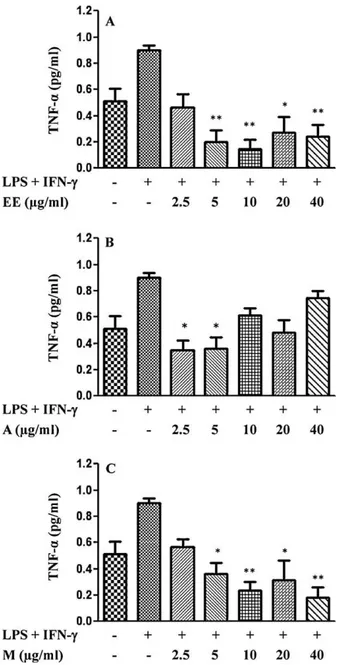
3.3. Effects of crude ethanolic extract and its fractions on TNF-˛ production
To determine the inhibitory effects of crude EE and A and M fractions of L. passerina, on LPS/IFN-␥-stimulated TNF-␣
pro-duction, J774.A1 macrophages were treated with LPS (1g/ml)
and IFN-␥(10 UI/ml) in the presence or absence of different
con-centrations of EE, A and M. The macrophages stimulated only with LPS/IFN-␥exhibited an appreciable increase of TNF-␣
lev-els, while in the absence of any stimulus the cells secreted basal levels of TNF-␣ (Fig. 2). The strong inhibition of TNF-␣
pro-duction was observed for almost all evaluated concentrations of EE (Fig. 2A) and M (Fig. 2C) and a lower effect was observed for the A fraction (Fig. 2B). The EE at 5, 10, 20 and 40g/ml
decreased the LPS/IFN-␥-stimulated TNF-␣production. The M
Fig. 2.(A) Effect of the ethanolic extract (EE), (B) effect of acetyl acetate fraction (A) and (C) effect of methanolic fraction (M) on TNF-␣production by LPS/IFN-␥
stimu-lated J774.A1 macrophages. The cells were stimustimu-lated with LPS (1g/ml) and IFN-␥
(10 UI/ml) for 1 h before the treatment with EE, A and M dissolved in DMSO at dif-ferent non-cytotoxic concentrations (2.5–40g/ml) for 24 h. The supernatants were collected and TNF-␣concentration was determined by specific ELISA kit. Data are represented as mean±S.E.M. values. *P< 0.05, **P< 0.01 and ***P< 0.001 compared with LPS/IFN-␥-stimulated macrophages alone.
showed better inhibitory activity at concentrations of 2.5g/ml
and 5g/ml.
3.4. Effects of crude ethanolic extract and its fractions on IL-10 production
The effects of EE and A and M ofL. passerinaon IL-10 produc-tion were investigated in J774.A1 macrophages stimulated with LPS (1g/ml) and IFN-␥ (10 UI/ml) in the presence or absence
of different concentrations. The cells stimulated with
LPS/IFN-␥exhibited an increase of IL-10 production, while unstimulated
J774.A1 cells secreted significant smaller basal levels of IL-10 (Fig. 3). EE increased the LPS/IFN-␥-induced IL-10 production in
almost all evaluated concentrations, except for 20g/ml. A
signif-icantly increased the IL-10 folds, mainly at 2.5g/ml, 5g/ml and
Fig. 3. (A) Effect of the ethanolic extract (EE), (B) effect of acetyl acetate fraction (A) and (C) effect of methanolic fraction (M) on IL-10 production by LPS/IFN-␥ stimu-lated J774.A1 macrophages. The cells were stimustimu-lated with LPS (1g/ml) and IFN-␥
(10 UI/ml) for 1 h before the treatment with EE, A and M dissolved in DMSO at dif-ferent non-cytotoxic concentrations (2.5–40g/ml) for 24 h. The supernatants were
collected and IL-10 concentration was determined by specific ELISA kit. Data are rep-resented as mean±S.E.M. values. *P< 0.05, **P< 0.01 and ***P< 0.001 compared with LPS/IFN-␥-stimulated macrophages alone.
10g/ml. M did not induce alteration of IL-10 production in all
evaluated concentrations (Fig. 3C).
3.5. Effects of crude ethanolic extract and its fractions on carrageenan-induced paw oedema
Table 1
Effect of topically applied ointments ofL. passerinaethanolic extract (EE) and its ethyl acetate (A) and methanolic (M) fractions evaluated using carrageenan-induced paw oedema.
Treatment after carrageenan Swelling thickness (%)
Control condition (without carrageenan) 2.7±0.35***
Base ointment 29.9±1.86
Diclofenac gel 6.6±1.22***
EE 10% 12.6±2.61***
M 20% 20.1±1.60*
A 20% 16.9±1.27***
Results represent the mean±S.E.M. of the percentage variation. *P< 0.05; **P< 0.01; ***P< 0.001, when compared to the group treated with base ointment (ANOVA followed Bonferroni’s test). The measurements were taken before and 3 h after administration of carrageenan.
4. Discussion
The present study assessed the anti-inflammatory potentialin vitroof EE in J774.A1 macrophages stimulated by LPS (1g/ml) and IFN-␥(10 UI/ml). The assays quantified the release of pro-inflammatory mediators NO and TNF-␣and the IL-10 production, an anti-inflammatory cytokine. The NO is produced by activated macrophages as a result of various stimuli, including TNF-␣, IFN-␥ and LPS, and it contributes to the pathological process of differ-ent acute and chronic inflammatory conditions (Nathan, 1992). The TNF-␣also plays an important role in inflammation and may act
on the monocytes and macrophages by an autocrine activity to increase diverse functional responses and to induce an over expres-sion of inflammatory mediators. (Baugh and Bucala, 2001). IL-10 is an anti-inflammatory cytokine, well known for its inhibitory effects on TNF-␣and inflammatory reactions. Its main activity is
to inhibit the cytokine production by macrophages (Bortesi et al., 2009).
EE, A and M fractions presented inhibitory activity on NO and TNF-␣production, by LPS/IFN-␥-induced J774.A1 macrophages.
Moreover, EE was able to significantly increase the IL-10 production by J774.A1 macrophages activated with LPS/IFN-␥, the model of in vitroinflammation used in the present work. However, it is inter-esting to note that inhibition of TNF-␣production and the induction
of IL-10 production by A decreased at the highest concentrations evaluated. The lack or decreasing of plant extracts activity at higher concentrations is also frequently cited in the literature (Punturee et al., 2004; Hammer et al., 2008). Concerning to IL-10, this may be due to post-transcriptional effects of metabolites present in EE, since it is known that production of this anti-inflammatory cytokine may be regulated by post-transcriptional mechanisms in macrophages (Nemeth et al., 2005). Adittionally, M strongly inhib-ited the TNF-␣production, but showed a weak inhibition of NO
production and weak activity on the IL-10 production by LPS/IFN-␥
-stimulated macrophages. The significant basal secretion of TNF-␣,
observed in the inhibition of production assay for this cytokine, has also been reported in other studies that showed greater produc-tion of this pro-inflammatory mediator by LPS/IFN-␥-stimulated
J774.A1 macrophages and by the same cells stimulated only with LPS (Herath et al., 2003; Fan et al., 2010).
Previous phytochemical investigations of the aerial parts ofL. passerinaresulted in the isolation of the triterpenoids, sesquiter-penes, including sesquiterpene lactones, steroid and of the flavonoids (Bohlmann et al., 1981; Oliveira et al., 1996; Chicaro et al., 2004).
Several terpenoids have inhibitory properties on the produc-tion of inflammatory cytokines (Bremner and Heinrich, 2002; Ríos, 2010) and among the terpenoids present in plant kingdom, the sesquiterpene lactones are specially produced and accu-mulated by species of Lychnophora genus, as well as by other
species of Asteraceae family (Bohlmann and Jakupovic, 1990). The sesquiterpene lactones goyazensolide and 15-desoxigoyazensolide showed pharmacological activities, including anti-inflammatory and trypanocidal activity (Chiari et al., 1996; Oliveira et al., 1996; Rüngeler et al., 1999). Several sesquiterpene lactones showed anti-inflammatory activity in cellular model of inflammation (Lyb et al., 1998; Rüngeler et al., 1999; Koo et al., 2001) and the action mecha-nism proposed has been the suppression of nuclear factor-kappa B (NF-
к
B) activation (Palladino et al., 2003; Ríos, 2010), which regulates the transcription of inflammatory cytokines and other molecules involved in inflammation (Cho et al., 2009; Lu et al., 2009). Thus, the inhibitory activity of NO and TNF-␣productionand induction of IL-10 production by A may be probably related to sesquiterpene lactones and triterpenoids presences and inhibition of the nuclear factor NF-B activity.
Some flavonoids are able to inhibit TNF-␣ production, the
iNOS expression and the NO production, as well as stimulation of the IL-10 expression, effects that have been associated to
NF-B activity inhibition (Herath et al., 2003; Comalada et al., 2006;
Hämäläinen et al., 2007). However, in this study, M did not show significant activity on NO and IL-10 production, but inhibited the TNF-␣ secretion. Since TNF-␣ is produced early in the
inflam-matory process (Jin et al., 2003; Hammer et al., 2008) and IL-10 is produced later (Jin et al., 2003; Jung et al., 2004), the polar flavonoids present in M can act at the initial phase diminishing the production of pro-inflammatory cytokines, but was not able to increase the secretion of cytokines that act in the late phase of inflammation, such as the IL-10. EE inhibited NO and
TNF-␣ and increased the IL-10 production by LPS/IFN-␥-stimulated
macrophages. The EE fractionation was able to concentrate more active substances in the A fraction than in the M fraction, resulting better activity to A fraction. The EE, characterized by presence of terpenoids and flavonoids, can exert immunomodulatory activity by synergistic ways, resulting in a remarkable anti-inflammatory activity.
Since the topical application of alcoholic macerates of aerial parts of L. passerina is used in traditional medicine to treat inflammation and pain, this route of administration was chosen in order to evaluate the in vivo anti-inflammatory proper-ties of EE, A and M. The in vivo anti-inflammatory activity for EE at 10% and A and M at 20% ointments was observed. These results confirmed the anti-inflammatory effects observed
in vitro.
Therefore, the remarkablein vivoactivity observed for EE may be attributed to synergism of several bioactive molecules. The activ-ity of different substances on different inflammation targets, as well as the possibility to improve the bioavailability of molecules, may result in a grater activity of crude extracts (Ji et al., 2009), rather than semi purified fractions. There is growing evidence showing that the medicinal plants may exhibit synergistic com-binations (Phillipson, 2003; Gilani and Rahman, 2005; Ma et al., 2009; Wagner and Ulrich-Merzenich, 2009; Graz et al., 2010).
5. Conclusion
The results obtained showed that possible anti-inflammatory effects of EE and its A and M fractions may be attributed to inhibi-tion pro-inflammatory cytokines producinhibi-tion, TNF-␣and NO and to
increased IL-10 production. EE, A and M ointments showed topical
Acknowledgements
The authors would like to thank FAPEMIG/CDS – APQ-01355-08 (Fundac¸ão de Amparo à Pesquisa do Estado de Minas Gerais), Rede TOXIFAR/FAPEMIG and Universidade Federal de Ouro Preto for financial support. The authors would like to thank Dr. Luís Car-los Crocco Afonso, for supplying J774.A1 macrophages, and other members of the Laboratório de Imunoparasitologia – NUPEB of Uni-versidade Federal de Ouro Preto for technical support.
References
Baugh, J.A., Bucala, R., 2001. Mechanisms for modulation TNF-␣in immune and
inflammatory disease. Current Opinion in Drug Discovery Development 4, 635–650.
Bohlmann, F., Müller, L., King, R.M., Robinson, H., 1981. Naturally occurring ter-pene derivates. Part 328. A guaianolide and other constituents fromLychnophora
species. Phytochemistry 20, 1149–1151.
Bohlmann, F., Jakupovic, J., 1990. Progress in the chemistry of theVernoniae (Com-positae). Plant Systematics and Evolution, 3–43.
Bortesi, L., Rossato, M., Schuster, F., Raven, N., Stadlmann, J., Avesani, L., Falorni, A., Bazzoni, F., Bock, R., Schillberg, S., Pezzotti, M., 2009. Viral and murine interleukin-10 are correctly processed and retain their biological activity when produced in tobacco. BMC Biotechnology 9, 22.
Bremner, P., Heinrich, M., 2002. Natural products as targeted modulators of the nuclear factor-kappaB pathway. Journal of Pharmacy and Pharmacology 54, 453–472.
Cerqueira, M.B.S., Souza, J.T., Júnior, R.A., Peixoto, A.B.F., 1987. Ac¸ão analgésica do extrato bruto aquoso liofilizado do caule e folhas daLychnophora ericoidesMart. (arnica). Ciência e Cultura 39, 551–553.
Chiari, E., Duarte, D.S., Raslan, D.S., Saúde, D.A., Perry, K.S.P., Boaventura, M.A.D., Grandi, T.S.M., Stehmann, J.R., Anjos, A.M.G., Oliveira, A.B., 1996. In vitro
screening of Asteraceae plant species againstTrypanossoma cruzi. Phytotherapy Research 10, 636–638.
Chicaro, P., Pinto, E., Colepicolo, P., Lopes, J.L.C., Lopes, N.P., 2004. Flavonoids from
Lychnophora passerina(Asteraceae): potential antioxidants and UV-protectants. Biochemical Systematics and Ecology 32, 239–243.
Cho, W., Nam, J.-W., Kang, H.-J., Windono, T., Seo, E.-K., Lee, K.-T., 2009. Zedoarondiol isolated from the rhizoma of Curcuma heyneana is involved in the inhibition of iNOS, COX-2 and pro-inflammatory cytokines via the downregulation of NF-B pathway in LPS-stimulated murine macrophages. International Immunophar-macology 9, 1049–1057.
Comalada, M., Ballester, I., Bailón, E., Sierra, S., Xaus, J., Gálvez, J., de Medina, F.S., Zarzuelo, A., 2006. Inhibition of pro-inflammatory markers in primary bone marrow-derived mouse macrophages by naturally occurring flavonoids: analysis of the structure–activity relationship. Biochemical Pharmacology 72, 1010–1021.
Fan, J., Liu, K., Zhang, Z., Luo, T., Xi, Z., Song, J., Liu, B., 2010. Modified si-miao-san extract inhibits the release of inflammatory mediators from lipopolysaccharide-stimulated mouse macrophage. Journal of Ethnopharmacology 129, 5–9.
Ferraz Filha, Z.S., Vitolo, I.F., Fietto, L.G., Lombardi, J.A., Saúde-Guimarães, D.A., 2006. Xanthine oxidase inhibitory activity ofLychnophoraspecies from Brazil (“Arnica”). Journal of Ethnopharmacology 107, 79–82.
Gautam, R., Jachak, S.M., 2009. Recent development in anti-inflammatory natural products. Medicinal Research Reviews 29, 767–820.
Gilani, A.H., Rahman, A.-U., 2005. Trends in ethnopharmacology. Journal of Ethnopharmacology 100, 43–49.
Gobbo-Neto, L., Santos, M.D., Kanashiro, A., Almeida, M.C., Lucisano-Valim, Y.M., Lopes, J.L.C., Souza, G.E.P., Lopes, N.P., 2005. Evaluation of the anti-inflammatory and antioxidant activities of di-C-glucosylflavones fromLychnophora ericoides
(Astearaceae). Planta Medica 71, 3–6.
Graz, B., Falquet, J., Elisabetsky, E., 2010. Ethnopharmacology, sustainable develop-ment and cooperation: the importance of gathering clinical data during field surveys. Journal of Ethnopharmacology 130, 635–638.
Green, L.C, Wagner, D.A., Glogowski, J., Skipper, P.L., Wishnok, J.J., Tannebaum, S.R., 1982. Analysis of nitrate, nitrite and (15 N) nitrate in biological fluids. Analytical Biochemistry 126, 131–138.
Guzzo, L.S., Saúde-Guimarães, D.A., Silva, A.C.A., Lombardi, J.A., Guimarães, H.N., Grabe-Guimarães, A., 2008. Antinociceptive and anti-inflammatory activities of ethanolic extracts ofLychnophoraspecies. Journal of Ethnopharmacology 116, 120–124.
Hämäläinen, M., Nieminen, R., Vuorela, P., Heinonen, M., Moilanen, E., 2007. Anti-inflammatory effects of flavonoids: genistein, kaempferol, quercetin, and daidzein inhibit STAT-1 and NF-B activations, whereas flavones,
isorham-netin, naringenin, and pelargonidin inhibit only NF-B activation along with
their inhibitory effect on iNOS expression and NO production in activated macrophages. Mediators of Inflammation, 45673–45683.
Hammer, K.D.P., Matthew, Hillwig, M.L., Neighbors, J.D., Sim, Y.-J., Kohut, M.L., Wiemer, D.F., Wurtele, E.S., Birt, D.F., 2008. Pseudohypericin is necessary for the light-activated inhibition of prostaglandin E2 pathways by a 4 compo-nent system mimicking anHypericum perforatumfraction. Phytochemistry 69, 2354–2362.
Herath, H.M.T., Takano-Ishikawa, Y., Yamaki, K., 2003. Inhibitory effect of some flavonoids on tumor necrosis factor-␣ production in lipopolysaccharide-stimulated mouse macrophage cell line J774.1. Journal of Medicinal Food 6, 365–370.
Ji, H.F., Li, X.J., Zhang, H.Y., 2009. Natural products and a drug discovery. European Molecular Biology Organization 10, 194–200.
Jin, M., Jung, H.J., Choi, J.J., Jeon, H., Oh, J.H., Kim, B., Shin, S.S., Lee, J.K., Yoon, K., Kim, S., 2003. Activation of selective transcription factors and cytokines by water-soluble extract fromLentinus lepideus. Society for Experimental Biology and Medicine 228, 749–758.
Jung, M., Sabat, R., Kratzschmar, J., Seidel, H., Wolk, K., Schonbein, C., Schütt, S., Friedrich, M., Döcke, W.-D., Asadullah, K., Volk, H.-D., Gerald, G., 2004. Expres-sion profiling of IL-10-regulated genes in human monocytes and peripheral blood mononuclear cells from psoriatic patients during IL-10 therapy. European Journal of Immunology 34, 481–493.
Koo, T.H., Lee, J.H., Park, Y.J., Hong, Y.S., Kim, H.S., Kim, K.W., Lee, J.J., 2001. A sesquiter-pene lactone, costunolide fromMagnolia grandiflora, inhibits NF-B by targeting
IB phosphorylation. Planta Medica 67, 103–107.
Lu, H., Shi, J.-X., Zhang, D.-M., Wang, H.-D., Hang, C.-H., Chen, H.-L., Yin, H.-X., 2009. Inhibition of hemolysate-induced iNOS and COX-2 expression by genis-tein through suppression of NF-кB activation in primary astrocytes. Journal of the Neurological Sciences 278, 91–95.
Lyb, G., Knorre, A., Schimidt, T.J., Nasman-Glaser, B., Ericsson, I., Lindgren, J.A., 1998. The anti-inflammatory sesquiterpene lactone helenalin inhibits the transcrip-tion factor NF-B by directly targeting p65. Journal of Biology Chemistry 273, 33508–33516.
Ma, X.H., Zheng, C.J., Han, L.Y., Xie, B., Jia, J., Cao, Z.W., Li, Y.X., Chen, Y.Z., 2009. Synergistic therapeutic actions of herbal ingredients and their mechanisms from molecular interaction and network perspectives. Drug Discovery Today 14, 579–588.
Nathan, C., 1992. Nitric oxide as a secretory product of mammalian cells. The FASEB Journal 6, 3051–3064.
Nemeth, Z.H., Lutz, C.S., Csoka, B., Deitch, E.A., Leibovich, S.J., Gause, W.C., Tone, M., Pacher, P., Vizi, E.S., Haskó, G., 2005. Adenosine augments IL-10 produc-tion by macrophages through na A2B receptor-mediated posttranscripproduc-tional mechanism. Journal of Immunology 175, 8260–8270.
Oliveira, A.B., Saúde, D.A., Perry, K.S.P., Duarte, D.S., Raslan, D.S., Boaventura, M.A.D., Chiari, E., 1996. Trypanocidal sesquiterpenes fromLychnophoraspecies. Phy-totherapy Research 10, 292–295.
Palladino, M.A., Bahjat, F.R., Theodorakis, E.A., Moldawer, L.L., 2003. Anti-TNF-␣
therapies: the next generation. Nature Reviews Drug Discovery 2, 736–746. Palmer, R.M., Rees, D.D., Ashton, D.S., Moncada, S., 1988. l-Arginine is the
physiological precursor for the formation of nitric oxide in endothelium depen-dent relaxation. Biochemical and Biophysical Research Communications 153, 1251–1256.
Phillipson, J.D., 2003. 50 years of medicinal plant research – every progress in methodology is a progress in science. Planta Medica 69, 491–495.
Punturee, K., Wild, C.P., Vinitketkumneun, U., 2004. Thai medicinal plants modulate nitric oxide and tumor necrosis factor-␣in J774.2 mouse macrophages. Journal
of Ethnopharmacology 95, 183–189.
Ríos, J.-L., 2010. Effects of triterpenes on the immune system. Journal of Ethnophar-macology 128, 1–14.
Rüngeler, P., Castro, V., Mora, G., Gören, N., Vichnewski, W., Pahl, H.L., Merfort, I., Schimidt, T.J., 1999. Inhibition of transcription factor NF-B by
sesquiter-pene lactones: a proposed molecular mechanism of action. Bioorganic Medicinal Chemistry 7, 2343–2352.
Sabat, R., Grütz, G., Warszawska, K., Kirsch, S., Witte, E., Wolk, K., Geginat, J., 2010. Biology of interleukin-10. Cytokine and Growth Factor Reviews 21, 331–344.
Stichtenoth, D.O., Frolich, J.C., 1998. Nitric oxide and inflammatory joint diseases. British Journal of Rheumatology 37, 246–257.
Verma, N., Tripathi, S.K., Sahu, D., Das, H.R., Das, R.H., 2010. Evaluation of inhibitory activities of plant extracts on production of LPS-stimulated pro-inflammatory mediators in J774 murine macrophages. Molecular Cell Biochemical 336, 127–135.
Wagner, H., Ulrich-Merzenich, G., 2009. Synergy research: approaching a new gen-eration of phytopharmaceuticals. Phytomedicine 16, 97–110.
Williams, R.O., Paleolog, E., Feldmann, M., 2007. Cytokine inhibitors in rheuma-toid arthritis and other autoimmune disease. Current Opinion Pharmacology 7, 412–417.
Winter, C.A., Risley, E.A., Nuss, G.W., 1962. Carragenan-induced oedema in hind paw of the rats as an assay for anti-inflammatory drugs. Proceedings of the Society for Experimental Biology and Medicine 111, 544–547.