CAIO ROBERTO SOARES BRAGANÇA
CONSTRUÇÃO DE UMA LINHAGEM RECOMBINANTE DE
Kluyveromyces marxianus
UFV-3 PARA EXPRESSÃO DA PROTEÍNA
NÃO ESTRUTURAL (NS1) DO VÍRUS DA DENGUE-1
Dissertação apresentada à Universidade
Federal de Viçosa, como parte das
exigências
do
Programa
de
Pós-Graduação em Microbiologia Agrícola,
para obtenção do título de
Magister
Scientiae.
VIÇOSA
CAIO ROBERTO SOARES BRAGANÇA
CONSTRUÇÃO DE UMA LINHAGEM RECOMBINANTE DE
Kluyveromyces marxianus
UFV-3 PARA EXPRESSÃO DA PROTEÍNA
NÃO ESTRUTURAL (NS1) DO VÍRUS DA DENGUE-1
Dissertação apresentada à Universidade
Federal de Viçosa, como parte das
exigências
do
Programa
de
Pós-Graduação em Microbiologia Agrícola,
para obtenção do título de
Magister
Scientiae.
APROVADA: 15 de março de 2013.
______________________________ ____________________________
Sérgio Oliveira de Paula Silvia Almeida Cardoso
(Coorientador)
_______________________________
Wendel Batista da Silveira
ii
iii
A Deus, minha essência. Aos meus amados pais, Osvaldo e Marlene, que
com carinho, amizade e compreensão me encorajaram e me incentivaram
em todas as situações ao longo do mestrado. À minha irmã Sabrina pelo
imenso apoio. À minha sobrinha Lana, pelo seu sorriso inesquecível. À
minha vó e à memória de meu avô pelo amor incondicional. Aos meus tios e
primos pela confiança. À minha família de viçosa (PIBV) pela lealdade de
uma amizade genuína. Aos amigos LABFIS, pelo companheirismo e
ensinamentos compartilhados, e à minha orientadora Flávia e coorientadores
por ter acreditado e confiado em mim.
iv
BIOGRAFIA
v
RESUMO
BRAGANÇA, Caio Roberto Soares, M.Sc., Universidade Federal de Viçosa,
março de 2013.
Construção de uma linhagem recombinante de
Kluyveromyces marxianus
UFV-3 para expressão da proteína não
estrutural (NS1) do vírus da dengue-1
. Orientadora: Flávia Maria Lopes
Passos. Coorientadores: Wendel Batista da Silveira e Sérgio Oliveira de
Paula.
A levedura
Kluyveromyces marxianus tem sido considerada uma candidata
hospedeira para a síntese industrial de biomoléculas. Apesar de seu potencial,
são poucos os estudos que relatam a expressão de proteínas heterólogas
utilizando esta levedura. Neste trabalho, foi relatado pela primeira vez, a
expressão da proteína do vírus da dengue em
K. marxianus. O gene que
codifica a proteína não estrutural (NS1) do vírus da dengue-1 foi integrado no
genoma da levedura K. marxianus UFV-3 no locus LAC4, utilizando um vetor
integrativo adaptado projetado para expressão de proteínas recombinantes
em Kluyveromyces lactis. O gene de dengue-1 NS1 foi otimizado utilizando os
códons preferenciais para aumentar os níveis de expressão de proteínas em
leveduras. O gene sintético foi clonado
“
in frame
”
com o peptídeo sinal
(mating-
α
-factor) de K. lactis e o plasmídeo recombinante obtido foi utilizado
para transformar
K. marxianus UFV-3 por eletroporação. As células
transformantes selecionadas em YPD (yeast extract peptone dextrose)
contendo 200 ug mL
-1de geneticina foram mitoticamente estáveis. A análise
vi
ABSTRACT
BRAGANÇA, Caio Roberto Soares, M.Sc., Universidade Federal de Viçosa,
March, 2013.
Construction of recombinant
Kluyveromyces marxianus
UFV-3 to express dengue virus type 1 nonstructural protein 1 (NS1)
.
Adviser: Flávia Maria Lopes Passos. Co-advisers: Wendel Batista da Silveira
and Sérgio Oliveira de Paula.
vii
SUMÁRIO
Construction of recombinant Kluyveromyces marxianus UFV-3 to express dengue
virus type 1 nonstructural protein 1 (NS1) ... 1
ABSTRACT ... 2
1. Introduction ... 3
2. Material and Methods ... 5
2.1 Microorganism and maintenance ... 5
2.2 Isolation and manipulation of nucleic acids and dengue NS1 nucleotide sequences ... 5
2.3 Yeast transformation... 8
2.4 Total DNA extraction and yeast transformants screening ... 8
2.5 Total RNA extraction from yeast and RT-PCR ... 9
2.6 Evaluating the genetic stability of the recombinant strains ... 9
2.7 Induction of protein expression ... 9
2.8 Fractionation of cultures for rNS1 SDS-page analysis and immunogenic detection using positive human serum for dengue virus ...10
2.9 rNS1 purification ...11
3. Results ...12
3.1 Construction of expression vector with kanamicin marker and harboring the NS1 gene to function in Kluyveromyces UFV-3 ...12
3.2 Transformation of K. marxianus UFV-3 with pKMCL-rNS1/Denv1 and mitotic stability of recombinants...13
3.3 Selection of K. marxianus UFV-3 recombinant strain and expression and function of the NS1 protein ...15
3.4 Growth of recombinant K. marxianus UFV-3 and production of rNS1 protein in shake-flask and bioreactor ...16
3.5 Detection and purification of rNS1 on supernatant culture ...19
4. Discussion ...22
5. Acknowledgements ...25
1
Construction of recombinant
Kluyveromyces
marxianus
UFV-3 to express dengue virus type
1 nonstructural protein 1 (NS1)
11Formatação realizada conforme as instruções da revista Applied Microbiology and
2
Construction of recombinant
Kluyveromyces marxianus
UFV-3 to
express dengue virus type 1 nonstructural protein 1 (NS1)
ABSTRACT
The yeast Kluyveromyces marxianus has been considered a candidate host for
industrial synthesis of biomolecules. Despite its potential, there are few studies
reporting the expression of heterologous proteins using this yeast. Here, it was
reported for the first time a dengue viral protein expression in K. marxianus. The
dengue virus type 1 nonstructural protein 1 (NS1) was integrated into the K.
marxianus UFV-3 genome at the LAC4 locus using adapted integrative vector
designed for high-level expression of recombinant protein in Kluyveromyces lactis.
The gene of dengue-1 NS1 was optimized using preferential codons to increase the
levels of proteins expression in yeast. The synthetic gene was cloned in frame with
K. lactis mating-α-factor signal peptide and the recombinant plasmid obtained was
used to transform K. marxianus UFV-3 by electroporation. The transformants cells selected in Yeast Extract Peptone Dextrose (YPD) containing 200 μg mL-1 Geneticin
were mitotically stable. The analysis of recombinant strains by RT-PCR technique and
the protein detection using blot analysis have confirmed both transcription and
expression of the extracellular peptides. After induction with galactose, the NS1
protein was analyzed by SDS-PAGE and immunogenic detection. The protein
production was investigated under two conditions: with galactose and biotin pulse at
24 hours intervals during 96 hours of induction and without galactose and biotin pulse.
Protease activity was not detected into the medium. Our results indicate that the
constructed recombinant K. marxianus can be considered good host for the
production of dengue virus proteins, which have a potential for diagnostic
applications.
Keywords: Kluyveromyces marxianus · Heterologous expression · dengue virus ·
3
1. Introduction
The yeast Kluyveromyces marxianus is phylogenetically related to the
common species of Saccharomyces cerevisiae and it is closer to Kluyveromyces
lactis, but it has particular characteristics which are differential for its biotechnological
applications. The most common feature among K. lactis and K. marxianus is the ability
to assimilate lactose and use this carbohydrate as sole carbon and energy source.
Because of this feature, which is absent in Saccharomyces cerevisiae, these yeasts
are common in dairy sources such as fermented milks, cheeses and yogurts (Lane
and Morrissey 2010) which gives this yeast the status of Generally Recognized as
Safe (GRAS) allowing its use at pharmaceutical and food industry. This yeast has
been also isolated from several other environments, which explain its high metabolic
diversity. As a consequence of this, several biotechnological applications have been
investigated in this yeast, as for example the production of aromatic compounds and
bioingredientes from cheese whey (Fonseca et al. 2008), ethanol formation (Dos
Santos et al. 2013; Silveira et al. 2005) and most recently as a host for heterologous
protein synthesis (Rocha et al. 2010; Rocha et al. 2011). In addition the potential of
K. marxianus for industrial purposes has been highlighted be some characteristics
such as thermotolerance, high growth rate, and a broad substrate spectrum (Fonseca
et al. 2008).
Despite the potential for biotechnological application, there is no commercial
cloning and expression system available for K. marxianus and little knowledge has
accumulated about its genetic manipulation. An interesting feature of K. marxianus
strains as a host system is that vectors that replicate extrachromosomally (episomal
vectors) or that integrate into the genome (integrative vectors) can be used to
transform cells. Some studies have shown that the use of episomal vectors showed
low copy number and low stability in nonselective medium (Fonseca et al. 2008).
Therefore, the option is to use the commercial available integrative expression vector,
pKLAC2 (New England Biolab®) which was designed for high-level expression of
recombinant protein in K. lactis yeast cells. Yeast transformants with pKACL2 vector
can be selected using the acetamidase selectable marker (amdS), which is expressed
from the yeast ADH1 promoter. Acetamidase expressed from pKLAC2 permits
transformed cells to utilize acetamide as a sole nitrogen source on defined medium.
When we try this system, we found a problem on the selection of recombinant K.
marxianus UFV-3. The wild type strains were able to utilize acetamide a sole nitrogen
4
to be replaced by a LoxP-KanMX-LoxP cassete that would confers resistance to the
geneticin for correct selection of transformants cells. Thus we expect to have K.
marxianus be usefull for recombinant protein expression and secretion. In order to
evaluate the potential of K marxianus as a host for recombinant protein synthesis, a
protein with immunogenic or vaccinal function was tested.
Dengue is a tropical mosquito-borned viral disease caused by infection with 1
of 4 serotypes of dengue virus (Coller et al. 2011), for which there is no preventive
vaccine or effective treatment available currently (Swaminathan and Khanna 2009).
Dengue virus contains a single positive-stranded RNA genome of about 11 kb that
encodes three structural proteins and seven nonstructural proteins (Guzman et al.
2010). Dengue nonstructural protein (NS1) is a glycoprotein of approximately 40 to
46 KDa, with two N-linked glycosylation sites, that may form homodimers and is
detected in serum of infected individuals and also in vitro infected cells (Noisakran et
al. 2007). The NS1 is known to be protective antigen (Athmaram et al. 2012), and
several studies conducted revealed the importance of dengue NS1 antigen as a
biomarker, because can be detected before the formation of antibodies (Alcon et al.
2002; Singh et al. 2010). The use of NS1 antigen has been suggested for early
diagnosis of dengue infection after the onset of fever (Chaiyaratana et al. 2009;
McBride 2009; Ramirez et al. 2009).
There are several reports on the use of heterologous expression systems like
vaccinia virus, cell lines, Pichia pastoris and Escherichia coli for expressing NS1
protein (Athmaram et al. 2012; Noisakran et al. 2007; Zhao et al. 1987; Zhou et al.
2006). However, no systematic studies have been carried out using Kluyveromyces
yeasts for protein synthesis from synthetic genes encoding viral proteins. The ideal is
to establish a system for large scale synthesis of the rNS1 protein performed in
standard yeast medium, which does not require the explosion-proof fermentation
equipment necessary for large-scale growth of methylotrophic yeasts such as P.
pastoris and to drive secretion of the protein into the culture supernatant, facilitating
downstream operations. With this background, the current study was focused on
construction of K. marxianus UFV-3 strains to produce recombinant NS1 (rNS1)
5
2. Material and Methods
2.1 Microorganism and maintenance
The yeast strain used in this work is designated as Kluyveromyces marxianus
UFV-3. It was isolated from Brazilian southeast regional dairy industry, and
taxonomically identified by the Centraalbureau voor Schimmelcultures (Utrecht, The
Netherlands) as Candida kefir (Beijerinck) Van Uden & Buckey [non-ascospore
forming state of Kluyveromyces marxianus (Hansen) Van der Walt]. The yeast strain
was maintained on YPD plates [1% (w/v) yeast extract, 2% (w/v) peptone, 2% (w/v)
glucose, 2% (w/v) agar]. For storage of the long-term, cells were maintained frozen at
-80 °C with 20% (v/v) glycerol. All K. marxianus UFV-3 strains (wild type and selected
mutants) were precultured in YPD medium [1% (w/v) yeast extract, 2% (w/v) peptone,
2% (w/v) glucose]. YPD agar containing 200 μg mL-1 of geneticin (G418, SIGMA®)
was used to select transformants. Escherichia coli strain DH5αTM was used for routine
recombinant plasmid manipulations. The wild type and recombinant E. coli cells were
sub-cultured regularly in Luria-Bertani (LB) medium at 37 °C, supplemented with ampicillin (50 μg mL-1) as appropriate.
2.2 Isolation and manipulation of nucleic acids and dengue NS1 nucleotide
sequences
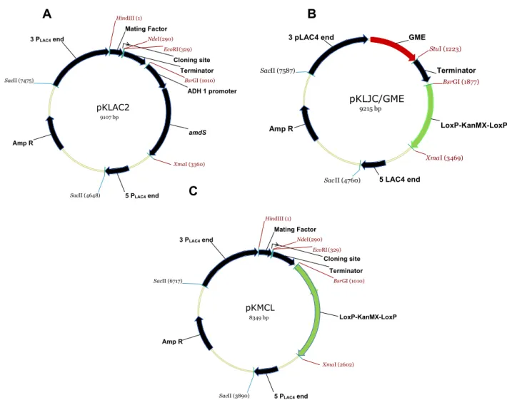
Maps of the plasmids used on this study are shown on Figure 1. The vector
pKLAC2 (New England Biolab®) was taken as a base. The construction pKLJC/GME
(ROSA, 2011) was used to obtain the LoxP-KanMX-LoxP cassete that confers
resistance to the aminoglycosid antibiotic G418 (geneticin) and Kanamycin, and was
subcloned into BsrGI and XmaI sites by replace of acetamidase selectable marker
(amdS) from pKLAC2 vector, now designated as pKMCL. The construction
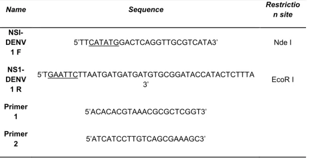
pTZ57R/T+NS1-Denv1 was used as template for recombinant NSI/Denv1
(rNS1/Denv1) fragment amplification with the primers ns1/denv1-F and ns1/denv1-R
(Table 1). The rNS1 fragments were obtained in amplification cycle: 30 seconds 98
°C, 10 seconds 98°C, 1 minute 70 °C, 1 minute 72 °C (35 cycles) and were subcloned
into pGEM T Easy Vector (Promega®) and further transferred to pKMCL generating
the plasmid pKMCL/NS1-Denv1. All ligation reactions were performed with T4 DNA
6
Figure 1. Maps of the plasmid vectors used for rNS1 gene expression. pKLAC2 (A). pKlJC/GME (B) and
pKMCL (C) vectors are derived from pKLAC1 and pKLAC2 respectively. pKMCL was modified with the
cassette LoxP-KanMX-LosP, between the restriction sites BsrGI e XmaI. The cassette
LoxP-KanMX-LosP confers resistance to geneticin. pKLAC2
9107 bp
amdS
ADH 1 promoter Cloning site Mating Factor
Terminator
5 PLAC4 end Amp R
3 PLAC4 end
Hin dIII (1)
Xma I (3360)
BsrGI (1010)
Nde (290)I
EcoRI(329)
SacII (4648)
SacII (7475)
pKLJC/GME
9215 bp
Terminator
5 LAC4 end Amp R
3 pLAC4 end
LoxP-KanMX-LoxP GME
Bsr GI (1877)
Stu I (1223)
Xma I (3469)
Sac II (4760)
Sac II (7587)
pKMCL
8349 bp LoxP-KanMX-LoxP
Cloning site Mating Factor
Terminator
5 PLAC4 end Amp R
3 PLAC4 end
Hin dIII (1)
Xma I (2602)
BsrGI (1010)
NdeI (290)
EcoRI(329)
SacII (3890)
SacII (6717)
A
B
7
Table 1: Primers sequences used in this study.Name Sequence Restrictio
n site
NSI-DENV
1 F
5’TTCATATGGACTCAGGTTGCGTCATA3’ Nde I
NS1-DENV
1 R
5’TGAATTCTTAATGATGATGATGTGCGGATACCATACTCTTTA
3’ EcoR I
Primer
1 5’ACACACGTAAACGCGCTCGGT3’
Primer
2 5’ATCATCCTTGTCAGCGAAAGC3’
Underlined are represented the restriction sites.
Nucleotide sequences encoding the viral protein were designed from the
primary sequence of the NS1/Denv1 peptide considering the preferential yeast
codons (Figure 2). The two N-linked glycosylation sites were maintained. This
sequence constitutes the ORF (open reading frame) of 1056 base pairs encoding the
NS1 protein from dengue virus type 1 and was cloned into the pTZ57 R / T vector.
The construction pTZ57R/T+NS1-Denv1 was gently ceded by Dr. Eduardo Rezende
Honda.
Figure 2. Protein alignment encoding the NS1/Denv1 peptide. The sequences were designed
8
2.3 Yeast transformation
K. marxianus UFV-3 transformation was carried out according to Sanchez et
al. 1993, with some modifications. Fresh K. marxianus UFV-3 cells were plated on
YPD agar medium and incubated overnight at 37°C. An isolated colony was
innoculated in 50 mL YED medium [1% (w/v) yeast extract, 1% (w/v) glucose] culture
at 30°C, 200 rpm overnight. 50 mL YED were inoculated with this pre-cultured cells
to start O.D600 0,0025 per mL (0,1 O.D.). When O.D600 reached approximately 0,8,
the cells were harvested at 3000 g for 5 minutes at 4°C and washed with 20 mL sterile
distilled water ice-cold. A volume of 20 mL of pretreatment buffer (YED, 25 mM DTT
and 20 mM HEPES-Tris pH 8.0) were added and further incubated at 30°C for 30
minutes and 100 rpm. Cells were collected at 3000 g for 5 minutes at 4°C and
resuspended in 0,3 mL electroporation buffer EB (10mM Tris-HCl, pH 7.5, 270 mM
sucrose and 1mM MgCl2) and splitted on 60 µL aliquots of competent cells in
Eppendorf tube on ice. On each aliquot were added 50 µg SS-DNA (Salmon Sperm
DNA) plus 2 µg transforming DNA and kept on ice for 15 minutes. The mixture was
transferred to chilled electroporation cuvette (2 mm) on ice and eletroporated at 1KV,
25 µF and 400 Ohm. Immediately, 1 mL YED ice-cold was added and incubated on
ice for 15 minutes and at 37°C for 1 hour, 200 rpm. The cells were plated on YPD
agar plates containing 200 μg mL-1 of geneticin and kept at 37°C for 2 days.
2.4 Total DNA extraction and yeast transformants screening
K. marxianus UFV-3 cells were grown in 5 mL YPD containing selectable
marker at 37°C to saturation. The cell mass were collected by centrifugation,
resuspended in 0,2 mL lysis buffer (2% Triton X-100, 1% SDS, 100 mM NaCl, 10 mM
Tris pH8, 1 mM EDTA) and transferred to 2 mL screwcap tube. Afterwards, was
added 0,2 mL PCI [phenol pH 6.7- chloroform-isoamylalcohol (25:24:1)] and 0,3 g
glass beads. The cells were broken at fastprep machine, speed 6 for 20 seconds
followed by centrifugation at 10000 g for 10 minutes. The supernatant were
transferred to new tube, 0,5 mL ethanol was added and kept on -20°C at least 20
minutes. The total DNA was pelleted by centrifugation, 14000 g, 10 minutes and
washed with 70% ethanol and dried at room temperature. The DNA samples were
dissolved in 20 µL nuclease-free water and kept on -20°C. A aliquot of 1 µL from total
9
used to detect the single cassette insertion into the LAC4 promoter locus are shown
on table 1. The amplification cycles comprised 5 minutes 98°C, 10 seconds 98°C, 30
seconds 70°C, 1 minute 72°C (35 cycles) and 5 minutes 72°C.
2.5 Total RNA extraction from yeast and RT-PCR
The transformants yeast cells were grown in 20 mL YPD medium containing
selectable marker at 37°C, 200 rpm overnight. The cells were pelleted by
centrifugation at 9000 g for 5 minutes at 4°C and the supernatant was discarded. The
total RNA from recombinant K. marxianus UFV-3yeast cells was extracted using the
hot acid phenol method as described by Collart and Oliviero (2001). The cDNA
synthesis from the total RNA extracted was achieved using the Reverse Transcription
System from Promega®. A 2 µL cDNA aliquot from each sample were used in a 50
µL PCR reaction in order to qualitatively detect mRNA expression of the rNS1 gene
inserted into K. marxianus UFV-3 genome. The RT-PCR was achieved using the
same primers and amplification cycles used for viral gene amplification.
2.6 Evaluating the genetic stability of the recombinant strains
Recombinant strains were transferred to YPD agar and incubated at 37°C until
the appearance of isolated colonies. Next, five colonies of each transformant were
successively transferred to nonselective complete medium, YPD, in a total of five
replating. Each replating was incubated at 37°C for 48 hours. At the end of the fifth
replating, the colonies were transferred to selective medium (YPD agar plates
containing 200 μg mL-1 of geneticin) and incubated at 37 ° C for over 48ºC. The
transformants cells were subjected to an induction phase test.
2.7 Induction of protein expression
The recombinant yeast cells were precultured overnight in 50 mL YPD, 200
rpm at 37°C for obtaining cell mass. The culture was washed with peptone water
0,01% (w/v) and used to inoculate 1 L Yeast Nitrogen Base with amino acids and 5
10
(inducer), yeast extract 0,5 % (w/v) supplemented with biotin 4 x 10⁻⁵% (w/v). The
cells were cultured at 37°C (optimum temperature determined for growth), 250 rpm,
pH was maintained at 5.0 by adding of either 10% (w/v) HCl or 10% (w/v) NaOH as
and when required in the 2,5 L - Bioflo® & Celligen® 310 – Fermentor/Bioreactor
(New Brunswick). Samples were collected every 24 hours to specific protease activity,
SDS-PAGE and rNS1 detection. An induction experiment was carried out in parallel,
in which every 24 hours of culture was added a pulse of 4 g·L-1 galactose solution
and 4 x 10⁻⁵% (w/v) biotin. Foaming was prevented by addition of sterile antifoam
solution (Sigma Chemicals) as required. Shake-flask experiments were performed
under the same conditions described above for comparing.
Total proteins content was measured according to Bradford (1975), using
bovine serum albumin (BSA) as standard. The specific protease activity was carried
out according to Ray et al. 1992 with some modifications, and was assayed with
cell-free culture supernatants, using azocasein as a substrate at a concentration of 1%
(w/v), pH 7.5. Enzymatic hydrolysis of azocasein produces stable dye-labelled
peptides and aminoacids into the reaction mixture which can be measured easily.
Azocasein protease activity was measured by incubating 100 µL of culture
supernatant and 100 µL of 0.5% (w/v) azocasein for 12 hours at 30ºC. The reaction
was stopped by adding 1 ml of 10% (w/v) trichloroacetic acid and allowed to stand for
15 minutes at room temperature. The mixture was centrifuged at 10000 g for 10
minutes to remove a yellow precipitate. The absorbance of the supernatant was
measured at 440 nm using a spectrophotometer. The activity of the protease was
expressed in arbitrary units, where 1 unit of activity was defined as the absorbance
obtained divided by the total protein concentration in mg·L-1 per time unit.
2.8 Fractionation of cultures for rNS1 SDS-page analysis and immunogenic
detection using positive human serum for dengue virus
Both cells and culture supernatants were routinely assayed for rNS1 detection.
The fractionation of cultures into supernatant, cell wall (retained inside the periplasmic
space) and cell-bound protein were separated according to the method described by
Rouwenhorst et al. 1988. Total protein content were collected during induction time
and precipitated with trichloroacetic acid and acetone, resuspended in SDS sample
buffer [150mM Tris-HCL pH 7.0, 200mM DTT, 12% (w/v) Sodium dodecyl sulfate
11
water) and store at -20°C. Samples were heated to 100°C for 5 minutes and proteins
were separated by SDS-polyacrylamide gel electrophoresis. Electrophoresed
proteins were transferred to nitrocellulose membrane and positive human serum for
dengue virus was used to detect the rNS1 in culture supernatant by western blot
analysis. In brief, for the western blot analysis, the proteins along with prestained
protein markers on adjacent lanes were transferred electrophoretically to
nitrocellulose membrane. The membrane was blocked overnight with 5% (w/v) bovine
serum albumin in TBS buffer (10 mM Tris-HCl, pH 7.6, 1,4 mM NaCl) at 4°C and then
washed with TBS buffer plus 0,1% (w/v) Tween 20 (TBS-T) for 20 minutes. Serum
samples were diluted 1:10.000 with blocking buffer and incubated with membranes
for two hours at room temperature. After incubation, membranes were washed with
TBS-T and incubated for 1 hour with human anti-IgG and anti-IgM (Sigma Chemicals).
The protein bands were visualized by incubating in substrate 3,3 diaminobenzidin
(Sigma Chemicals).
2.9 rNS1 purification
The culture supernatant containing rNS1 protein was rapidly adjusted to pH
8.0 using 5M NaOH, to minimize precipitation of rNS1 (pI: 5.7) and was freshly diluted
with an equal volume of binding buffer pH 8.0 (20 mM Tris-HCl). Subsequently was
clarified by filtration through a 0.45µm membrane, and then submitted to ion
chromatography (IC). The clarified supernatant was load onto a 1 mL HisTrap Capto
Q column, previously equilibrated with binding buffer and attached to an AKTA purifier
system (GE Healthcare Life Sciences, Uppsala, Sweden). Ten-column volume of
binding buffer was passed through the column to remove non-specifically bound
proteins, before the elution with 10, 25, 50 100% of elution buffer (20 mM Tris-HCl,
500 mM NaCl), Peak fractions were collected and dialyzed against PBS pH 8.0 using
centrifugal filtration devices with a 10,000-molecular weight cut-off (Millipore), for the
protein estimation using was determined with a BCA kit (Pierce Chemical Co.,
Rockford, USA). For the characterization the purified proteins were subjected to 15%
12
3. Results
3.1 Construction of expression vector with kanamicin marker and harboring the
NS1 gene to function in Kluyveromyces UFV-3
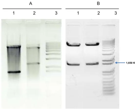
In order to replace the acetamidase selectable marker (amdS) from the
pKLAC2, the LoxP-KanMX-LoxP cassete of 1592 base pairs that confers resistance
to the geneticin from the vector pKLJC/GME was obtained by cleavage with BsrG I
and Xma I restriction endonucleases. The DNA fragments were recovered from the
agarose gel and subcloned in pKLAC2 vector now designed as pKMCL (Figure 3).
Figure 3. Construction of expression vector. (A) Agarose gel electrophoresis 1,2% (w/v) of double
digestion restriction fragments (BsrG I and Xma I) of the pKLJC/GME and pKLAC2 vectors (1,2)
respectively; (3) 1 Kb DNA ladder plus (Invitrogen). (B) Agarose gel electrophoresis 1,2% (w/v) of
subcloning confirmation by cleavage of pKMCL vector with BsrG I and Xma I restriction endonucleases
(1); (2) pKLJC/GME cleaved with the same enzymes (positive control); (3) 1 Kb DNA ladder plus
(Invitrogen).
After confirming the pKMCL vector construction, the nucleotide sequence
designed from the primary amino acid sequence of the NS1/Denv1 peptide was
A B
1 2 3 1 2 3
13
obtained by PCR amplification. The amplification resulted in fragment about of 1056
bp approximately, consistent with the expected size for the gene of NS1. This DNA
fragment obtained by PCR was used to the construction of expression vector
pKMCL-rNS1/Denv1. In the pKMCL-rNS1/Denv1 construct, the NS1 region from DENV-1 was
cloned in frame with K. lactis α-mating factor secretion domain (α-MF) to direct the
fusion protein to the general secretory pathway, resulting in secretion of the
recombinant protein into the medium. The construction was confirmed by cleavage
with the EcoR I and Nde I restriction endonucleases, resulting in DNA fragments with
the expected size of approximately 1056 base pairs (Figure 4).
Figure 4. Confirmation of construction vector. (A) pKMCL-rNS1/Denv1 expression vector map. (B)
Agarose gel electrophoresis 1,2% (w/v); (1) Insert released after cleavage the construction with the EcoR
I and Nde I restriction endonucleases; (2) 1 Kb DNA ladder plus (Invitrogen).
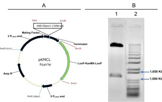
3.2 Transformation of K. marxianus UFV-3 with pKMCL-rNS1/Denv1and mitotic stability of recombinants
Before transformation of K. marxianus UFV-3, with the constructed and
checked vector pKMCL-rNS1/Denv1, this vector was linearized with the Sac II
enzyme. Cleavage resulted in a cassette containing the sequence of interest flanked
by regions 3'PLAC4 and 5'PLAC4, directing cassette integration by homologous
pKMCL
8349 bp LoxP-KanMX-LoxP Mating Factor
Terminator
5 PLAC4 end Amp R
3 PLAC4 end
Xma I
BsrGI
NdeI
EcoRI
SacII (3890)
SacII (6717)
A B
1 2
rNS1/Denv1 (1056 bp)1,650 Kb
14
recombination in the promoter region of the LAC4 locus. The NS1 gene was fused in
frame with K. lactisα-mating factor secretion domain (α-MF), which directs the fusion
protein to the general secretory pathway. We selected transformants which were grown in the presence of 200 μg mL-1 geneticin and further verified the presence of
the integrated NS1 gene by direct colony PCR. Transformation efficiency was 504
transformants per µg of plasmidial DNA. To analyze if the integration of the expression
fragment at the LAC4 locus in the K. marxianus UFV-3 genome was in the correct
location, DNA extraction from clones was performed to PCR amplification with the
primers 1 and 2 (Table 1), which resulted in amplification of a diagnostic DNA
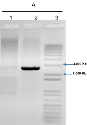
fragment of 2.4 Kb product (Figure 5) from a putative clone. No nonspecific
amplification band was found. As the next step, we chose several of these
PCR-positive NS1 gene-harboring clones, and tested them for mitotic stability. All
recombinants analyzed were stable (data not shown).
Figure 5. Agarose gel electrophoresis 1,2% (w/v) of genomic DNA PCR product from wild type (1) and
recombinant strain. The fragment inserts into the promoter of the LAC4 locus. Single-copy integration at
the LAC4 locus was detected using primers 1 and 2 to amplify a 2.4 kb diagnostic fragment (2), 1 Kb
DNA ladder plus (Invitrogen) (3).
3,000 Kb
2,000 Kb
15
3.3 Selection of K. marxianus UFV-3 recombinant strain and expression and function of the NS1 protein
In order to analyze the expression of the rNS1/Denv1 protein, an induction test
of the transcript and recognition of proteins secreted by dot-blot immunoassay with
positive human serum for dengue virus was performed. It was chosen several of
recombinant clones, and tested them in a small-scale expression assay in induction
medium. DNA fragments of approximately 1056 bp obtained by RT-PCR (Figure 6 A)
confirmed the transcription of the inserted sequences into the genome of K. marxianus
UFV-3. Cell-free culture supernatants from recombinant cells was used to perform
dot-blot immunoassay, which revealed that the recombinant strain was able to secrete
rNS1/Denv1 (Figure 6 B), demonstrating that the signal sequences cloned upstream
the ns1 gene indeed targeted the recombinant protein to secretion.
To identify the best expressing clone, logarithmically growing test tube cultures
of 10 clones were galactose-induced for 72 hours, and supernatants were assayed
for rNS1 SDS-page (data not shown). One clone, #1 (a putative multi-copy clone),
which expressed maximal levels of rNS1 among all clones tested, was used for further
study. Since it was our objective to develop costeffective production of the viral
protein, we sought to explore culture conditions that would help maximize rNS1
16
Figure 6. Induction test of the transcript and recognition of proteins secreted by dot-blot immunoassay.
(A) Agarose gel electrophoresis 1,2% (w/v) of RT-PCR product of mRNA from recombinant strains
induced with galactose. Detecting the mRNA encoding the peptides of interest confirmed the
rNS1transcription introduced into the genome of K. marxianus UFV-3. (1) 1 Kb DNA ladder plus
(Invitrogen), (2) wild type, negative control, (3-10) K. marxianus UFV-3 transformed with the rNS1 gene.
(B) Dot-blot analysis of rNS1 secreted by K. marxianus UFV-3. (1-4) culture supernatant (100 µL) of
recombinant strains and wild type (C-) were spotted onto a nitrocellulose membrane and
immunodetected with positive human serum for dengue virus. All the recombinants analyzed were able
to secrete the rNS1 peptide.
3.4 Growth of recombinant K. marxianus UFV-3 and production of rNS1 protein in shake-flask and bioreactor
Recombinant protein expressing in laboratory-scale is generally performed
using complex medium at shake-flasks culture. However, for producing protein in
bioreactor, the composition of media seems to be an important strategy to ensure
both cell growth and rNS1 protein production. To synthesize the rNS1 protein by K.
marxianus UFV-3 recombinant strain, the culture was carried out in two stages. In the
B
1 2 3 4 C-
1,650Kb
1,000Kb
A
17
first, a cell mass of approximately 10 g·L-1, corresponding to OD
600 of 20 was obtained.
This stage was conducted in YPD medium for 16 hours of a culture starting with OD600
of 2. In the second stage, the recombinant cell mass was harvested by centrifugation
and resuspended in induction medium [YNB buffered at pH 5.0, containing 4 g·L-1 of
galactose, yeast extract 0,5 % (w/v) supplemented with biotin 4 x 10⁻⁵% (w/v)]. The
experiments were conducted in Bioflo® & Celligen® 310 – Fermentor/Bioreactor (New
Brunswick). Shake-flask experiments were performed. In 250 mL-Erlenmeyer flasks
containing 50 mL of the same cultivation medium. Samples of the supernatants of
induction medium were evaluated by the total proteins and proteolytic activity. Total
protein was analyzed during the induction phase from culture aliquots, withdrawn at
various time points. It was observed that the recombinant strains secreted a greater
amount of total proteins when galactose pulse was applied every 24 hours (data not
shown). The quantitation measured according to Bradford revealed that their
concentrations reached a maximum of ∼1,2 mg mL−1 after 72 hours of induction. The
accumulation of total proteins in the culture medium revealed the stability of secreted
proteins and the absence of proteases, which was confirmed by analysis of protease
activity assay analyzed in the supernatant of both recombinant and wild type strains
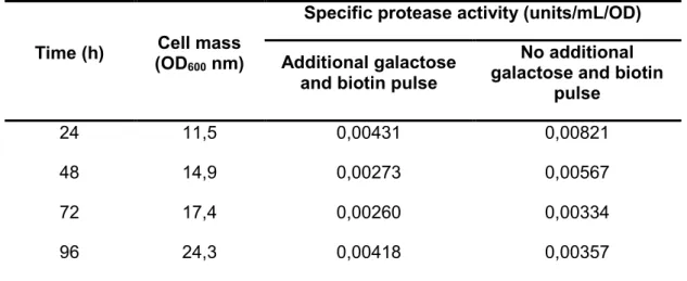
cultured in bioreactor and shake flasks (Tables 2-5).
Table 2. Protease activity assay of the recombinant strain supernatant in bioreactor with additional/no
additional 4% (w/v) galactose and 4 x 10⁻⁵% (w/v) biotin pulse at 24 hours intervals. The cell mass (OD600
nm) corresponds only the cells cultured on induction medium with additional galactose and biotin pulse.
Time (h) Cell mass (OD600 nm)
Specific protease activity (units/mL/OD)
Additional galactose and biotin pulse
No additional galactose and biotin
pulse
24 11,5 0,00431 0,00821
48 14,9 0,00273 0,00567
72 17,4 0,00260 0,00334
96 24,3 0,00418 0,00357
U=A630 divided by the total protein concentration in mg·L-1 per unit time.
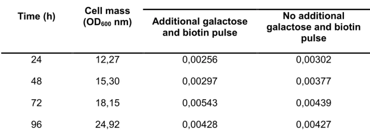
Table 3. Protease activity assay of wild type strain supernatant in bioreactor with additional/no additional
4% (w/v) galactose and 4 x 10⁻⁵% (w/v) biotin pulse at 24 hours intervals. The Cell mass (OD600 nm)
18
Time (h) Cell mass (OD600 nm)
Specific protease activity (units/mL/OD)
Additional galactose and biotin pulse
No additional galactose and biotin
pulse
24 12,27 0,00256 0,00302
48 15,30 0,00297 0,00377
72 18,15 0,00543 0,00439
96 24,92 0,00428 0,00427
U=A630 divided by the total protein concentration in mg·L-1 per unit time.
Table 4. Protease activity assay of the recombinant strain supernatant in shake-flasks with additional/no
additional 4% (w/v) galactose and 4 x 10⁻⁵% (w/v) biotin pulse at 24 hours intervals. The Cell mass (OD600
nm) corresponds only the cells cultured on induction medium with additional galactose and biotin pulse.
Time (h) Cell mass (OD600 nm)
Specific protease activity (units/mL/OD)
Additional galactose and biotin pulse
No additional galactose and biotin
pulse
24 9,13 0,00812 0,00562
48 12,66 0,00331 0,00596
72 13,5 0,00259 0,00403
96 13,64 0,00593 0,00391
U=A630 divided by the total protein concentration in mg·L-1 per unit time.
Table 5. Protease activity assay of wild type supernatant in shake-flasks with additional/no additional 4%
(w/v) galactose and 4 x 10⁻⁵% (w/v) biotin pulse at 24 hours intervals. The Cell mass (OD600 nm)
corresponds only the cells cultured on induction medium with additional galactose and biotin pulse.
19
Cell mass (OD600 nm)
Additional galactose and biotin pulse
No additional galactose and biotin
pulse
24 9,27 0,00812 0,00562
48 12,82 0,00331 0,00596
72 13,12 0,00259 0,00403
96 13,56 0,00593 0,00391
U=A630 divided by the total protein concentration in mg·L-1 per unit time.
3.5 Detection and purification of rNS1 on supernatant culture
To analyze the distribution of the rNS1 protein, the culture were fractionated
in supernatant, cell-bound and cell wall protein and separated by SDS-polyacrylamide
gel electrophoresis (Figure 7). The results confirmed the rNS1 secretion (Figure 7A)
and that the rNS1 appear in the three fractions of the recombinant strain culture and
it was a consequence of the expression of the cloned synthetic gene. The
concentration of rNS1 released by cell wall and cell-bound protein (Figure 7B) was
lower than the total protein secreted in the supernatant. In particular, the secretion
efficiency of rNS1 using the pKMCL expression vector adapted from pKLAC2 was
confirmed by western blot (Figure 8). No similar band appeared in negative control.
20
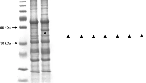
Figure 7. Coomassie blue stained SDS-PAGE 12% (w/v) for the analysis of the rNS1 expressed in K.
marxianus UFV-3. (A) K. marxianus supernatant. (1) Prestained protein ladder (Thermo Scientific); (2)
Wild type; (3) recombinant strain producing rNS1. (B) Fractionation of cultures for rNS1 detection in K.
marxianus. (1) Prestained protein ladder (Thermo Scientific); (2) negative control; (3-4) K. marxianus
cell-bound rNS1; (5-8) K. marxianus cell wall associated rNS1. The arrow and the arrowheads identify
rNS1.
Figure 8. Expression of dengue virus type 1 rNS1 in K. marxianus UFV-3. (1) prestained molecular weight
standard (Thermo Scientific). (2) Western blot analysis of rNS1 protein using positive human serum for
dengue virus.
The 96 hours cell-free culture supernatant from bioreactor cultures was diluted
with an equal volume of binding buffer pH 8.0 (20 mM sodium phosphate, 500 mM
NaCl) and clarified by filtration through a 0.45 µm membrane, and then loaded onto
an ion chromatography which yielded four distinct peaks (Figure 9 A). The
55 kDa
38 kDa
55 kDa
21
recombinant protein emerged on peak 2 with other proteins found in supernatant
(Figure 9 B).
Figure 9. Ion exchange chromatography profile of 10 mL K. marxianus UFV-3 bioreactor supernatant
containing rNS1. (A) Chromatogram of the IC. Four distinct peaks are shown. Peaks 1 to 4 were eluted
with 10, 25, 50 100% of elution buffer respectively. (B) SDS-page gel electrophoresis analysis of peaks
fractions collected. (1) Prestained protein ladder (Thermo Scientific); (2-5) peaks 1 to 4 respectively; (6)
non-specifically bound proteins. The arrowhead indicates the position of the rNS1 protein.
.
A
B
1 2 3 4 5 6
Time / min
C
o
n
d
u
c
ta
n
c
e
/
µ
22
4. Discussion
In the last years, successful production of dengue virus protein has been
reported in several vector systems (Batra et al. 2010; Blaney et al. 2005; Block et al.
2010; Jaiswal et al. 2004; Saxena et al. 2008; Zhou et al. 2006). Although prokaryotic
expression system such as E. coli is simple to perform, lack in this system the
modification mechanism of eukaryotic expression, like post-translational
modifications that many eukaryotic proteins require for optimal biological activity and
stability (Coller et al. 2011). Furthermore, it is difficult to purify recombinant protein
from inclusion bodies (Wei et al. 2003). Therefore, the heterologous expression
system of eukaryotic proteins in prokaryotes led a search for more suitable hosts. In
the present study, the K. marxianus UFV-3 yeast was used to produce dengue-1 NS1
protein because of the simplicity of techniques needed for molecular genetic
manipulation, its ability to express foreign proteins either intra or extracellularly, its
high growth rate and capacity to produce cell mass and its status of generally
regarded as safe (GRAS), which convey for biological synthesis of protein for public
health purposes.
The first difficult found on utilizing the K. marxianus as a host strains to
synthetize recombinants proteins is due to the absence of commercial cloning and
expression system available for this strains. When the system was used on the
selection of recombinant K. marxianus UFV-3, the wild type strains were able to utilize
acetamide, the selective marker, a sole nitrogen source. We have tested several
concentrations of acetamida (5 mM to 40 mM) and in all concentrations tested was
observed growth of the K. marxianus strains. In order to use this vector in K.
marxianus UFV-3 cells, it was necessary to replace the acetamidase selectable
marker (amdS) from pKLAC2 by a LoxP-KanMX-LoxP cassete that would confers
resistance to the geneticin. To transform the K. marxianus UFV-3 cells several
protocols were tested but only the protocol (with modifications) described in material
and methods was successful with transformation efficiency of 504 transformants per
µg of plasmidial DNA.
An interesting feature of K. marxianus and K. lactis strains as a host system is
that vectors that replicate extrachromosomally (episomal vectors) or that integrate into
the genome (integrative vectors) can be used to transform cells. Both vector systems
have been used to direct expression of recombinant proteins as describe in OOYEN
et al., (2006) and ROCHA, SAUL N et al., (2010). Episomal vectors provide higher
23
selection. This instability leads to expression problems of some heterologous
proteins, especially in large industrial applications which cells are often subjected to
prolonged growth in the absence of selection. ROCHA, SAUL N et al., (2010)
employed different episomal and integrative constructs and demonstrated that
according to plasmid stability studies, after 48 hours of cultivation, both cells of K.
marxianus and K. lactis nearly lost their episomal expression plasmids. In other
studies, after growth of a K. lactis strain secreting human lysozyme expressed from a
pKD1 vector, only 17.3 % of cells retained the vector contrasted with > 91.5% of cells
retaining an integrated expression vector producing the same enzyme under the
same growth conditions (Iwata et al. 2004). The integration cassette used in this work
has regions of homology with the LAC4 promoter, which reduces but does not
eliminate the possibility of ectopic integration. In order to identify the correct genomic
integration of a linear expression cassette into the promoter region of the LAC4 locus
in the K. marxianus genome was used specific primers designed to K. lactis. Our
results demonstrates that these primers can be used to K. marxianus, because
nonspecific bands were not amplified. The analysis of K. marxianus UFV-3
recombinant strains by RT-PCR and the protein detection using positive human
serum for dengue virus confirmed the transcription and secretion of rNS1. A major
benefit of the using integrative expression vector was the increased genetic stability
of recombinant strain, confirmed by mitotic stability test, since recombinant cells
mitotically stable reduces the difficulties with maintaining of selective pressure in the
culture medium for the large-scale industrial application.
Different conditions to maximize rNS1 production were evaluated. Early
experiments at our laboratory showed that of the different media tested, highest yields
were obtained using YNB containing 4 g·L-1 of galactose and yeast extract 0,5 %
(w/v). In this work, a variety of induction conditions were evaluated, including pH and
galactose and biotin pulse to enhance the cell mass and level of rNS1 induction in
YNB, rather than in YPGal [1% (w/v) yeast extract, 2% (w/v) peptone, 2% (w/v)
galactose]. We observed that the induction performed in YNB [containing 5 g·L-1
ammonium, 4 g·L-1 of galactose, yeast extract 0,5 % (w/v) supplemented with biotin 4
x 10-5%] buffered at pH 5.0, increased cell mass and rNS1 levels approximately
50-fold, compared to YPGal medium. Periodic monitoring of the cell density during the
induction phase revealed that the cells grew better at the pH 5.0. Since in this pH
value, cell viability increased and no proteolysis was detected, there was a higher
accumulation of recombinant protein in K. marxianus UFV-3 cultured in bioreactor.
24
reached during 96 hours of induction in buffered medium. In contrast, when the
recombinant cells were performed in bioreactor under the same conditions described
above for comparing, the maximum DO600 24 was reached. These results suggest that
the use of bioreactors for recombinant proteins production is recommended, since the
production of recombinant protein is directly related to the cell mass in the medium.
We also evaluated the effect of pH on the rNS1 production from K. marxianus
UFV-3. As the pI (isoelectric point) of NS1 is predicted to be ∼5.7, we used YNB at pH 3,
4 and 5, to preclude isoelectric precipitation of the recombinant protein. The YNB
buffered at pH 5.0 was selected. The effect of galactose concentration on the
induction of gene expression and cell mass was also evlauated. Several studies used
2% (w/v) galactose or lactose as inducer for heterologous proteins production
(Bartkeviciute and Sasnauskas 2003; Donnini et al. 2004; Ganatra et al. 2011;
Kooistra et al. 2004; Li et al. 2011), but we have detected that the use of 4% (w/v)
galactose increased the concentration of cell mass and hence the recombinant
protein in bioreactor.
rNS1 synthesis by K. marxianus UFV-3 recombinant strain was performed in
two stages and conducted in bioreactor and shake-flasks containing the same
cultivation medium. In the first stage, glucose was used as carbon source for obtaining
cell mass. A culture complex medium provides high rates for K. marxianus growth.
However, this culture medium can compromise the recovery of recombinant peptides
due to the presence of self-peptides in this culture medium. Set a culture medium for
maximum cell mass production is crucial in order to improve rNS1 yields. In this
sense, it was defined a culture medium to maximize production of cell mass and
consequently increasing the production of rNS1. In the second step, the induction of
rNS1synthesis in 4 g·L-1 of galactose showed that the induction parameters are also
important and can directly affect the yield of the recombinant protein. Two induction
conditions were tested: pulse and no pulse of galactose and biotin every 24 hours
during 96 hours of induction, since biotin is an essential vitamin for the growth of many
microorganisms (Jungo et al. 2007). The concentration of extracellular proteins in the
step of galactose and biotin pulse was higher compared to the condition without pulse
of galactose and biotin. Therefore, it can be inferred that the inductor is consumed
over time and the addition of galactose maintains the synthesis and secretion of
proteins. In this way, the galactose and biotin pulse must be considered as a strategy
for increasing rNS1 yields. The accumulation of total proteins in the supernatant
revealed the stability of secreted proteins and absence of proteases, which was
25
the process, because the absence of proteases in the supernatant prevents higher
expenses for the proteases inactivation, since they could compromise the obtaining
of the interest proteins. Sugrue et al. (1997) showed that expression of the dengue
protein in P. pastoris was accompanied by extensive proteolytic degradation,
difficulting downstream operations.
Currently, most P. pastoris fed-batch cultivations for the production of
recombinant proteins, using AOX1 expression system, consist of three or four distinct
phases, which include glycerol batch, glycerol fed batch, starvation and methanol fed
batch. However, we have recently demonstrated a simple two-step strategy, which
only includes a glucose batch phase, to generate sufficient cell mass, directly followed
by the induction phase with a galactose for the recombinant protein synthesis. In this
work, we have successfully employed this two-step strategy for the secretion of rNS1.
Recombinant NS1 protein was obtained at the first 24 hours of cultivation. SDS-Page
analysis showed that large quantities of secreted rNS1 were present in the
supernatant than retained inside the periplasmic space or cell-bound. The cell-free
culture supernatant obtained was loaded onto an ion chromatography for rNS1
isolation, but other proteins were also isolated in the same fractions.
Our rNS1 protein exhibited the same molecular size as authentic dengue virus
protein. A novel approach to generate recombinant NS1 protein was performed using
K. marxianus UFV-3. To the best of our knowledge, this is the first study about the
viral protein production with immunogenic potential of pharmaceutical interest using
the yeast K. marxianus. The rNS1 produced has a potential application for detection
of anti-dengue IgM and IgG antibodies as well a further studies in vaccine
development for dengue virus infection. By using this strategy, other recombinant viral
proteins can also be produced. Further study of the structure and functions of the
rNS1 protein will be performed.
26
We thank the Brazilian Agency CNPq (National Science and Technology
Development Council) for the scholarship and CAPES (Coordination for the
Improvement of Higher Education Personnel) and FAPEMIG (Foundation for
27
References
Alcon S, Talarmin A, Debruyne M, Falconar A, Deubel V, Flamand M (2002) Enzyme-Linked Immunosorbent Assay Specific to Dengue Virus Type 1 Nonstructural Protein NS1 Reveals Circulation of the Antigen in the Blood during the Acute Phase of Disease in Patients Experiencing Primary or Secondary Infections. 40:376–381. doi: 10.1128/JCM.40.2.376
Athmaram TN, Saraswat S, Misra P, Shrivastava S, Singh AK, Verma SK, Gopalan N, Behara PK, Rao PVL (2012) Optimization of Dengue-3 recombinant NS1 protein expression in E. coli and in vitro refolding for diagnostic applications. Virus genes. doi: 10.1007/s11262-012-0851-5
Bartkeviciute D, Sasnauskas K (2003) Studies of yeast Kluyveromyces lactis mutations conferring super-secretion of recombinant proteins. Yeast (Chichester, England) 20:1–11. doi:
10.1002/yea.935
Batra G, Gurramkonda C, Nemani SK, Jain SK, Swaminathan S, Khanna N (2010) Optimization of conditions for secretion of dengue virus type 2 envelope domain III using Pichia pastoris. Journal of bioscience and bioengineering 110:408–14. doi: 10.1016/j.jbiosc.2010.05.001
Blaney JE, Matro JM, Murphy BR, Whitehead SS (2005) Recombinant , Live-Attenuated Tetravalent Dengue Virus Vaccine Formulations Induce a Balanced , Broad , and Protective Neutralizing Antibody Response against Each of the Four Serotypes in Rhesus Monkeys. 79:5516–5528. doi: 10.1128/JVI.79.9.5516
Block OKT, Rodrigo WWSI, Quinn M, Jin X, Rose RC, Schlesinger JJ (2010) A tetravalent recombinant dengue domain III protein vaccine stimulates neutralizing and enhancing antibodies in mice. Vaccine 28:8085–94. doi: 10.1016/j.vaccine.2010.10.004
Chaiyaratana W, Chuansumrit A, Pongthanapisith V, Tangnararatchakit K, Lertwongrath S, Yoksan S (2009) Evaluation of dengue nonstructural protein 1 antigen strip for the rapid diagnosis of patients with dengue infection. Diagnostic microbiology and infectious disease 64:83–4. doi: 10.1016/j.diagmicrobio.2009.01.004
Collart MA, Oliviero S (2001) Preparation of yeast RNA. Current protocols in molecular biology / edited by Frederick M Ausubel . [et al] Chapter 13:Unit13.12. doi:
10.1002/0471142727.mb1312s23
Coller B-AG, Clements DE, Bett AJ, Sagar SL, Ter Meulen JH (2011) The development of
recombinant subunit envelope-based vaccines to protect against dengue virus induced disease. Vaccine 29:7267–75. doi: 10.1016/j.vaccine.2011.07.021
Donnini C, Farina F, Neglia B, Compagno MC, Uccelletti D, Goffrini P, Palleschi C (2004) Improved Production of Heterologous Proteins by a Glucose Repression-Defective Mutant of
Kluyveromyces lactis. Society 70:2632–2638. doi: 10.1128/AEM.70.5.2632
Fonseca GG, Heinzle E, Wittmann C, Gombert AK (2008) The yeast Kluyveromyces marxianus and its biotechnological potential. Applied microbiology and biotechnology 79:339–54. doi: 10.1007/s00253-008-1458-6
Ganatra MB, Vainauskas S, Hong JM, Taylor TE, Denson J-PM, Esposito D, Read JD, Schmeisser H, Zoon KC, Hartley JL, Taron CH (2011) A set of aspartyl protease-deficient strains for improved expression of heterologous proteins in Kluyveromyces lactis. FEMS yeast research 11:168–78. doi: 10.1111/j.1567-1364.2010.00703.x
28
(2010) Dengue: a continuing global threat. Nature reviews Microbiology 8:S7–16. doi: 10.1038/nrmicro2460
Iwata T, Tanaka R, Suetsugu M, Ishibashi M, Tokunaga H, Kikuchi M, Tokunaga M (2004) Efficient secretion of human lysozyme from the yeast, Kluyveromyces lactis. Biotechnology letters 26:1803–8. doi: 10.1007/s10529-004-4614-9
Jaiswal S, Khanna N, Swaminathan S (2004) High-level expression and one-step purification of recombinant dengue virus type 2 envelope domain III protein in Escherichia coli. Protein expression and purification 33:80–91. doi: 10.1016/j.pep.2003.09.009
Jungo C, Urfer J, Zocchi A, Marison I, Von Stockar U (2007) Optimisation of culture conditions with respect to biotin requirement for the production of recombinant avidin in Pichia pastoris. Journal of biotechnology 127:703–15. doi: 10.1016/j.jbiotec.2006.08.001
Kooistra R, Hooykaas PJJ, Steensma HY (2004) Efficient gene targeting in Kluyveromyces lactis. Yeast (Chichester, England) 21:781–92. doi: 10.1002/yea.1131
Lane MM, Morrissey JP (2010) Kluyveromyces marxianus: A yeast emerging from its sister’s shadow. Fungal Biology Reviews 24:17–26. doi: 10.1016/j.fbr.2010.01.001
Li S, Shen W, Chen X, Shi G, Wang Z (2011) Secretory expression of Rhizopus oryzae -amylase in Kluyveromyces lactis. Journal of Biotechnology 10:4190–4196.
McBride WJH (2009) Evaluation of dengue NS1 test kits for the diagnosis of dengue fever. Diagnostic microbiology and infectious disease 64:31–6. doi:
10.1016/j.diagmicrobio.2009.01.002
Noisakran S, Dechtawewat T, Rinkaewkan P, Puttikhunt C, Kanjanahaluethai A, Kasinrerk W, Sittisombut N, Malasit P (2007) Characterization of dengue virus NS1 stably expressed in 293T cell lines. Journal of virological methods 142:67–80. doi: 10.1016/j.jviromet.2007.01.008
Ooyen A Van, Dekker P, Huang M (2006) Heterologous protein production in the yeast Kluyveromyces lactis. FEMS yeast 6:381–392. doi: 10.1111/j.1567-1364.2006.00049.x
Ramirez AH, Moros Z, Comach G, Zambrano J, Bravo L, Pinto B, Vielma S, Cardier J, Liprandi F (2009) Evaluation of dengue NS1 antigen detection tests with acute sera from patients infected with dengue virus in Venezuela. Diagnostic Microbiology and Infectious Disease 65:247–253.
Ray MK, Devi KU, Kumar GS, Shivaji S (1992) Extracellular protease from the antarctic yeast Candida humicola. Applied and environmental microbiology 58:1918–23.
Rocha SN, Abrahão-Neto J, Cerdán ME, Gombert AK, González-Siso MI (2011) Heterologous expression of a thermophilic esterase in Kluyveromyces yeasts. Applied microbiology and biotechnology 89:375–85. doi: 10.1007/s00253-010-2869-8
Rocha SN, Abrahão-Neto J, Cerdán ME, González-Siso MI, Gombert AK (2010) Heterologous expression of glucose oxidase in the yeast Kluyveromyces marxianus. Microbial cell factories 9:4. doi: 10.1186/1475-2859-9-4
Rouwenhorst RJ, Visser LE, Van Der Baan a a, Scheffers W a, Van Dijken JP (1988) Production, Distribution, and Kinetic Properties of Inulinase in Continuous Cultures of Kluyveromyces marxianus CBS 6556. Applied and environmental microbiology 54:1131–7.
29
Dos Santos VC, Bragança CRS, Passos FJV, Passos FML (2013) Kinetics of growth and ethanol formation from a mix of glucose/xylose substrate by Kluyveromyces marxianus UFV-3. Antonie van Leeuwenhoek 103:153–61. doi: 10.1007/s10482-012-9794-z
Saxena P, Tripathi NK, Rao PVL, Jana AM (2008) Heterologous expression of envelope Protein ( Domain III ) of Dengue Virus Type 2 for Serodiagnosis. 16–20.
Silveira WB, Passos FJV, Mantovani HC, Passos FML (2005) Ethanol production from cheese whey permeate by Kluyveromyces marxianus UFV-3: A flux analysis of oxido-reductive metabolism as a function of lactose concentration and oxygen levels. Enzyme and Microbial Technology 36:930–936. doi: 10.1016/j.enzmictec.2005.01.018
Singh MP, Goyal K, Ratho RK (2010) Nonstructural protein NS1: giving a new structure to dengue diagnosis. Journal of clinical microbiology 48:4688; author reply 4688–9. doi:
10.1128/JCM.01668-10
Sugrue RJ, Cui T, Xu Q, Fu J, Chan YC (1997) The production of recombinant dengue virus E protein using Escherichia coli and Pichia pastoris. Journal of virological methods 69:159–69.
Swaminathan S, Khanna N (2009) Dengue: recent advances in biology and current status of translational research. Curr Mol Med 9:152–173.
Wei H, Jiang L, Xue Y, Fang D, Guo H (2003) Secreted expression of dengue virus type 2 full-length envelope glycoprotein in Pichia pastoris. Journal of Virological Methods 109:17–23. doi: 10.1016/S0166-0934(03)00039-9
Zhao BT, Prince G, Horswood R, Eckels K, Summers P, Chanock R, Lai CJ (1987) Expression of dengue virus structural proteins and nonstructural protein NS1 by a recombinant vaccinia virus. Journal of virology 61:4019–22.