Anti-Inflammatory Properties and Chemical
Characterization of the Essential Oils of Four
Citrus
Species
Jorge Luis Amorim1, Daniel Luiz Reis Simas2, Mariana Martins Gomes Pinheiro1, Daniela Sales Alviano Moreno3, Celuta Sales Alviano3, Antonio Jorge Ribeiro da Silva2,
Patricia Dias Fernandes1*
1Universidade Federal do Rio de Janeiro, Instituto de Ciências Biomédicas, Laboratório de Farmacologia da Dor e da Inflamação, Rio de Janeiro, Brasil,2Universidade Federal do Rio de Janeiro, Instituto de Pesquisa de Produtos Naturais, Laboratório de Análise Fitoquímica, Rio de Janeiro, Brasil,3Universidade Federal do Rio de Janeiro, Instituto de Microbiologia Paulo de Góes, Laboratório de Estruturas de Superfície de Micro-organismos, Rio de Janeiro, Brasil
*patricia.dias@icb.ufrj.br;patricia.dias.icbufrj@gmail.com
Abstract
Citrus fruits have potential health-promoting properties and their essential oils have long been used in several applications. Due to biological effects described to some citrus spe-cies in this study our objectives were to analyze and compare the phytochemical composi-tion and evaluate the anti-inflammatory effect of essential oils (EO) obtained from four differentCitrusspecies. Mice were treated with EO obtained fromC.limon,C.latifolia,C. aurantifoliaorC.limonia(10 to 100 mg/kg, p.o.) and their anti-inflammatory effects were evaluated in chemical induced inflammation (formalin-induced licking response) and carra-geenan-induced inflammation in the subcutaneous air pouch model. A possible antinoci-ceptive effect was evaluated in the hot plate model. Phytochemical analyses indicated the presence of geranial, limonene,γ-terpinene and others. EOs fromC.limon,C.aurantifolia andC.limoniaexhibited anti-inflammatory effects by reducing cell migration, cytokine pro-duction and protein extravasation induced by carrageenan. These effects were also obtained with similar amounts of pure limonene. It was also observed thatC.aurantifolia induced myelotoxicity in mice. Anti-inflammatory effect ofC.limonandC.limoniais proba-bly due to their large quantities of limonene, while the myelotoxicity observed withC. auran-tifoliais most likely due to the high concentration of citral. Our results indicate that these EOs fromC.limon,C.aurantifoliaandC.limoniahave a significant anti-inflammatory effect; however, care should be taken withC.aurantifolia.
Introduction
As rich sources of dietary fiber, vitamin C, phenols, and flavonoids,Citrusfruits are believed to have potential health-promoting properties [1]. Essential oils ofCitrusspecies have long been used for insecticidal, medicinal and cosmetic applications [2]. The peel (pericarp) essential oils of various species ofCitrusare widely used as flavoring agents in food, beverages and
a11111
OPEN ACCESS
Citation:Amorim JL, Simas DLR, Pinheiro MMG, Moreno DSA, Alviano CS, da Silva AJR, et al. (2016) Anti-Inflammatory Properties and Chemical Characterization of the Essential Oils of FourCitrus Species. PLoS ONE 11(4): e0153643. doi:10.1371/ journal.pone.0153643
Editor:Partha Mukhopadhyay, National Institutes of Health, UNITED STATES
Received:February 11, 2016
Accepted:April 1, 2016
Published:April 18, 2016
Copyright:© 2016 Amorim et al. This is an open access article distributed under the terms of the Creative Commons Attribution License, which permits unrestricted use, distribution, and reproduction in any medium, provided the original author and source are credited.
Data Availability Statement:All relevant data are within the paper and its Supporting Information files.
Funding:This work received support from Conselho Nacional de Pesquisa (CNPq): 303033/2013-4 (fellowship) to PDF, 473106/2012-4 (grant) to PDF and from Fundação de Amparo a Pesquisa do Estado do Rio de Janeiro (FAPERJ): E-26/210.141/ 2014 (grant) to PDF, E-26/010.002566/2014 (grant) to PDF, E-26/203.018/2015 (fellowship) to PDF.
confections [3]. Recent studies point to the possibility of employingCitrusoils or their active principles to prevent or treat several pathological conditions, where they can be used as antimi-crobial [4], antifungal [5], neuroprotective [6], anxiolytic, anticonvulsant and sedative [7], anti-nociceptive [8], anti-inflammatory [9] and antioxidant [10] agents.
The chemical composition of the peel essential oils ofCitrusfruits is complex and rich in limonene and other monoterpene components, such asβ-pinene andγ-terpinene, as well as some sesquiterpenes generally present in low amounts [11]. According to Mehl et al. [12], the volatile fraction (85% to 99% of liquid oil) is composed predominantly of terpenes (monoter-penes and sesquiter(monoter-penes) and their oxygenated derivatives, as well as aliphatic alcohols, esters and aldehydes. Limonene is the main component of the essential oils ofCitrus(30% to 70% in different varieties). Other significant components includeαandβ-pinene,γ-terpinene, terpi-nolene, sabinene.
Due to the great variety of species distributed all over the planet and the large number of widely used species in Brazil this study seeks to assess the chemical composition and the anti-inflammatory properties of the essential peel oils of the fruitsC.limon,C.aurantifolia,C.limonia
andC.latifolia. We also investigate possible existing toxic effects after the use of these species.
Methods
Plant material
C.limon[(L.) Osbeck,“siciliano”lemon],C.latifolia(Tanaka ex Q. Jimenez,“tahiti”lime),C.
aurantifolia[(Christm.) Swingle,“mirim”lime] andC.limonia[(L.) Osbeck,“cravo”lime) were collected in the farm of Instituto Vital Brazil, Rio de Janeiro, Brazil, located in Cachoeira de Macacu, Road Rio-Friburgo Km 32.5. Via Magé, RJ 122. GPS coordenatiors are 22° 20’753”
S, 42° 42’858”W. The botanical identification was provided by Dr. Rosana Conrado Lopes (Biology Institute, Federal University of Rio de Janeiro/Brazil), and voucher specimens are deposited in the RFA Herbarium, Federal University of Rio de Janeiro/Brazil. The exsiccate numbers are:C.limonia(RFA-39493),C.aurantifolia(RFA-39492),C.limon(RFA-39494) andC.latifolia(RFA-39491).
The fruit peels were manually removed and homogenized with water in a blender, then immediately submitted to hydro distillation in a modified Clevenger-type apparatus for 2 hours. After extraction, the oils were dried over anhydrous Na2SO4and stored at -18°C.
Typi-cally, 150 g of fruit peels were used and average yields of 1.4% were obtained.
GC/FID analysis
GC analyses were carried out using a Shimadzu GC 2010 (Tokyo, Japan) equipped with a flame ionization detector on a DB5 fused silica capillary column (30 m, 0.25 mm i.d., film thickness 0.25μm). The oven temperature was programmed to rise from 60°C to 246°C at 3°C/
min, then hold at 246°C for 20 min. Injector and detector temperatures were maintained at 220°C and 290°C, respectively. The oil samples were dissolved in CHCl3, and 1μL aliquots
were injected in split mode with split ratio of 1:33 using H2as the carrier gas (1.44 mL/min).
Kovats retention indices (KI) of the compounds were determined relative to the retention times of a series of n-alkanes (C7–C30) with linear interpolation. The relative amounts of the components were calculated based on GC peak areas without correction factors.
GC-MS analysis
to rise from 60°C to 246°C at 3°C/min and then to hold at this temperature for 20 min. The car-rier gas was He (99.999%) with a flow rate of 1.03 mL/min and the injector temperature was 220°C. Oil samples were diluted in CHCl3and 1μL aliquots were injected in split mode (split
ratio 1:50). Mass spectra were obtained under electroionization at 70 eV with a mass range m/z of 40 to 1000 D.
Compound identification
The identification of individual components was based on (i) a comparison of the mass spectral fragmentation patterns with those stored in the NIST Mass Spectral Library and (ii) a compari-son of the GC Kovats retention indices (KI) on a DB-5 column with those of authentic com-pounds and from literature data [13]. Geranial and neral were additionally identified by co-injection of authentic standards obtained by preparative liquid chromatography RP18 column (Supelco, 150 x 10 mm; isocratic condition, mobile phase: methanol:water 60/40 and UV detec-tor at 254 nm) from citral. The characterization of the isolated compounds was made by com-parison of their13C NMR spectra (CDCl3, 100 MHz) with literature data [14].
Animals
Male Swiss Webster mice (2 months old, 18–25 g), kindly donated by Instituto Vital Brazil, were used in this study. Animals were housed in a temperature-controlled room at 22 ± 2°C with a 12 h light/dark cycle and free access to pelleted food (Nutrilab, Brazil) and water. Twelve hours before each experiment, the animals received only water in order to avoid food interference with substances absorption. The animals were acclimatized to the laboratory for at least 1 h before testing and were used only once throughout the experiments. Physical condition of animals was daily monitored and animals with any signs of suffering were euthanized. Also none of animals used became severely ill or died at any time prior to the experimental endpoint.
The research was conducted in accordance with the internationally accepted principles for laboratory animal use and care. The experimental protocols used in this work followed the rules advocated by Law 11,794, of October 8, 2008 by the National Council of Animal Experi-mentation Control (CONCEA) and were approved by the Ethics Committee of Animal Use (CEUA), Science Center Health/UFRJ and received the number DFBCICB015-04/16.
Drugs, reagents and treatments
All solvents were with chromatographic grade (Tedia, Brazil). Acetylsalicylic acid (ASA), carra-geenan, dexamethasone, citral and limonene were purchased from Sigma-Aldrich (St. Louis, MO, U.S.A.). Formalin was purchased from Merck (Germany). Morphine sulfate was kindly provided by Cristália (São Paulo, Brazil). Essential oils (EO) were dissolved in oil (Sigma-Aldrich, USA) to prepare a stock solution at 100 mg/ml. They were administered by oral gavage, at doses of 10 to 100 mg/kg 60 min prior to experiments. Morphine (5 mg/kg, p.o.), ASA (100 mg/kg, p.o.) and dexamethasone (1.5 mg/kg, i.p.) were used as reference drugs. All drugs were diluted in phosphate buffer saline (PBS) just before use. The control group was composed by vehicle (PBS with the same amount of oil used in the highest dose). The final concentration of oil did not exceed 0.5%, and had no effectper se. The doses of ASA and morphine were chosen based on previous experi-ments performed by our group and caused a 50% reduction on each protocol (IC50).
Hot plate test
55 ± 1°C. At successive intervals of 30 min after oral administration of EOs or vehicle, the reac-tion time was recorded when the animals licked their fore- and hind-paws and jumped. Base-line was considered the mean reaction time obtained at 60 and 30 min before administration of the compounds, vehicle, or morphine and was defined as the normal reaction of the animal to the temperature. When animals were kept on the hot plate for a period of time greater than three times the baseline (cut-off), they were removed to avoid possible damage to the paws. Antinociception was quantified as either the increase in baseline (%) calculated by the formula (reaction time x 100/baseline)–100.
Formalin test
The licking behavior was examined immediately after injection of formalin into the hind paw. The procedure was similar to the method described by Hunskaar and Hole [17] and adapted by Gomes et al. [18]. Mice received an injection of 20μL of formalin (2.5% v/v) into the dorsal
surface of the left hind paw. The time that the animal spent licking the injected paw was imme-diately recorded. The nociceptive and inflammatory response consists of the following two phases: the first phase lasts until 5 min after the formalin injection (first phase, neurogenic pain response), and the second phase occurs 15–30 min after the formalin injection (second phase, inflammatory pain response). The animals were pre-treated with oral doses of EOs, vehicle or ASA for 60 min before the administration of formalin.
Subcutaneous air pouch (SAP) model
Animals were used as described by Sedgwick et al. [19] with modifications done by Raymundo and colleagues [20]. After subcutaneous injection of 10 mL of sterile air in the dorsal region in three alternate days, an injection of sterile carrageenan suspension (1%; 1 mL) was done in the SAP formed. Mice were divided in the following groups: vehicle, EOs (10, 30 or 100 mg/kg, p. o.), dexamethasone (1.5 mg/kg, i.p.) treated groups 1 h before carrageenan injection. Another vehicle-treated group was used in mice that received PBS (phosphate buffer saline, 1 mL) in SAP. After 24 h all groups were sacrificed, SAP was washed with 1 mL of sterile PBS and exu-dates collected. Bone marrow cells were obtained by flushing the femoral cavity with 1 mL of phosphate buffer saline (PBS). Peripheral blood was collected in a heparinized tube. Cells counts in from aliquot of bone marrow cells, blood cells suspension or exudates were deter-mined in a CellPocH-100iV Diff (Sysmex) haematology analyser. The exudates were also cen-trifuged (5,000 xg, 10 min, 4°C) and aliquots of the supernatants were stored at -20°C until the assays.
Quantification of TNF-
α
, IL-1
β
, IFN-
γ
and protein
Supernatants from the exudates collected from the SAP were used to measure tumor necrosis factor-α(TNF-α), interleukin 1β(IL-1β), interferon-γ(IFN-γ) by enzyme-linked immunosor-bent assay (ELISA) according to the manufacturer’s instructions (B&D, USA) and extravasated protein determined using the BCA method (BCA™Protein Assay Kit, Pierce).
Nitrate measurement
Devices, USA) was used to measure the absorbance at 540 nm. Nitrite concentration was mea-sured by comparison with a standard curve of sodium nitrite.
Behavioral and stomach observations
To evaluate a possible toxic effect we adapted the method used by Lorke [23]. Briefly, different groups of mice were treated with 500 mg/kg of each EO. During 5 consecutive days several parameters (i.e., convulsion, sedation, respiration, and food and water intake) were observed. After five days the animals were euthanized with an overdose of ketamine/xilazine (150 mg/10 mg/kg). The stomachs of the animals were removed and opened to observe any signs of hyper-emia and the presence or absence of ulcer.
Statistical analysis
All experimental groups consisted of 6–10 mice. The results are presented as the mean ± S.D. Statistical significance between groups was performed by applying analysis of variance (ANOVA) followed by Bonferroni’s test.Pvalues less than 0.05 (p<0.05) were considered significant.
Results
Chemical characterization of essential oils
Table 1show retention indices and percentages of each compound identified in the essential
oils of theCitrusspecies studied. Twenty-one to thirty-one compounds were identified, com-prising 94.8 to 99.4% of the oils. Limonene (31.1 to 65.7%),β-pinene (5.1 to 13.1%) andγ -ter-pinene (10.8 to 12.2%) were the compounds found in highest concentration. The oils of these species are notable for possessing relatively low levels of limonene compared to oranges (less than 70%) and relatively high levels ofα-pinene,β-pinene, sabinene andγ-terpinene [24]. The sesquiterpenesβ-bisabolene, trans-α-bergamotene andβ-caryophyllene were present in the essential oils of all the citrus studied in low amounts. The majority of compounds identified are hydrocarbon monoterpenes. Kovats indexes (KI) were compared with literature [25].
Anti-inflammatory activity
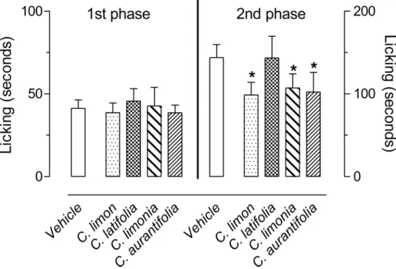
In order to evaluate possible anti-inflammatory or antinociceptive effects of the EOs obtained fromC.limon,C.aurantifolia,C.latifoliaandC.limonia, the first model used was the forma-lin-induced paw licking. After subplantar injection of formalin (2%), an intense licking behav-ior divided in two distinct phases was observed. The first phase developed during the first 5 minutes after formalin injection, with mice in the control group continuing to lick the paw for 41.3 ± 5 seconds. The second phase developed between 15 and 30 minutes after formalin injec-tion, with mice continuing to lick the injected paw for 143.8 ± 15.7 seconds. None of EO inhib-ited the 1stphase, while pre-treatment of the mice with 100 mg/kg of each EO resulted in a significant inhibition of the 2ndphase of formalin-induced licking for theC.limon,C.limonia
andC.aurantifoliaEOs (Fig 1). To rule out a possible central antinociceptive activity from the EOs, we also evaluated their effects in the hot plate model. Even the highest dose (100 mg/kg) of each EO did not demonstrate any antinociceptive activity in this model, indicating that there is no central effect (S1 Fig).
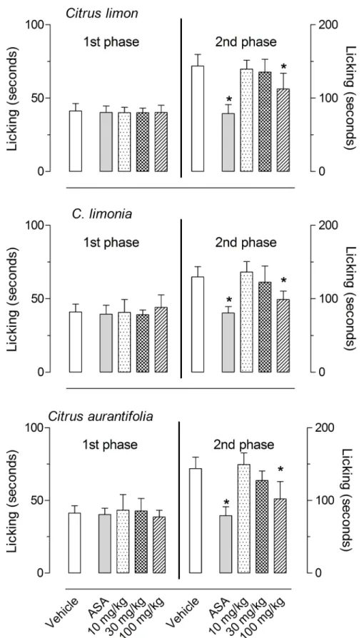
Based on the results of the formalin-induced licking behavior, we decided to further test smaller doses of those EOs that demonstrated a significant effect (i.e.,C.limon,C.limoniaand
response. The positive control group used (acetylsalicylic acid, ASA) significantly reduced lick-ing-response at the 2ndphase.
Next, we analyzed the capacity of the EOs to reduce cell migration into the subcutaneous air pouch (SAP) after the injection of carrageenan. The results obtained in this model show thatC.
Table 1. Chemical composition of peel essential oils of fourCitrussamples.
Compound KI cal. KI lit. C.limon C.aurantifolia C.limonia C.latifolia
Percent relative area
α-thujene 930 932 0.7 0.1 0.4 0.2
α-pinene 939 939 0.7 0.6 2 1.5
camphene 954 954 0.1 - - 0.3
sabinene 975 978 3.4 1.3 1.2
-β-pinene 979 982 13.1 8.5 9 5.1
myrcene 991 992 2.7 1 1.9 1.6
α-phellandrene 1003 1004 - - - 0.2
α-terpinene 1017 1019 0.5 0.3 0.4 0.6
r-cymene 1026 1028 0.3 0.5 0.7 0.1
limonene 1029 1033 53.9 31.1 65.7 35.4
β-cymene 1050 1051 - - 0.1 0.2
γ-terpinene 1060 1062 12.2 10.8 12.3 12.1
terpinolene 1089 1090 0.8 0.8 0.7 1.4
linalool 1097 1099 0.3 1.1 0.2 1.7
nonanal 1101 - 0.2 - -
-citronelal 1153 1153 - - 0.3
-terpinen-4-ol 1177 1178 0.5 1.6 1 2.3
α-terpineol 1189 1190 0.8 1.6 1.7 6.5
n-decanal 1202 1204 - 0.9 0.1
-nerol 1230 1229 0.2 1.1 - 1.1
neral 1238 1238 1.7 7.1 - 2.6
geraniol 1253 1256 0.3 1.8 - 1.3
geranial 1267 1271 2.2 9.6 - 3.6
δ-elemene 1338 1339 0.5 - 0.2
neryl acetate 1362 1365 0.8 0.2 - 2.3
geranil acetate 1381 1381 0.4 - - 0.8
β-elemene 1391 1392 - 0.3 -
-cis-α-bergamotene 1413 1415 - - - 0.2
β-caryophyllene 1419 1420 0.3 2.4 0.2 2
trans-α-bergamotene 1435 1437 0.4 2.1 0.4 3.4
α-humulene 1455 1454 - 0.3 - 0.2
(E)-β-farnesene 1458 1458 - 0.2 -
-germacrene D 1485 1481 - 0.8 0.2
-α-bisabolene 1506 1506 - - - 0.6
β-bisabolene 1506 1509 0.6 6.8 0.7 6.5
δ-cadinene 1523 1523 - 0.6 0.1
-germacrene B 1561 1557 - 1.8 - 0.6
α-bisabolol 1685 1682 - - - 0.3
Total 97.2 95.7 99.4 94.8
KI cal.: Calculated Kovats indexes; KI lit.
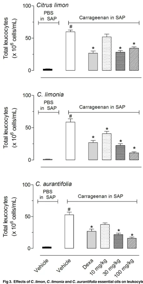
limonandC.aurantifoliasignificantly reduced cell migration after pre-treatment of animals with 30 or 100 mg/kg, whileC.limoniademonstrated a significant inhibitory effect with all three doses tested (10, 30 and 100 mg/kg). The effects observed with 30 or 100 mg/kg doses were similar to those obtained with the positive group, dexamethasone (Dexa, 1.5 mg/kg, i.p.)
(Fig 3). The differential cell count of exudates demonstrated that more than 90% of leukocytes
that migrate to the SAP was composed by polymorphonuclear neutrophils. Reduction in cells number inhibited both mononuclear and polymorphonuclear leukocytes without distinction between then (data not shown).
It seems that a maximal inhibitory effect occurs with 30 and 100 mg/kg doses since there were any statistical differences between these doses. For this reason we did not increase the doses more than 100 mg/kg.
Because both EOs significantly reduced cell migration into the SAP, we decided to further analyze other parameters present in the inflammatory processes induced by carrageenan. We therefore measured the amount of nitric oxide (NO) produced and the amount of protein extravasated to the exudate in the cavity. BothC.limon,C.limoniaandC.aurantifoliaEOs sig-nificantly reduced the amount of protein extravasated and NO produced in all three doses eval-uated with results similar to those obtained after pretreatment of animals with dexamethasone (Fig 4).
Fig 5shows the results of the cytokine quantification in the SAP exudate. Carrageenan
injec-tion in the SAP induced 11.6, 22 and 15.4-fold increases in cytokines levels. Pretreatment with 30 or 100 mg/kg dose ofC.limonsignificantly reduced the amount of IL-1βin the SAP, and all three doses inhibited TNF-αand IFN-γproduction.C.limoniapretreatment reduced IL-1β
and TNF-αproduction at 30 or 100 mg/kg doses, while IFN-γproduction was reduced with all
Fig 1. Effects ofC.limon,C.latifolia,C.limoniaandC.aurantifoliaessential oils on the formalin-induced licking response in mice.Animals were pre-treated with oral doses of 100 mg/kg dose of each essential oil or vehicle. The results are presented as the mean±S.D. (n = 6 per group) of the time that the animal spent licking the formalin-injected paw. Statistical significance was calculated by ANOVA followed by Bonferroni's test.*P<0.05 when compared to vehicle-treated mice.
Fig 2. Effects ofC.limon,C.limoniaandC.aurantifoliaessential oils on the formalin-induced licking response in mice.Animals were pre-treated with oral doses (10, 30 or 100 mg/kg) of each essential oil, acetylsalicylic acid (ASA, 100 mg/kg) or vehicle. The results are presented as the mean±S.D. (n = 7 per group) of the time that the animal spent licking the formalin-injected paw. Statistical significance was calculated by ANOVA followed by Bonferroni's test.*P<0.05 when compared to vehicle-treated mice.
three doses and almost totally blocked with the highest doses (30 and 100 mg/kg). Levels of IL-1βin the exsudates were reduced with pretreatment of mice with higher dose ofC.aurantifolia
EO (100 mg/kg). While, doses of 30 and 100 mg/kg of this EO significantly reduced levels of TNF-αand IFN-γ. Pretreatment of mice with dexamethasone also significantly reduce cytokine levels.
As the EOs significantly reduced cell migration into the SAP, we decided to evaluate a possi-ble cytotoxic effect against blood and bone marrow leukocytes. There was no alteration in the number of leukocytes counted, even 24 h after oral administration of dexamethasone or 100 mg/kgC.limonorC.limonia. However, a significant reduction in leukocytes counts in blood and bone marrow were observed after treatment with the same dose ofC.aurantifoliaEO, sug-gesting cell toxicity (Fig 6).
The unexpected effect observed inC.aurantifolialed us to investigate the possible causes. As previously shown, all three EOs contain citral.C.aurantifoliaEO contain 9.6% of the trans isomer (or geranial) and 7.1% of the cis isomer (or neral), for a total of 16.7% citral.C.limon
EO contains 2.2% geranial and 1.7% neral for a total of 3.9% citral. To confirm that citral is responsible for the observed cytotoxicity, we tested purified citral obtained from the EO in doses of 2, 5 and 20 mg/kg. These doses corresponded to the amount obtained fromC. auranti-foliaEO. As can be observed inFig 6, all three doses of citral significantly reduced the number of leukocytes in blood and bone marrow.
Due to the cytotoxic effect against the blood and bone marrow cells we decided to evaluate the possible toxicity in behavioral parameters in mice. After treatment with 500 mg/kg of EO we did not observe neither behavioral alterations nor stomachs lesions in the animals up to the oral dose of 500 mg/kg of body weight (data not shown).
After finding high amounts of limonene in the EOs (53.9% inC.limon, 65.7% inC.limonia
and 31.1% inC.aurantifolia), we decided to evaluate pure limonene. The doses used were simi-lar to the amount found inC.aurantifoliaEO.Fig 7show that pure limonene significantly reduced the formalin-induced licking behavior only in high doses (55 mg/kg). In the SAP model, 16.5 and 55 mg/kg doses reduced cell migration to and IFN-γproduction in the pouch, whereas protein extravasation, NO and IL-1βproduction were reduced only with a 55 mg/kg dose. TNF-αlevels were inhibited by all three doses (5.5, 16.5 and 55 mg/kg). None of the doses tested influenced the total leukocyte counts in blood or bone marrow (data not shown).
Discussion
In this study we demonstrated that essential oils from several citrus species presented signifi-cant anti-inflammatory effects in different models in mice. To confirm the hypothesis that essential oil (EO) fromC.limon,C.latifolia,C.aurantifoliaandC.limoniahave antinocicep-tive effects, we tested each one on the formalin-induced licking behavior. This model is a widely-used pain model in evaluating antinociceptive and anti-inflammatory drugs. There are two phases, with the first (neurogenic phase) occurring peripherally and resulting from forma-lin activation of nociceptors located in the tissue and the second (inflammatory phase) occur-ring after the release of a multitude of molecules, such as histamine, serotonin, and bradykinin, developing an inflammatory response [25]. Our results suggest that Eos fromC.limon,C. aur-antifoliaandC.limoniahave an anti-inflammatory effect because they reduced the second
significance was calculated by ANOVA followed by Bonferroni's test.#P<0.05 when comparing vehicle treated group that received carrageenan in the SAP with vehicle-treated animals that received PBS in SAP;
*P<0.05 when comparing essential oils-treated animals with that received carrageenan in the SAP with the group that only received carrageenan in the SAP.
Fig 4. Effects ofC.limon,C.limoniaandC.aurantifoliaessential oils on carrageenan-induced protein extravasation and nitric oxide (NO) production in a subcutaneous air pouch (SAP).Animals were pre-treated with various doses (10, 30 or 100 mg/kg, p.o.) of EO, dexamethasone (5 mg/kg, i.p.) or vehicle. The results are presented as the mean±S.D. (n = 10 per group). Statistical significance was calculated by ANOVA followed by Bonferroni’s test.#P<0.05 when comparing the carrageenan-injected group with the PBS-injected group and
*P<0.05 when comparing EO or dexamethasone-treated groups with the vehicle-treated group.
phase response to formalin. This may occur through a reduction in inflammatory mediator lib-eration in mice paws or a direct action on one or more mediator receptors.
The formalin model alone cannot be used to ascertain the anti-inflammatory effects of the
Citrusspecies. We therefore used the carrageenan-induced inflammation in a subcutaneous air pouch (SAP) model. This model is characterized by a drastic increase in leukocytes, cytokines, inflammatory mediators and protein after the carrageenan injection [20]. The mechanism by which carrageenan induces the inflammatory response is complex and involves the liberation of several mediators and an increase in vascular permeability. Our results indicate that EOs induce an anti-inflammatory effect by reducing several of the parameters observed in carra-geenan-induced inflammation. These results cannot guarantee the exact local action of the EOs, but do suggest that EOs can be acting in one or more mediator systems involved in the inflammation.
During the inflammatory event there is also an increase in cytokine production. Tumor necrosis factor-α(TNF-α), interleukin-1β(IL-1β), and interferon-γ(IFN-γ) have important roles in the maintenance of the inflammatory profile [26,27]. Our results indicate that EOs have significant anti-inflammatory effects in reducing all parameters evaluated. Since IL-1β
and TNF-αregulate leukocyte migration, we can infer that a reduction in leukocyte number
Fig 5. Effects ofC.limon,C.aurantifoliaandC.limoniaessential oils on carrageenan-induced IL-1β, TNF-αand IFN-γproduction in a
subcutaneous air pouch (SAP).Animals were pre-treated with various doses (10, 30 or 100 mg/kg, p.o.) of EO, dexamethasone (5 mg/kg, i.p.) or vehicle. The results are presented as the mean±S.D. (n = 10 per group). Statistical significance was calculated by ANOVA followed by Bonferroni’s test.#P<0.05 when comparing the carrageenan-injected group with the PBS-injected group and*P<0.05 when comparing EO or dexamethasone-treated groups with the vehicle-treated group.
may have been a direct effect of EOs as well as an indirect effect resulting from the reduction in the levels of those cytokines. Taken together, these results indicate that these EOs act similarly to immunomodulators in reducing cell migration and inflammatory mediator production. Our results are in agreement with others that showed various essential oils or their constituents inhibit cytokine production. For example, 1,8-cineol inhibited TNF-αand IL-1βin human lymphocytes,α-humulene reduced TNF-αproduction, and terpinen-4-ol suppressed the pro-duction of TNF-α, IL-1β, IL-8, IL-10, and PGE2 by LPS-activated monocytes [28].C.latifolia
reduced TNF-αand IL-10 levels in zymosan-induced peritonitis [9].
Despite all EOs present limonene in there constitution a difference occurred between the inhibitions observed in the formalin-induced licking and carrageenan-induced cell migration models. A possible explanation to the differences could be the fact that both models are very different. In the first one we observe liberation of inflammatory mediators such as histamine, serotonin and bradykinin in mice paws [17]. In the second model we have the involvement of several other mediators and systems, liberation of nitrogen reactive species (ROS) and cyto-kines [19]. In this regard, one possible explanation to the fact that EOs did not demonstrate a significant inhibitory effect in the 2ndphase of formalin-induced licking model despite its con-tent of limonene could be explained by the possibility that limonene has a more pronounced effect in cells activities, cytokine and/or ROS production and not in mediators that are mainly released in the formalin-induced licking response.
Additionally,C.aurantifoliaEO demonstrated some toxicity, likely due to the presence of high amounts of citral. Others have demonstrated a toxic effect of citral. However, those groups used different models such as embryofetal [29] and carcinogenesis induction [30]. Although very different models and doses were used, our data, in our experimental conditions, indicate that citral has toxic effect in other model and indicate that this effect is not a model-specific effect. With our acute assays it is not possible to definitively conclude that there is a myelotoxi-city given that such effects are usually studied in repeated dose toximyelotoxi-city studies, as e.g. given in the FDA guideline. It seems premature to conclude definitively that there is myelotoxicity also in other conditions. The observed phenomena could also be a temporary reaction which would resume later, so that further studies in models for repeated dose toxicity and e.g. also in another animal species seem adequate. In this regard, further studies are necessary before drawing so far reaching conclusions, given the wide spread occurrence of citral in many plants as in food as in medicinal use.
We further showed thatC.limonandC.limoniahave significant effects, likely due to the presence of high amounts of limonene (53.9% and 31.1%, respectively). The fact that EO from
C.limonandC.limoniapresented significant anti-inflammatory effect probably due to the presence of limonene (53.9% and 31.1%, respectively) and EO fromC.latifoliadid not present this effect even with 35.4% limonene could be explained, at least in part, by an assumption that some chemical interactions may be occurring in intestinal tract in such a way that this EO did not present a good absorption. Or it could be that any other substance present in a different amount than in others EO could interfere with an effect. Several pharmacological effects for limonene have been documented, such asin vitroinhibition of NO and PGE2 production [31], antineoplastic [32] and anti-inflammatory in a colitis model [33], but to the best of our knowl-edge this work is the first to describe its effect as an inhibitor of cell migration and cytokine production.
when comparing citral-treated animals with carrageenan injected in the SAP with the group that received carrageenan in the SAP.
Conclusions
Our results indicate that the essential oils obtained fromCitrus limon,Citrus limoniaand Cit-rus aurantifoliademonstrate a significant anti-inflammatory effect. We also draw attention to the fact thatC.aurantifoliais rich in citral, which gives it a toxic and myelotoxic effects. In this regard, care should be taken withC.aurantifoliadue to its potential toxic effect. The essential oils compositions found for the species included in this report are inside the range of composi-tions already published for those species [3]. Additionally all the species are cultivated, mean-ing that we have homogenous genetic matrices as oil sources. However, due to the well-recognized variability in the composition of secondary metabolites caused by the susceptibility of plants to, for example, seasonal, geographical and geographical influence the potential appli-cability of these oils to treat inflammatory conditions would gain efficacy and safety after the standardization of production of these oils focusing on the best composition for therapeutic applications.
Supporting Information
S1 Fig. Effects ofC.limon,C.latifolia,C.limoniaandC.aurantifoliaessential oils on the hot plate model.Animals were orally pretreated with different doses of each essential oil or vehicle. The results are presented as mean ± S.D. (n = 6 per group) of the increase in response time relative to baseline levels. Statistical significance was calculated by ANOVA followed by Bonferroni's test.
(TIF)
Acknowledgments
We would like to thank Mr. Alan Minho for technical assistance, Dr. Graciela Donald for English revision and Instituto Vital Brazil (Niteroi, Brazil) for donation of mice.
Author Contributions
Conceived and designed the experiments: AJRS PDF. Performed the experiments: JLA DLRS MMGP DSAM CSA. Analyzed the data: JLA AJRS PDF. Contributed reagents/materials/analy-sis tools: DSAM CSA AJRS PDF. Wrote the paper: CSA DSAM AJRS PDF.
References
1. Gorinstein S, Martin-Belloso O, Park YS, Haruenkit R, Lojek A,C^ížM, et al. Comparison of some
bio-chemical characteristics of different citrus fruits. Food Chem. 2001; 74: 309–315.
2. Ariasa BA, Ramon-Lacab L. Pharmacological properties of citrus and their ancient and medieval uses in the Mediterranean region. J Ethnopharmacol. 2005; 97: 89–95. PMID:15652281
3. Bisignano G, Cimino F, Saija A. Biological activities of citrus essential oils. In:Citrus Oils:Composition,
Advanced Analytical Techniques,Contaminants,and Biological Activity, Giovanni D., Luigi M., Eds.; CRC Press (Taylor and Francis): Florida, USA, 2011.
4. Belletti N, Ndagijimana M, Sisto C, Guerzoni ME, Lanciotti R, Gardini F. Evaluation of the antimicrobial activity of citrus essences onSaccharomyces cerevisiae. J Agric Food Chem. 2004; 52: 6932–6938. PMID:15537299
5. Romano L, Battaglia F, Masucci L, Posteraro SB, Plotti G, Zanetti S, et al. In vitro activity of bergamot natural essence and furocoumarin-free and distilled extracts, and their associations with boric acid, against clinical yeast isolates. J Antimic Chemother. 2005; 55: 110–114.
7. Pultrini AM, Galindo LA, Costa M. Effects of the essential oil fromCitrus aurantiumL. in experimental anxiety models in mice. Life Sciences 2006; 78: 1720–1725. PMID:16253279
8. Berliocchi L, Russo R, Levato A, Fratto V, Bagetta G, Sakurada S, et al. (–)-Linalol attenuates allodynia in neuropathic pain induced by spinal nerve ligation in c57/bl6 mice. Int Rev Neurobiol. 2009; 85: 221–
35. doi:10.1016/S0074-7742(09)85017-4PMID:19607973
9. Kummer R, Queiroz FCF, Estevão-Silva CF, Grespan R, Silva EL, Amado CAB, et al. Evaluation of anti-Inflammatory activity ofCitrus latifoliaTanaka essential oil and limonene in experimental mouse models. Evidence-Based Complem Altern Med. 2013; Article ID 859083. 1–8.
10. Misharina TA, Samusenko AL. Antioxidant properties of essential oils from lemon, grapefruit, coriander, clove, and their mixtures. Prikl Biokhim Mikrobiol. 2008; 44: 482–486. PMID:18924419
11. Lota ML, Serra DR, Tomi FL, Jacquemond C, Casanova J. Volatile components of peel and leaf oils of lemon and lime species. J Agric Food Chem. 2002; 50: 796–805. PMID:11829647
12. Mehl F, Marti G, Boccard J, Debrus B, Merle P, Delort E, et al. Differentiation of lemon essential oil based on volatile and non-volatile fractions with various analytical techniques: a metabolomic approach. Food Chem. 2014; 143: 325–335. doi:10.1016/j.foodchem.2013.07.125PMID:24054247 13. Adams RP. Identification of Essential Oil Components by Gas Chromatography/Mass Spectrometry,
4th ed. Allured: Carol Stream, IL, USA, 2007.
14. Wehrli FW, Nishida T. The use of carbon-13 nuclear magnetic resonance spectroscopy in natural prod-ucts chemistry. In: Progress in the Chemistry of Organic Natural Prodprod-ucts, Herz W., Grisebach H., Kirby G.W., Eds.; Springer-Verlag: Wien, 1979, 36, pp. 1–229.
15. Sahley TL, Berntson GG. Antinociceptive effects of central and systemic administration of nicotine in the rat. Psychopharmacol. 1979; 65: 279–283.
16. Matheus ME, Berrondo LF, Vieitas EC, Menezes FS, Fernandes PD. Evaluation of the antinociceptive properties fromBrillantaisia palisotiiLindau stems extracts. J Ethnopharmacol. 2005; 102: 377–381. PMID:16076537
17. Hunskaar S, Hole K. The formalin test in mice: dissociation between inflammatory and non-inflamma-tory pain. Pain 1987; 30: 103–114. PMID:3614974
18. Gomes NM, Rezende CM, Fontes SP, Matheus ME, Fernandes PD. Antinociceptive activity of amazo-nian copaiba oils. J Ethnopharmacol. 2007; 109: 486–492. PMID:17029841
19. Sedgwick AD, Sin YM, Mackay AR, Al-Duaij A, Willoughby DA. Studies into the mode of action of non-steroidal anti-inflammatory drugs using a model of facsimile synovium. J Pharm Pharmacol 1984; 36: 171–174. PMID:6144751
20. Raymundo LJRP, Guilhon CC, Alviano DS, Matheus ME, Antoniolli AR, Cavalcanti SCH, et al. Charac-terization of the anti-inflammatory and antinociceptive activities of theHyptis pectinata(L.) Poit essen-tial oil. J Ethnopharmacol. 2011; 134: 725–732. doi:10.1016/j.jep.2011.01.027PMID:21277967 21. Bartholomew B. A rapid method for the assay of nitrate in urine using the nitrate reductase enzyme of
E.coli. Food Chem Toxicol. 1984; 22: 541–543. PMID:6378739
22. Green LC, Wagner DA, Glogowski J, Skipper PL, Wisnok JS, Tannenbaum SR. Analysis of nitrate, nitrite, and [15N] nitrate in biological fluids. Anal Biochem. 1982; 126: 131
–138. PMID:7181105 23. Lorke D. A new approach to practical acute toxicity testing. Arch Toxicol. 1983; 54: 275–287. PMID:
6667118
24. Rouseff R. Citrus flavour in flavours and fragrances: Chemistry, bioprocessing and sustainability. In: Berger R.G. (Ed.), Springer, Hannover, 2011.
25. Parada CA, Tambeli CH, Cunha FQ, Ferreira SH. The major role of peripheral release of histamine and 5-hydroxytryptamine in formalin-induced nociception. Neuroscience 2001; 102: 937–944. PMID:
11182255
26. Parodi A, Sanguineti R, Catalano M, Penco S, Pronzato MA, Scanarotti C, et al. A comparative study of leukaemia inhibitory factor and interleukin-1αintracellular content in a human keratinocyte cell line after exposure to cosmetic fragrances and sodium dodecyl sulphate. Toxicol Lett. 2010; 192: 101–107. doi:
10.1016/j.toxlet.2009.10.013PMID:19878710
27. Moermans C, Heinen V, Nguyen M, Henket M, Sele J, Manise M, et al. Local and systemic cellular inflammation and cytokine release in chronic obstructive pulmonary disease. Cytokine 2011; 56: 298–
304. doi:10.1016/j.cyto.2011.07.010PMID:21880505
28. Hirota R, Roger NN, Nakamura H, Song HS, Sawamura M, Suganuma N. Anti-inflammatory effects of limonene from yuzu (citrus junos tanaka) essential oil on eosinophils. J Food Sci. 2010; 75: H87–H92. doi:10.1111/j.1750-3841.2010.01541.xPMID:20492298
30. Ress NB, Hailey JR, Maronpot RR, Bucher JR, Travlos GS, Haseman J, et al. Toxicology and carcino-genesis studies of microencapsulated citral in rats and mice. Toxicol Sci. 2003; 71: 198–206. PMID:
12563105
31. Rehman MU, Tahir M, Khan AQ, Khan R, Lateef OOH, Hassan SK, et al. D-limonene suppresses doxo-rubicin-induced oxidative stress and inflammation via repression of COX-2, iNOS, and NFκB in kidneys of Wistar rats. Exp Biol Med. 2014; 239: 465–476.
32. Kapoor S. D-Limonene: an emerging antineoplastic agent. Hum Exp Toxicol. 2013; 32: 1228. doi:10. 1177/0960327113489053PMID:23716733