Submitted12 September 2015
Accepted 3 November 2015
Published22 December 2015
Corresponding author
Steffen Harzsch, [email protected]
Academic editor
Darren Williams
Additional Information and Declarations can be found on page 39
DOI10.7717/peerj.1433
Copyright
2015 Krieger et al.
Distributed under
Creative Commons CC-BY 4.0
OPEN ACCESS
Comparative analyses of olfactory
systems in terrestrial crabs (Brachyura):
evidence for aerial olfaction?
Jakob Krieger1,*, Philipp Braun1,*, Nicole T. Rivera2, Christoph D. Schubart2,
Carsten H.G. Müller3and Steffen Harzsch1
1Zoological Institute and Museum, Department of Cytology and Evolutionary Biology, Ernst-Moritz-Arndt Universität Greifswald, Greifswald, Germany
2Institute for Zoology, Department of Zoology & Evolution, Universität Regensburg, Regensburg, Germany
3Zoological Institute and Museum, Department of General and Systematic Zoology, Ernst-Moritz-Arndt
Universität Greifswald, Greifswald, Germany *These authors contributed equally to this work.
ABSTRACT
Adaptations to a terrestrial lifestyle occurred convergently multiple times during the evolution of the arthropods. This holds also true for the ‘‘true crabs’’ (Brachyura), a taxon that includes several lineages that invaded land independently. During an evolutionary transition from sea to land, animals have to develop a variety of physiological and anatomical adaptations to a terrestrial life style related to respiration, reproduction, development, circulation, ion and water balance. In addition, sensory systems that function in air instead of in water are essential for an animal’s life on land. Besides vision and mechanosensory systems, on land, the chemical senses have to be modified substantially in comparison to their function in water. Among arthropods, insects are the most successful ones to evolve aerial olfaction. Various aspects of terrestrial adaptation have also been analyzed in those crustacean lineages that evolved terrestrial representatives including the taxa Anomala, Brachyura, Amphipoda, and Isopoda. We are interested in how the chemical senses of terrestrial crustaceans are modified to function in air. Therefore, in this study, we analyzed the brains and more specifically the structure of the olfactory system of representatives of brachyuran crabs that display different degrees of terrestriality, from exclusively marine to mainly terrestrial. The methods we used included immunohistochemistry, detection of autofluorescence- and confocal microscopy, as well as three-dimensional reconstruction and morphometry. Our comparative approach shows that both the peripheral and central olfactory pathways are reduced in terrestrial members in comparison to their marine relatives, suggesting a limited function of their olfactory system on land. We conclude that for arthropod lineages that invaded land, evolving aerial olfaction is no trivial task.
SubjectsEvolutionary Studies, Neuroscience, Zoology
Keywords Land crabs, Terrestrialization, Decapods, Neuroanatomy, Immunohistochemistry, Morphometry, Sexual dimorphism, Crustaceans
INTRODUCTION
Land-living crustaceans are fascinating animals that adapted during a relatively short evolutionary time period to a number of highly diverse terrestrial habitats in which they
have become highly successful, and in some cases the predominant life forms (Hansson et al., 2011). Representatives in not less than five major malacostracan crustacean taxa have conquered the terrestrial habitat independently. Because the successful transition from a marine or freshwater habitat to terrestrial life requires a number of physiological adapta-tions which are important for survival out of water, terrestrial crustaceans constitute an excellent animal group to study evolutionary adaptations related to the invasion of land. Such adaptations include changes to gas exchange, salt and water balance, nitrogenous excretion, thermoregulation, molting, and reproduction (reviews inBliss & Mantel, 1968;Burggren & McMahon, 1988;Greenaway, 1988;Greenaway, 1999;Greenaway, 2003;
McMahon & Burggren, 1988;Powers & Bliss, 1983). The Brachyura (short-tailed crabs or ‘‘true crabs’’) include several lineages that invaded land. The degree of terrestrial adaptation in crustaceans has been categorized into five classes ranging fromT1toT5
depending on the degree of independence from immersion in water and the animal’s need to access water for reproduction (Hartnoll, 1988;Greenaway, 1999;Powers & Bliss, 1983; but seeSchubart et al., 2000for an alternative classification). In this traditional classification, several brachyuran taxa have been ranked within the two highest grades of terrestrial adaptation (e.g., Gecacinidae, and some representatives of the Sesarmidae, Potamidae, Gecarcinidae, Potamonautidae, Pseudothelphusidae, and Trichodactylidae), whereas many amphibious freshwater forms and supra-littoral species are ranked in less terrestrialized categories.
The phylogenetic relationships of Brachyura and the systematics of brachyuran taxa are the topic of ongoing research (Scholtz & Richter, 1995;Dixon, Ahyong & Schram, 2003;
Ahyong & O’Meally, 2004;Ng, Guinot & Davie, 2008;Tsang et al., 2014;Brösing, Richter & Scholtz, 2007;Fig. 1) but there is increasing evidence that the conquest of land occurred several times independently amongst Brachyura as suggested byPowers & Bliss (1983)
andHartnoll (1988). All members of the Gecarcinidae (with the exception of the genus
Epigrapsus) and representatives of the grapisd genusGeograpsushave achieved complete
terrestriality as adults, but larval development, which is not abbreviated, takes place in the oceans. The family Sesarmidae (sensu Schubart, Cuesta & Felder, 2002) includes crabs such asSesarma jarvisi,S. cookei, andS. verleyi, which radiated into a broad range of terrestrial habitats, including mountainous rain forest and caves on Jamaica (Wolcott, 1988;Schubart, Diesel & Hedges, 1998;Diesel & Schubart, 2000;Diesel, Schubart & Schuh, 2000). The bromeliad crabMetopaulias depressusraises its offspring in water-filled leaf axils of bromeliads and certainly has evolved one of the most notable reproductive adaptations to terrestrial habitats (Diesel & Schubart, 2000), but remains immersed in water for extended time periods. The Ocypodidae comprise the generaOcypode, Uca,
andUcides;and some of its representatives were qualified to reach terrestrial gradeT3by
Powers & Bliss (1983). In the phylogenetic analysis based on stomach ossicles byBrösing, Richter & Scholtz (2007), several taxa with terrestrial tendencies, the Potamonautidae, Ocypodidae, Gecarcinidae, Grapsidae, and Mictyridae cluster together with other taxa in the proposed taxon Neobrachyura, suggesting a close relationship of those brachyuran groups which include terrestrial forms, but this grouping is not recovered in the newest and most comprehensive phylogeny byTsang et al. (2014), so that it appears to be based
Figure 1 Simplified phylogenetic relationships among Brachyura afterTsang et al. (2014). For simpli-fication some major brachyuran clades are grouped. Note that brachyuran clades including representatives showing higher degrees of terrestrial adaptation (Ti>T2) are indicated by orange circles. Groups of
inves-tigated species are highlighted by indented and larger (bold) letters and are color-coded according to their lifestyles (pale blue, freshwater crabs; dark blue, marine; brown, terrestrial).
on convergences. The paraphyletic superfamily Grapsoidea (comprising 88 genera with over 480 species including the Gecarcinidae (6 genera with 19 species) include intertidal to supratidal as well as limnic forms in addition to terrestrial ones, so that there is increasing evidence that the colonization of inland habitats evolved in several lineages (Schubart et al., 2000;Schubart et al., 2006;Tsang et al., 2014).
An essential physiological adaptation to master a terrestrial lifestyle during and after an evolutionary transition from sea to land includes the need for sensory organs to function in air instead of in water (Greenaway, 1999;Greenaway, 2003;Hansson et al., 2011). Mechanosensory systems must detect stimuli that propagate in airversusin water, and visual systems must operate in media with different refractive properties. In olfaction, a transition from sea to land means that molecules need to be detected in or bound from gas phase instead of being transmitted directly from one water solution (e.g., sea water) into another one (receptor lymphs). Marine crustaceans live in a world full of chemical information. It is well established that they use chemical cues to locate mates, signal dominance, recognize individual conspecifics, find favored food and appropriate habitats, and assess threats such as the presence of predators (reviews e.g., Derby et al., 2001;Grasso & Basil, 2002;Derby & Sorensen, 2008;Hazlett, 2011;Thiel & Breithaupt, 2011;Wyatt, 2011;Derby & Weissburg, 2014). However, aquaticversusland-living animals must detect highly different semiochemicals, because the medium places different demands on the compounds used. In water, molecules have to be more or less water-soluble and stable
enough to travel from one individual to another. On land, semiochemicals have to be light enough to form a gas in the ambient temperatures where animals live (discussed inStensmyr et al., 2005). These molecules also have to be sufficiently chemically stable to reach the sensory receptor cells. These new selection pressures take part together in reshaping the sense of smell during the invasion of new, terrestrial habitats (reviews
Hansson et al., 2011;Hay, 2011;Weissburg, 2011).
Malacostracan crustaceans living in aquatic habitats use several systems for de-tecting chemicals, and these are distributed over their entire body surface, walking appendages, and mouthparts, but are also concentrated on two pairs of antennae (reviews e.g., Hallberg, Johansson & Elofsson, 1992;Hallberg & Skog, 2011;Schmidt & Mellon, 2011). The first antennal pair (the antennules) is equipped with specialized olfactory sensillae (aesthetascs) in addition to bimodal chemo- and mechanosensilla (contact chemoreceptors), whereas the second pair of antennae is only equipped with the latter. The tips of the first antennae (more specifically the lateral flagella) bear a tuft region with arrays of aesthetascs that house branched dendrites of olfactory sensory neurons (reviews byHallberg, Johansson & Elofsson, 1992;Hallberg & Hansson, 1999;Mellon Jr, 2007;Hallberg & Skog, 2011;Schmidt & Mellon, 2011;Derby & Weissburg, 2014).
Schmidt & Mellon (2011)pointed out that in aquatic crustaceans, chemical information is received and processed in two fundamentally different modes. The first mode is ‘‘olfaction’’ defined as chemoreception mediated by the aesthetasc pathway; the second mode is called ‘‘distributed chemoreception’’ defined as chemoreception mediated by contact chemoreceptors on all appendages (Schmidt & Mellon, 2011). Chemosensory neurons associated with the aesthetascsversusthe contact chemoreceptors on the first antenna of malacostracan crustaceans innervate distinct regions in the brain. The axons of the olfactory sensory neurons associated with the aesthetascs target the deutocerebral chemosensory lobes (DCLs; also called olfactory lobes), whereas the axons associated with non-aesthetasc sensilla innervate the lateral antenna 1 neuropil (LAN;Schachtner, Schmidt & Homberg, 2005;Schmidt & Mellon, 2011;Strausfeld, 2012;Loesel et al., 2013). As for the different functions of these two modes of aquatic chemoreception,Schmidt & Mellon, (2011)suggested that ‘‘the essence of olfaction’’ is to provide a detailed representation of the complex chemical environment integrating chemical signals from a variety of interesting sources (. . . ) without reference to the location of stimuli (. . . ). In contrast, the essence of ‘‘distributed chemoreception’’ is to form representations of only few key chemicals (food-related chemicals, pheromones) within a somatotopic context provided by mechanoreception. The integration of chemo- and mechanosensory information permits pinpointing the location of chemical stimuli . . . ’’
Independently of insects, chelicerates, and myriapods, terrestrial Crustacea provide a fascinating chance to look on a wonderful evolutionary experiment by analyzing which potential alternative solutions arthropods have evolved to explore the terrestrial olfactory landscape (Hansson et al., 2011). We have previously analyzed the olfactory system of land hermit crabs (Anomala, Coenobitidae) including their peripheral (Stensmyr et al., 2005;
Tuchina et al., 2014) and central olfactory pathway (Harzsch & Hansson, 2008;Krieger et al., 2010;Polanska et al., 2012;Wolff et al., 2012;Tuchina et al., 2015), in addition to
behavioral and physiological aspects (Stensmyr et al., 2005;Krång et al., 2012). These studies provided evidence for coenobitids having a superb sense of aerial olfaction. In this paper, we ask whether terrestrial brachyuran crabs also evolved the neuronal basis for aerial olfaction. Therefore, we compare the anatomy of the central olfactory pathway of selected species of brachyuran crustaceans featuring a rather terrestrial lifestyle to that of their marine relatives.
MATERIAL AND METHODS
Experimental animalsWe analyzed representatives of several different species of brachyurans representing aquatic species (four exclusively marine and one freshwater crab species) as well as four brachyuran species featuring different grades of terrestrial adaptation (Table 1; Figs. 2and3). For simplification, the four latter brachyurans are referred as terrestrial brachyurans throughout this text, although all nine species feature terrestrial adaptions at various degrees (Table 1). After shipping, living specimens ofCardisoma armatum,
Geosesarma tiomanicum, andUca tangeriwere kept in tanks providing both a water and
a land part. Husbandry as well as observation and documentation of these species were conducted between 5 and 14 days until dissection in the laboratory. Before dissection, animals were sexed, and the carapace width as well as the wet weight of each animal was measured. The collection of specimens ofGecarcoidea nataliswas permitted by Christmas Island National Park (Australian Government; Department of the Environment; Parks Australia; Permit No.: AU_COM 2010-090-1).
Analysis of antennae and aesthetascs
The first pairs (antennules) and the second pairs (antennae) of post-ocular appendages were cut off prior to brain dissection and were transferred into 70% ethanol (G.
na-talisand two animals ofG. tiomanicum) or in 2% glutaraldehyde in 0.1 M phosphate
buffered saline (PBS; further specimens ofG. tiomanicum,C. armatumandU. tangeri). Micrographs of these appendages were documented in PBS with a Nikon Eclipse 90i microscope equipped with a digital camera (Nikon DS2-MBWc) and analyzed by the use of the software package NIS-Elements AR. Cuticular auto-fluorescence of the first and second antennae was excited with ultraviolet light (UV) with a wavelength of 340– 380 nm eliciting light emissions with a wavelength of 435–485 nm. Aesthetascs of marine specimens such as ofPagurus bernhardus(Linnaeus, 1758),Carcinus maenas,Xantho hydrophilus,X. poressaandPercnon gibbesiwere cut off from the lateral flagella with a razor blade and counted on an object slide using UV-excitation as well as bright field illumination.
Histochemistry, immunohistochemistry, and microscopy
The animals were anaesthertized by cooling on ice for 1 h before dissection. Follow-ing the protocol byOtt (2008), the dissected brains were fixedin totofor approxi-mately 20 h (room temperature) in 3.7% formaldehyde/zinc-fixative (the ready-to-use formaldehyde/zinc-fixative was obtainedviaElectron Microscopy Sciences. Cat. No.
Table 1 Investigated species including the sexes, numbers, grades of terrestriality (T1–T5, whereT1is the lowest grade andT5the highest), and origins of specimens.
Species Taxon Sex (n ind.) Grade(s) of terrestrial
adaptation
Source/origin;
Cardisoma armatum, Herklots, 1851 Gecarcinidae ♂(5) T3 https://www.interaquaristik.de
Gecarcoidea natalis, (Pocock, 1888) Gecarcinidae ♀(2);♂(2) T4 Christmas Island (Australia)
Geosesarma tiomanicum,Ng, 1986 Sesarmidae ♀(2);♂(2) T5 https://www.interaquaristik.de
Uca tangeri, (Eydoux, 1935) Ocypodidae ♀(2);♂(2) T2–T3 www.tropicwater.eu
Epilobocera sinuatifrons,Rathbun 1866 Pseudothelphusidae n.a. (4) T2–T3 Guajataca, Puerto Rico
Carcinus maenas, (Linnaeus, 1758) Portunidae ♀(4);♂(11) T1 Marine Science Center in Rostock (Germany)
Percnon gibbesi, (Milne-Edwards, 1853) Percnidae ♀(1);♂(1) T1 Mediterranean, Cala Llenya (Ibiza, Spain)
Xantho hydrophilus, (Herbst, 1790) Xanthidae ♀(4);♂(2) T1 Mediterranean, Cala Llenya (Ibiza, Spain)
Xantho poressa, (Olivi, 1792) Xanthidae ♀(2);♂(2) T1 Mediterranean, Cala Llenya (Ibiza, Spain)
Pagurus bernhardus(Linnaeus, 1758) Paguridae n.a. (1) T1 North Atlantic Ocean, Roscoff (France)
Krieg
er
e
t
al.
(2015),
P
eerJ
,
DOI
10.7717/peerj.1433
Figure 2 Brachyuran species analyzed.Portraits of individuals of investigated brachyuran species in living state. (A–C) Marine Brachyura: (A)Carcinus maenas(Boiensdorf, Baltic Sea, 1998), (B)Percnon gibbesi(infralittoral rock bottom, 1 m depth, Cala Llenya, Ibiza, Spain, 2013), (C)Xantho hydrophilus (in-fralittoral rock bottom, 5 m depth, Cala Llenya, Spain, 2012). (D–H) Terrestrial Brachyura: (D) Epilobo-cera sinuatifrons(Guajataca, Puerto Rico, 2004), (E)Gecarcoidea natalis(At the Pink House, Christmas Is-land, Australia, 2011), (F)Cardisoma armatum, (G)Geosesarma tiomanicum, (H)Uca tangeri. Portraits of (F–H) in living state were taken in the laboratory in Greifswald in 2013.
15675). For whole-mount preparations, the brains and eyestalk ganglia were dissected, and the retina including all pigments was removed. The whole-mounts were washed three times in HEPES-buffered saline (HBS) for 15 min, subsequently transferred to Dent’s fixative (80% methanol/20% DMSO), and post-fixated for two hours at room temperature. Specimens were then transferred to 100% methanol and stored overnight in the refrigerator and rehydrated stepwise for 10 min each in 90%, 70%, 50%, 30% methanol in 0.1 M Tris–HCl-buffer, and finally in pure 0.1 M Tris–HCl-buffer (pH 7.4). Alternatively, for preparing horizontal brain sections, after anaesthetizing the animals by cooling on ice for 1 h, the brains were dissected and fixed in 4% paraformaldehyde (PFA) in 0.1 M PBS overnight. The dissected brains were washed for 4 h in several changes of PBS and sectioned (80–100µm sections) horizontally at room temperature using a
vibratome (Zeiss Hyrax V590; Carl Zeiss, Oberkochen, Germany). For permeation of cell
Figure 3 Comparative draft of studied animals: their brains, first antennae, deutocerebral chemosen-sory lobes (DCLs) and olfactory glomeruli.Note that drawings are equally scaled in each line. Each col-umn corresponds to same species as follows:Carcinus maenas,Percnon gibbesi,Xantho hydrophilus, as rep-resentatives for marine brachyurans are given and opposed (separated by a dashed line) toGecarcoidea na-talis,Cardisoma armatum,Geosesarma tiomanicum, andUca tangerirepresenting brachyuran species fea-turing terrestrial lifestyles to different degrees. Note that for animals feafea-turing a markedly size-specific sex-ual dimorphism, only the males are drawn. (A) Dorsal view of habitus in all studied species. (B) Distal an-tennomeres of the first antennae (antennules) of all species featuring the minor median and major lateral flagella which bear the aeasthetascs. (C) Outlines of central brains based on the synapsin immunoreactiv-ity. The lateral protocerebrum and nerves are not displayed. (D) Outlines of DCLs and peripheral arrange-ment of olfactory glomeruli as they appear in horizontal sections. Note that the position of DCL within the brain is indicated inC. maenasin line C. (E) Examples of shape and organization of randomly chosen olfactory glomeruli of all studied species as they appear in horizontal sections.
membranes, both brain whole-mounts as well as brain sections were then preincubated for 90 min in PBS-TX (1% Bovine-Serum-Albumine, 0.3% TritonX-100, 0.05% Na-acide, in 0.1 M PBS; pH 7.4). In contrast to the protocol ofOtt (2008), PBS-TX was used instead of PBSd-NGS. Finally, the samples were incubated at 4◦C for 84 h (whole-mounts) or overnight (sections) in the primary antisera. The following sets of reagents were used (compareKrieger et al., 2012):
Set A:rabbit anti-Dip-allatostatin 1 (AST-A; final dilution 1:2,000 in PBS-TX; antibody provided by H Agricola, Friedrich-Schiller Universität Jena, Germany); monoclonal mouse anti-synapsin ‘‘SYNORF1’’ antibody (final dilution 1:30 in PBS-TX; antibody provided by E Buchner, Universität Würzburg, Germany) detected by anti- mouse Cy3 (CyTM3-conjugated AffiniPure Goat Anti-Mouse IgG Antibody; Jackson ImmunoRe-search Laboratories Inc., West Grove, PA, USA).
Set B:polyclonal rabbit anti-FMRFamid (in PBS-TX; final dilution 1:2,000; Acris/Im-munostar; Cat. No. 20091) detected by anti-rabbit Alexa Flour 488 (IgG Antibody, invitrogen, Molecular Probes); monoclonal mouse anti-synapsin ‘‘SYNORF1’’ antibody (in PBS-TX; final dilution 1:30; antibody provided by E Buchner, Universität Würzburg,
Germany) detected by anti- mouse Cy3 (CyTM3-conjugated AffiniPure Goat Anti-Mouse IgG Antibody; Jackson ImmunoResearch Laboratories Inc., West Grove, PA, USA).
Set C:monoclonal mouse anti-synapsin ‘‘SYNORF1’’ antibody (in PBS-TX; final dilution 1:30; antibody provided by E Buchner, Universität Würzburg, Germany) detected by anti- mouse Alexa Flour 488 (IgG Antibody; Invitrogen, Waltham, MA, USA; Molecular Probes, Eugene, OR, USA); counterstain: phallotoxins conjugated to Alexa Fluor 546 (concentration 200 units/ml; Molecular Probes, Eugene, OR, USA) as a high-affinity probe for f-actin.
In all three sets, the tissues were incubated in mixture containing the secondary antisera and the nuclear marker HOECHST (33,242; 0.1µg/ml) for 2.5 days at 4◦C
(whole-mounts) or for 4 h at room temperature (sections). Finally, the brain sections were washed for at least 2 h in several changes of PBS at room temperature and mounted in MowiolR (Calbiochem) between two coverslips. After secondary antibody incubation,
the whole-mounts were dehydrated in changes of ascending glycerol concentrations (1%, 2%, 4% (2 h each), 8%, 15%, 30%, 50%, 60%, 70%, and 80% (1 h each) glycerol diluted in Tris–HCl buffer, with DMSO to 1% final concentration). After the last step of dehydration, the whole-mounts were washed twice for 30 min in 99.6% denatured ethanol. The ethanol was then underlyed by the same volume of methylsalicylate for clearing of the whole-mount brains. After the brains were cleared, the supernatant liquid was removed and the samples were and then mounted in customized chambers (a custom washer from the hardware store was glued between two coverslips as spacer) filled with methylsalicylate and sealed with MowiolR. The triple-labeled and sectioned tissues were
analyzed using a Nikon eclipse 90i microscope equipped with a digital camera (Nikon DS2-MBWc). The whole-mounts were analyzed by using a confocal laser scanning microscope (clsm; Leica TCS SP5II; Leica, Wetzlar, Germany). The pictures were then processed using the NIS-Elements AR software and Adobe Photoshop CS4. Only global picture enhancement features of Photoshop elements (black to white inversion, brightness, and contrast) were used for all experiments. Three-dimensional (3D) brain reconstructions in addition to volumetric analysis based on optical section series of clsm data were performed using the reconstruction software AmiraR (FEI Visualization
Sciences Group, Mérignac, France).
Raw data of brain section series is available fromhttps://www.morphdbase.deunder the ‘‘media" tab under a combination of the short title ‘‘Krieger’’ and an identifier according to the species and ID of the specimen.
Three-dimensional reconstructions of brains and substructures are based on tomogra-phies of three specimens per species forC. armatum,G. tiomanicumandU. tangeri. For each specimen, surfaces of one DCL including the corresponding olfactory glomeruli and the ipsilateral AcN were generated by manual labeling. Finally, the computed 3D surfaces were slightly smoothed and resulting parameters such as the glomerular number and volume as well as the volume of the whole DCL were analyzed.
Antibody specificity Synapsin
The monoclonal mouse anti-Drosophilasynapsin ‘‘SYNORF1’’ antibody (provided by E Buchner, Universität Würzburg, Germany) was raised against aDrosophilaGST-synapsin fusion protein and recognizes at least four synapsin isoforms (ca. 70, 74, 80, and 143 kDa) in western blots ofDrosophilahead homogenates (Klagges et al., 1996). In western
blot analysis of crayfish homogenates, this antibody stains a single band at ca. 75 kDa (seeSullivan et al., 2007).Harzsch & Hansson (2008)conducted western blot analysis comparing brain tissue ofDrosophilaand the hermit crabCoenobita clypeatuswhich is closely related to the species studied in this contribution. The antibody provided identical results for both species, staining one strong band around 80–90 kDa and a second weaker band slightly above 148 kDa (seeHarzsch & Hansson, 2008). Their analysis strongly suggests that the epitope which SYNORF 1 recognizes is strongly conserved between the fruit fly and the hermit crab. Similar toDrosophila, the antibody consistently labels brain structures in representatives of all major subgroups of the malacostracan crustaceans (seeBeltz et al., 2003;Harzsch, Anger & Dawirs, 1997;Harzsch et al., 1998;Harzsch et al., 1999;Harzsch & Hansson, 2008;Vilpoux, Sandeman & Harzsch, 2006;Krieger et al., 2010;Krieger et al., 2012) in a pattern that is consistent with the assumption that this antibody labels synaptic neuropils in Crustacea. In the crustacean first visual neuropil (the lamina), synapsin labeling is weak compared to the other brain neuropils (Harzsch, Anger & Dawirs, 1997;Harzsch & Hansson, 2008). Similarly, inDrosophila melanogaster
labeling of the lamina is weak, because photoreceptors R1–R6 which have their synapses in the lamina contain very little of the presently known synapsin isoforms (Klagges et al., 1996). The antibody also labels neuromuscular synapses both inDrosophilaand in Crustacea (Harzsch, Anger & Dawirs, 1997). These close parallels in the labeling pattern of SYNORF1 betweenDrosophilaand various Crustacea strengthen the claim that it also recognizes crustacean synapsin homologues. This antibody even labels synaptic neuropil in an ancestral clade of protostomes, the Chaetognatha (Harzsch & Müller, 2007) suggesting that the epitope recognized by this antiserum is conserved over wide evolutionary distances.
Allatostatin A
The A-type allatostatins (A-ASTs; synonym dip-allatostatins) constitute a large family of neuropeptides that were first identified from the cockroachDiploptera punctataand that share the C-terminal motif-YXFGLamide (reviewsStay, Tobe & Bendena, 1995;
Nässel & Homberg, 2006;Stay & Tobe, 2007). In decapod crustaceans, almost 20 native A-ASTs and related peptides were initially identified from extracts of the thoracic ganglia of the shore crabCarcinus maenas(Duve et al., 1997), and shortly after several other A-ASTs were isolated from the freshwater crayfishOrconectes limosus(Dircksen et al., 1999). Meanwhile, the family of crustacean A-ASTs has substantially grown to several dozens of representatives (reviewChristie, Stemmler & Dickinson, 2010) with additional members being discovered in the prawnsPenaeus monodon(Duve et al., 2002) andMacrobrachium rosenbergii(Yin et al., 2006), in the brachyuran crabsCancer borealis(Huybrechts et al.,
2003) andCancer productus(Fu, Christie & Li, 2005),Carcinus maenas(Ma et al., 2009a), the crayfishProcambarus clarkii(Yasuda-Kamatani & Yasuda, 2006), the lobsterHomarus americanus(Cape et al., 2008;Ma et al., 2008;Ma et al., 2009b) the shrimpsLitopenaeus
vannamei(Ma et al., 2010) as well as a non-malacostracan crustacean, the copepod
Calanus finmarchicus(Christie et al., 2008).
We used an antiserum that was raised against theDiploptera punctata(Pacific beetle cockroach) A-type Dip-allatostatin I, APSGAQRLYGFGLamide, coupled to bovine thyroglobulin using glutaraldehyde (Vitzthum, Homberg & Agricola, 1996) that was kindly provided by H Agricola (Friedrich-Schiller Universität Jena, Germany) and that previously has been used to localize A-ASTs in crustacean and insect nervous systems (e.g., Vitzthum, Homberg & Agricola, 1996; Dircksen et al., 1999;Skiebe, 1999; Utting et al., 2000;Kreissl, Strasser & Galizia, 2010). Competitive ELISA with DIP-allatostatin I, II, III, IV and B2 showed that the antiserum is two orders of magnitude more sensitive to Dip-allatostatin I than to Dip-allatostatins II, III, IV, and B2 (Vitzthum, Homberg & Agricola, 1996).Vitzthum, Homberg & Agricola (1996)have reported that the antiserum displays no cross-reactivity with corazonin, CCAP, FMRFamide, leucomyosuppression, locustatachykinin 11, perisulfakinin, and proctolin as tested by non-competitive ELISA. Preadsorption of the diluted antisera against Dip-allatostatin I, GMAP andManduca sexta
allatotropin with 10 µM of their respective antigens abolished all immunostaining in
brain sections ofSchistocerca gregaria (Vitzthum, Homberg & Agricola, 1996). A sensitive competitive enzyme immunoassay (EIA) confirmed the high specificity of the antiserum for A-type Dip-allatostatin I (Dircksen et al., 1999). In the brains of the honey beeApis
mellifera, preadsorption controls with AST I and AST VI completely abolished all staining
of the antiserum (Kreissl, Strasser & Galizia, 2010).Sombke, Harzsch & Hansson (2011)
repeated a preadsorption test in Scutigera coleoptrataand preincubated the antiserum with 200µg/ml A-type allatostatin I (A9929; 16 h 4◦C; Sigma-Aldrich, St. Louis, MO,
USA) and this preincubation abolished all staining. Preadsorption of the antiserum with AST-3 was reported to abolish all labeling in the stomatogastric nervous system of the crab
Cancer pagurus, the lobsterHomarus americanusand the crayfishCherax destructor and
Procambarus clarki(Skiebe, 1999). It seems safe to assume that this antiserum most likely
binds to all A-ASTs that share a -YXFGLamide core. However, the term ‘‘allatostatin-like immunoreactivity’’ is used throughout this work, because it may possible that the antibody also binds related peptides.
RFamide-related peptides
The tetrapeptide FMRFamide and FMRFamide-related peptides (FaRPs) are prevalent among invertebrates and vertebrates and form a large neuropeptide family with more than 50 members all of which share the RFamide motif (Price & Greenberg, 1989;Greenberg & Price, 1992;Nässel, 1993;Homberg, 1994;Dockray, 2004;Nässel & Homberg, 2006;Zajac & Mollereau, 2006). In malacostracan Crustacea, at least twelve FaRPs have been identified and sequenced from crabs, shrimps, lobsters, and crayfish (Huybrechts et al., 2003;Mercier, Friedrich & Boldt, 2003), which range from seven to twelve amino acids in length and most of them share the carboxy-terminal sequence Leu-Arg-Phe-amide. The utilized
antiserum was generated in rabbit against synthetic FMRFamide (Phe-Met-Arg-Phe-amide) conjugated to bovine thyroglobulin (DiaSorin, Cat. No. 20091, Lot No. 923602). According to the manufacturer, immunohistochemistry with this antiserum are completely eliminated by pretreatment of the diluted antibody with 100µg/ml of FMRFamide.Harzsch & Hansson (2008)repeated this experiment in the anomalanCoenobita clypeatuswhich is closely related to the species studied here, specifically to the hermit crabs, and preincubated the antiserum with 100µg/ml FMRFamide (16 h, 4◦C; Sigma-Aldrich, St. Louis, MO,
USA) resulting in a complete abolishment of all staining. Because the crustacean FaRPs know so far all share the carboxy-terminal sequence LRFamide, we conclude that the DiaSorin antiserum that we used most likely labels any peptide terminating with the sequence RFamide. Therefore, we will refer to the labeled structures in our specimens as ‘‘RFamide-like immunoreactivity’’ throughout the paper.
Nomenclature
The neuroanatomical nomenclature used in this manuscript is based onSandeman et al. (1992) andRichter et al. (2010)with some modifications adopted fromHarzsch & Hansson (2008)) andLoesel et al. (2013). In favor of a consistent terminology, here we suggest avoiding the term ‘‘optic neuropils’’ (Hanström, 1925;Sombke & Harzsch, 2015) as well as ‘‘optic lobes’’ (Kenyon, 1896). Even if the Greek ‘‘optikos’’ and the Latin term ‘‘visus’’ have the identical meaning, nowadays, ‘‘optic’’ in the field of visional anatomy and physiology refers to the physically refractive components of the eye for the reception of light. To emphasize the perceptive character of these neuropils, we suggest using the term ‘‘visual neuropils’’ which is consistent with, e.g., the visual cortex in mammals, formerly also termed ‘‘optic’’ cortex (Spiller, 1898). All post-retinal components that are related to vision, such as the ‘‘optic tract’’ and the ‘‘inner’’ as well as the ‘‘outer optic chiasm’’ should be consequently renamed, too. Here, we suggest to use ‘‘visual tract’’ (VT) and the ‘‘inner’’ (iCh) as well as the ‘‘outer visual chiasm’’ (oCh) accordingly. However, for all pre-retinal components that are related to vision, the term ‘‘optic,’’ as for example in the dioptric apparatus of the ommatidia, should be maintained. We also discourage the commonly used terms ‘‘eyestalk neuropils’’ (Bliss & Welsh, 1952;Polanska, Yasuda & Harzsch, 2007), ‘‘optic ganglia’’ (Medan et al., 2015), or ‘‘eyestalk ganglia’’ (Harzsch & Dawirs, 1996;Techa & Chung, 2015), usually summarizing the visual neuropils as well as the neuropils of the TM/HN-complex, because these neuropils together can be located more proximal to the central brain and not in the eyestalk in some species, and thus are part of the central brain as exemplified below. Furthermore, the neuropils of the lateral protocerebrum (visual neuropils +TM/HN-complex) do not fulfill the definition of a ganglion (seeRichter et al., 2010). The traditional nomenclature of the visual neuropils lamina ganglionaris, medulla interna, and medulla externa has been modified as suggested byHarzsch (2002)
to lamina, medulla, and lobula. Because we could not detect any border between the cell body clusters (9) and (11) of olfactory interneurons as described inSandeman et al. (1992), we collectively refer to them as cluster (9/11) (seeKrieger et al. 2010). The term ‘‘oesophageal connective’’ and the corresponding abbreviation OC (British English) are maintained here for simplicity. The olfactory neuropil (ON or OL) is now named the
deutocerebral chemosensory lobe (DCL), and the olfactory globular tract (OGT) is now named the projection neuron tract (PNT) according toLoesel et al. (2013). Consequently, the olfactory globular tract neuropil OGTN is now named projection neuron tract neuropil (PNTN). For simplification, the neuroanatomical descriptions are kept restricted to only one hemisphere of the brain and hold true for all specimens studied if not stated otherwise.
The data presented in this study are drawn from different sets of triple-labeling immunofluorescence experiments as laid out above. The localizations of synapsins provides a general labeling of all neuropils in the brain whereas staining of actin is better suited to label neurite bundles and fiber tracts. The two antisera against allatostatin and FMRFamide label specific neuronal subsets and were chosen for a better comparison with other studies that have used the same markers (e.g.,Harzsch & Hansson, 2008;Krieger et al., 2010;Krieger et al., 2012). The following abbreviations (color-coded in the figures) identify the markers:
SYN synapsin-like immunoreactivity (magenta or black)
RFA RFamid-like immunoreactivity (green or black)
PHA actin labeling by the use of phalloidin (green or black)
AST allatostatin-like immunoreactivity (green or black)
NUC nuclear counterstain with HOECHST-dye H 33258 (cyan or black)
RESULTS
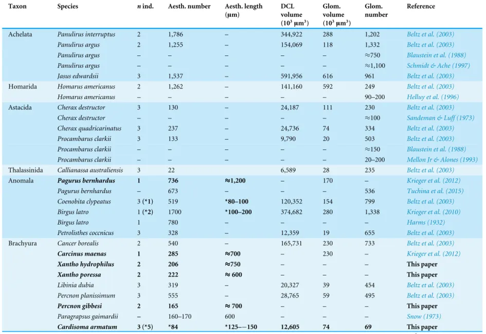
The antennaeIn general, the first antennae in brachyuran crustaceans each consists of two branches called the median and the lateral flagellum (Fig. 4). Both flagella are composed of several units, the flagellomeres. Each flagellomere of the lateral flagellum is equipped with one row of the typical unimodal chemosensory sensilla, the aesthetascs, in both marine and terrestrial brachyurans (Fig. 5). A quantification of aesthetasc numbers is provided inTable 2. The shape of the aesthetascs in marine versusterrestrial brachyurans displays marked differences. In the marine species, the aesthetascs are long and slender, whereas in all species featuring a rather terrestrial lifestyle, they are short and blunt (Fig. 5). The second antennae consist of one articulated branch only, composed of multiple antennomeres, and with the low-resolution light microscopic methods used here, we could not detect any striking differences in the sensillar equipment between the marine and terrestrial representatives (Fig. 6).
The brain
General arrangement of neuropils in the brachyuran brain
The general morphology of the brachyuran brain, as in other Malacostraca, is composed of three consecutive neuromeres, the proto-, deuto-, and tritocerebrum as extensively reported in previous studies (reviewed inHarzsch, Sandeman & Chaigneau, 2012;Schmidt, in press). In some anomalan species, such asBirgus latroorPetrolisthes lamarckiias well as in the axiid shrimpCallianassa australiensis, the bilaterally paired visual neuropils and the neuropils of the terminal medulla/hemiellipsoid body—complex (TM/HN-complex)
Table 2 Morphometric data of structures within the peripheral olfactory pathway and of the primary olfactory centers in the brain of decapod crustaceans.
Taxon Species nind. Aesth. number Aesth. length
(µm)
DCL volume (103µm3)
Glom. volume (103µm3)
Glom. number
Reference
Achelata Panulirus interruptus 2 1,786 – 344,922 288 1,202 Beltz et al. (2003)
Panulirus argus 2 1,255 – 154,069 118 1,332 Beltz et al. (2003)
Panulirus argus – – – – – ≈750 Blaustein et al. (1988)
Panulirus argus – – – – – ≈1,100 Schmidt & Ache (1997)
Jasus edwardsii 3 1,537 – 591,956 616 961 Beltz et al. (2003)
Homarida Homarus americanus 2 1,262 – 141,160 592 249 Beltz et al. (2003)
Homarus americanus – – – – – 90–200 Helluy et al. (1996)
Astacida Cherax destructor 3 130 – 24,187 111 230 Beltz et al. (2003)
Cherax destructor – – – – – ≈100 Sandeman & Luff (1973)
Cherax quadricarinatus 3 237 – 24,736 74 334 Beltz et al. (2003)
Procambarus clarkii 3 133 – 9,790 20 503 Beltz et al. (2003)
Procambarus clarkii – – – – – ≈150 Blaustein et al. (1988)
Procambarus clarkii – – – – – 20–200 Mellon Jr & Alones (1993)
Thalassinida Callianassa australiensis 3 22 6,589 28 235 Beltz et al. (2003)
Anomala Pagurus bernhardus 1 736 ≈1,200 – 170 – Krieger et al. (2012)
Pagurus bernhardus – 673 – – – 536 Tuchina et al. (2015)
Coenobita clypeatus 3(*1) 519 *80–100 120,352 154 799 Beltz et al. (2003)
Birgus latro 1(*2) 1700 *100–200 374,682 280 1,338 Krieger et al. (2010)
Birgus latro 1 780 – – – – Harms (1932)
Petrolisthes coccnicus 3 328 – 12,359 19 655 Beltz et al. (2003)
Brachyura Cancer borealis 2 540 – 165,731 230 733 Beltz et al. (2003)
Carcinus maenas 1 285 ≈700 – 230 – Krieger et al. (2012)
Xantho hydrophilus 2 206 ≈750 – – – This paper
Xantho poressa 2 222 ≈600 – – – This paper
Libinia dubia 3 319 – 20,327 39 454 Beltz et al. (2003)
Percnon planissimum 3 555 – 28,765 59 495 Beltz et al. (2003)
Percnon gibbesi 2 165 ≈700 – – – This paper
Paragrapsus gaimardii – 160–170 600 – – – Snow (1973)
Cardisoma armatum 3 (*5) *84 *125–−150 12,605 74 69 This paper
(continued on next page)
Krieg
er
e
t
al.
(2015),
P
eerJ
,
DOI
10.7717/peerj.1433
Table 2(continued)
Taxon Species nind. Aesth. number Aesth. length
(µm)
DCL volume (103µm3)
Glom. volume (103µm3)
Glom. number
Reference
Sesarma sp. 3 33 – 6,617 15 446 Beltz et al. (2003)
Gecarcoidea natalis 1 (*3) *113 *100–125 9,432 49 193 This paper
Geosesarma tiomanicum 3 (*5) *26 *60–80 4,253 21 61 This paper
Uca tangeri(♀+♂) 3 (*6) *38 *90–110 5,355 42±29 64 This paper
Uca tangeri (♂) 2 (*3) *36 5,300 20±6 78 This paper
Uca tangeri (♀) 1 (*3) *40 5,400 86 36 This paper
Uca minax 3 39 – 4,558 18 284 Beltz et al. (2003)
Uca pugilator 3 28 – 3,115 13 234 Beltz et al. (2003)
Uca pugnax 3 26 – 3,012 8 374 Beltz et al. (2003)
Note that volumes of DCLs and olfactory glomeruli (glom.) are estimates based on a variety of different neuroanatomical methods (for further information see references). All volumes are averaged for
one single structure (one DCL per hemisphere or one average glomerulus), rounded to the nearest 1,000µm3and are thus slightly modified from the original literature. Note that for each individual
in-vestigated, the number of aesthetascs (aesth.) per antenna are based on one randomly chosen antenna per pair. The table is compiled afterBeltz et al. (2003),Schachtner, Schmidt & Homberg (2005)and
Krieger et al. (2010)and complemented with data of other authors (see reference column) as well as with our own data (in bold) in addition of aesthetasc lengths. Note that the aesthetasc lengths inB.
la-tro(upper range estimated fromStensmyr et al. (2005)and lower range from J Krieger, 2010, unpublished data) andC.clypeatus(unpublished data) are estimated based on scanning electron micrographs.
Associated subsets of morphometric data apart from the main data set are indicated by asterisks.
Krieg
er
e
t
al.
(2015),
P
eerJ
,
DOI
10.7717/peerj.1433
Figure 4 First antenna in studied brachyuran species.(A) UV-autofluorescence micrograph shows equally scaled first antenna (AI) from four marine speciesCarcinus maenas,Percnon gibbesi,Xantho hy-drophilusandXantho poressa; and (B) from four terrestrial speciesGecarcoidea natalis,Cardisoma arma-tum,Geosesarma tiomanicumandUca tangeri. Abbreviations: AS, aesthetascs; lFl, lateral flagellum; mFl, median flagellum.
are located anteriorly adjacent to the ‘‘central’’ brain as a consequence of elongated axons composing the optic nerve. In all brachyurans studied so far, however, these neuropils are located within the eyestalks, thus being situated at some distance from the central portion of the syncerebrum. Note that in the comparativeFig. 3, for simplicity, only outlines of the central portions of the brains, in the following simply termed the ‘‘central brain’’—are drawn, without the neuropils of the lateral protocerebrum. In horizontal sections, this central brain appears broader than elongated along the neuraxis (Fig. 3C). The species studied here displayed markedly different carapace widths ranging from 14 mm in
G.tiomanicumup to 90 mm inG.natalis. In contrast, the general brain dimensions are
rather similar across species as indicated by a range of brain width between 1.4 mm inG.
tiomanicumto 2.5 mm inG.natalisand 2.7 mm inC.maenas. Hence, there seems to be
only a weak correlation between brain size and body size.
Contrary to most other decapods analyzed so far (see e.g., Sandeman, Scholtz & Sandeman, 1993), a distinct compartmentalization of the brain neuropils is less obvious in brachyurans. For instance, the neuropil boundaries in true crabs are much less distinct than in Anomala (compareKrieger et al., 2012). However, the general organization of the brachyuran brain and arrangement of its subunits can be deduced from anatomical data by tracing nerves as well as interconnecting tracts between the corresponding neuropils as outlined below:
The protocerebral tract (PT) is composed of neurites originating in neuropils of the lateral protocerebrum (lPC). The PT interconnects these neuropils with the proximal part of the brain, the median protocerebrum (mPC). The median protocerebrum is composed
Figure 5 Flagella and aesthetascs on first antenna in different brachyuran species.(A) UV-autofluorescence micrograph shows lateral and median flagellum as well as the aesthetascs from four marine species:Carcinus maenas,Percnon gibbesi,Xantho hydrophilus, andXantho poressaand (B) from four terrestrial speciesCardisoma armatum, Gecarcoidea natalis,Geosesarma tiomanicum, andUca tangeri. A micrograph using transmitted light shows the lateral flagellum and aesthetascs fromGeosesarma tiomanicum.Asterisks identify single annuli of the lateral flagellum. Abbreviations: AS, aesthetascs; lFl, lateral flagellum; mFl, median flagellum.
of the anterior (AMPN) and the posterior medial protocerebral neuropil (PMPN), which together resemble the shape of a butterfly in horizontal brain sections (compareFigs. 3, 9D,9E,11D,11E,13C–13G,15A–15D,16A1–16C1and17). Both neuropils are almost completely fused anterioposteriorly as well as across the midline with their contralateral counterparts into one single neuropil mass in the brachyuran brain, but they appear separated in horizontal sections at the level of the central body (Figs. 11E–11F, 13D, 15Band15F). Furthermore, the median protocerebrum includes neuropils of the central complex, namely from anterior to posterior: the unpaired protocerebral bridge (PB), the unpaired central body (CB), and the bilaterally paired lateral accessory lobes (Lals).
In all brachyuran species investigated, the neuropils of the deutocerebrum (DC) that extend posteriorly adjacent to the median protocerebrum consist of the unpaired median antenna I neuropil (MAN), the bilaterally paired antenna I neuropils (LANs), the deutocerebral chemosensory lobes (DCLs; formerly referred as olfactory lobes or olfactory neuropils), the accessory lobes (AcNs) and the projection neuron tract neuropils (PNTNs; formerly referred as olfactory globular tract neuropils or OGTNs). Each DCL
Figure 6 Second antenna of studied brachyuran species.(A) UV-autofluorescence micrograph shows the equally scaled second antenna (AII) from four marine speciesCarcinus maenas,Percnon gibbesi,Xantho hydrophilusandXantho poressaand (B) from four terrestrial speciesGecarcoidea natalis,Cardisoma arma-tum,Geosesarma tiomanicumandUca tangeri.
consists of several to hundreds of barrel-shaped subunits of synaptic neuropil, the olfactory glomeruli (OG) which are arranged in a radial, palisade-like array in the periphery of the lobe. Medially to each DCL, a cluster of somata (9/11) of hundreds of interneurons of varying sizes is present. These neurons extend neurites which enter the DCLviathe median foramen (mF), one of three gaps in the palisade-like array of olfactory glomeruli. Furthermore, several hundreds of somata of olfactory projection neurons are grouped in cell cluster (10) posteriorly to each DCL. Their neurites enter each DCLviathe posterior foramen (pF), innervate the olfactory glomeruli, and project axons that exit each lobeviaits median foramen (mF) in a large bundle that constitutes the projection neuron tract (PNT). The axons of the PNT interconnect each DCL with the ipsilateral as well as contralateral hemiellipsoid body within the lateral protocerebrum by forming a chiasm dorsally of the central body.
The tritocerebrum (TC) posteriorly adjoins the neuropils of the deutocerebrum and is composed of the bilaterally paired antenna II neuropils (AnNs) and further dorsally, of the tegumentary neuropils (TNs).
Lateral protocerebrum: the visual neuropils and the terminal medulla/hemiellipsoid body—complex (TM/HN-complex)
The eyestalks of most malacostracan crustaceans each contain three successive retinotopic neuropils. These three main visual neuropils process visual input and from distal to proximal are termed the lamina, medulla, and lobula. An additional (fourth) neuropil can be found adjacent to the lobula referred to as lobula plate. If present, the lobula plate adheres the lobula. The architecture of these visual neuropils which often are referred to as optic neuropils is best known in crayfish and lobsters (reviewHarzsch, Sandeman & Chaigneau,
Figure 7 Optical horizontal sections of lateral protocerebrum and central brain inCardisoma ar-matum. (A–E) Micrographs of triple-labeled optical horizontal sections showing visual neuropils and the lateral protocerebrum. Lamina was lost through dissection. (F–J) brain and details of specific brain areas such as, median protocerebrum and deutocerebrum in H and protocerebral bridge (PB) in I. The arrow with a dashed line marks a giant neuron in I. Note that (B, D, E, G and J) show inverted single-channel micrographs of different labelings (indicated by abbreviations). Abbreviations of immunhisto-chemical labelings and histoimmunhisto-chemical markers: NUC, nuclear marker (cyan); PHA, actin-labeling using phalloidin (green or black); RFA, labeling against RFamide (black); SYN, labeling against synapsin (ma-genta or black). Other abbreviations: 2, 3, 4/5, 6, and 9/11, cell clusters (2), (3), (4/5), (6), and (9/11);
AINv, antenna I nerve; AcN, accessory neuropil; AMPN, anterior medial protocerebral neuropil; CB, cen-tral body, DCL, deutocerebral chemosensory lobe; HN, hemiellipsoid body; iCh, inner visual chiasm; LAN, lateral antenna I neuropil; Lo, lobula; MAN, median antenna I neuropil; Me, medulla; oCh, outer visual chiasm; PMPN, posterior medial protocerebral neuropil; PNT, projection neuron tract; PT, proto-cerebral tract; TM, terminal medulla; VT, visual tract. Scale bars, 250µm.
2012) but was also analyzed in a number of marine and amphibious brachyurans including
Chasmagnathus granulatus,Hemigrapsus oregonensis(Sztarker, Strausfeld & Tomsic, 2005;
Sztarker et al., 2009; Berón de Astrada, Medan & Tomsic, 2011; Berón de Astrada et al., 2013), andCarcinus maenas(Elofsson & Hagberg, 1986;Krieger et al., 2012). Although the visual neuropils are not the focus of the present study, successful eyestalk preparations from
C.armatum(Figs. 7A–7D),G.natalis(Figs. 9A–9C),G.tiomanicum(Figs. 11Aand11B),
andU. tangeri(Fig. 13A) show that these terrestrial species display well developed visual
neuropils that show distinct synapsin-like immunoreactivity (SYN). Distinct clusters of
somata become clearly visible distal to each visual neuropil. According to their appearance from distal to proximal and based on nuclear counterstaining, we distinguish cluster (1) associated with the lamina; cluster (2) associated with the medulla; cluster (3) associated with the lobula (Figs. 7A,9A,11Aand13A). Their arrangement and layered architecture closely correspond to those of their marine relatives. In the lobula, we could resolve three main layers in all four species (Figs. 7B,9B,11Aand13A) suggesting that at the level of resolution we analyzed, the visual neuropils show a high level of similarity.
The most proximal neuropils of the lateral protocerebrum, the terminal medulla (TM; also termed medulla terminalis) and the hemiellipsoid body (HN), which are considered multimodal associative areas (Wolff et al., 2012), are located within the eyestalk and together constitute an almost spherical neuropil mass (TM/HN-complex) in the species studied. They are identifiable in preparations ofC.armatum(Figs. 7A–7E),G.natalis(Figs. 9A–9C),
G.tiomanicum(Figs. 11A–11C2), andU.tangeri(Figs. 13Aand13B) showing distinct SYN,
but were also described inChasmagnatus granulatus(Berón de Astrada & Tomsic, 2002),
Hemigrapsus oregonensis(Sztarker, Strausfeld & Tomsic, 2005), andC.maenas(Krieger et al., 2012). A clear distinction between these two neuropils is difficult because they are tightly adjoined. Therefore, a comparative volumetric analysis was impractical. Nevertheless, our preparations indicate that inUca, the TM/HN-complex is markedly smaller in diameter compared to all other crabs being analyzed (compareFig. 13A withFigs. 7A–7E,Figs. 9A–9Cand11A–11B). A compartmentalization of the hemiellipsoid body into one cap and 1–2 core neuropil masses is obvious inG.natalis(Fig. 9C) and inG.tiomanicum(Figs. 11B–11C2), whereas such a subdivision could not be resolved in the other crabs analyzed. According toSandeman et al. (1992), each of these neuropils is associated with a cluster of neurons, namely cluster (4), which is located closely to the terminal medulla and cluster (5), which is adjacent to the hemiellipsoid body in decapods and contains hundreds of interneurons of minute diameter. However, in the brachyurans studied, a clear separation of these two clusters was impossible. Rather, the TM/HN -complex is surrounded by a confluent cortex of somata which therefore will be referred to as cluster (4/5) here (Figs. 7C,9C,11Band13A–13B).
Median protocerebrum
The median protocerebrum is composed of the closely fused AMPN and PMPN and appears broader than long. In all brachyuran crabs studied, it has a butterfly-shape in horizontal sections (Fig. 3C). The AMPN and the PMPN are identifiable by showing distinct SYN (Figs. 7F,7H–7J,9D–9E,11D–11F,13C–13E,15A–15D,16A1–16C2and17), weaker RFA (Figs. 11D–11F,13D–13E,13G,16A1–16C2and17) and AST in the periphery (Figs. 9D–9E,15A–15Dand17). Although allatostaninergic and RFamidergic fibers innervate the whole brain, the V-shaped protocerebral bridge (PB) and especially the cylindrical or cigar-shaped central body (CB) further posteriorly show the densest RFA as well as AST (Figs. 7F–7I,9E,11D–11F,13D,13G,15A–15D,16A2–16C2and18) besides distinct SYN (Figs. 7H–7J,9E,11E–11F,13D–13E,15A–15D,16A2–16C2and17). In sections at the level of the CB, the separation of the AMPN from the PMPN becomes visible (Figs. 7H,9E, 11F,11G,13D,13F,15Band17). Anterior to the PB, the nuclear marker reveals hundreds
of somata of varying diameters (5–10µm in all species studied) that are grouped within
the cell cluster (6). This cluster also comprises a subset of few somata with a markedly larger diameter that display distinct RFA (diameters approx. 30µm inC.armatum, 25µm
inG.tiomanicum, and 20µm inU.tangeri) and AST (approx. 30µm inE.sinuatifrons
andG.natalis). The neuropils of the median protocerebrum are regularly pierced by blood
vessels of the circulatory system (e.g., the cerebral artery (CA);Figs. 9D–9E 11D,11F,13C and13E) and by large tracts of neurites (e.g., the projection neuron tract (PNT);Figs. 7H, 9D,11D–11G,13C–13E,13G,15B,15D,16A1and16B1–16B2) and can be inferred from the negative imprint due to the absence of immunoreactivity against the tested antisera. The projection neuron tract consists of neurites of olfactory projection neurons whose cell bodies are located in the somata clusters (10) situated posterior-lateral to each DCL. These neurites connect each DCL to the ipsilateral as well as the contralateral TM/HN-complex within the lateral protocerebrum and constitute a chiasm dorsally to the central body. The cerebral artery (CA) located between median protocerebrum (posterior to the PMPN) and deutocerebrum (anterior to the median antenna I neuropil) is identifiable by the nuclear counterstain of the perivascular cells (Figs. 9D–9E,11F,13Fand15A–15B) in horizontal sections. The dorsoventral course of the CA through the central brain could be confirmed in all crabs as it has been shown forC.maenas(Sandeman, 1967) but not its ramifications.
Deutocerebrum with special focus on structures of the primary olfactory pathway
Directly posterior-ventral to the PMPN and the CA, the unpaired median antenna I neuropil (MAN; Figs. 7F–7H,9D–9E,11D–11G, 13C–13G and15B) is present in all brachyuran crabs studied displaying distinct SYN as well as AST and RFA. The border between PMPN and MAN is rather confluent but is identifiable due to the clear position of the CA (compareFigs. 9Dand9E).
Besides the deutocerebral chemosensory lobe (DCL) and the accessory lobe (AcN), other neuropils of the lateral deutocerebrum can be found within the confluent mass of the central brain composed of parts of the proto-, deuto- as well as the tritocerebrum (compare Fig. 17) such as the lateral antenna I neuropil (LAN;Figs. 7F,9D,11D,13D–13Eand15B) and in a few preparations, the projection neuron tract neuropil (PNTN;
Figs. 15B and16A1). However, a complete, in-depth reconstruction of their definite outlines remains challenging. In all species investigated, the AcN and, in particular the DCL are the most delimited structures within the otherwise confluent brachyuran brain. The DCL is composed of several dozens (60–80 inG.sesarma,U.tangeriandC.armatum
up to almost 200 inG.natalis—seeTable 2for further information) of barrel-like to conical cardridges, termed olfactory glomeruli (OG), of varying sizes (seeFig. 18). From a limited number of investigated specimens ofU.tangeri, it appeared that in two males analyzed the number of olfactory glomeruli exceeded that of one female by a factor of ca. 2 (36 OG in
♀versus76–80 OG in♂), whereas the males featured approximately a third of the average
female glomerular volume (seeTable 2) resulting in an almost equal volume of the entire DCL in both sexes. In all marine brachyuran species studied, the numbers as well as the average volumes of olfactory glomeruli markedly exceed those of the co-studied terrestrial
brachyurans (Table 2andFig. 18), though the general brain dimensions are somewhat similar (seeFigs. 3and17). In brain sections of aquatic representatives of Brachyura (in the four exclusively marine; and to some degree, in the freshwater speciesE.sinuatifrons), the olfactory glomeruli are larger and more elongated compared to those of the terrestrial species studied here. A clear regionalization of each olfactory glomerulus into a cap, a subcap, and a base region (from the periphery of the DCL to its center) appears more pronounced in aquatic brachyurans than in the terrestrial species (Fig. 18). The cap and base regions show stronger SYN (Figs. 8A,8D1,14E,15E,16A1,16B1,17and18) than the subcap region in these species. In a subset of experiments, the subcap region shows distinct RFA, but RFA is weaker in the base region and is absent in the cap region (i.e., inP.gibbesi,
X.hydrophilus,X.poressa,U.tangeri;Figs. 18A and18B), whereas the subcap region
shows the most distinct AST and becomes absent towards the base region in each OG (in
E.sinuatifrons,Figs. 18Aand18B;G.natalis, not shown; and inC.maenas, seeKrieger et al., 2012). Anteriomedial to the median foramen of each DCL, the accessory lobe (AcN) becomes visible, consisting of dozens of microglomeruli that show distinct SYN but widely lack RFA as well as AST. The diameter of the almost spherical AcN ranges from 50µm (in
U.tangeri,X.hydrophilus,X.poressa, andP.gibbesi;Figs. 14A,14D–14Eand16A3–16C3) up to 100µm (75µm inC.armatum, 100µm inG.natalisas well asG.tiomanicum;Figs.
7F,8A,8E,9D,10C,12A,12Dand12F). Further medial and between the PMPN and the AcN, a somata cluster of hundreds of interneurons (ca. 5–8µm in diameter) appears. This
cell cluster (9/11) is clearly revealed by the nuclear counterstaining (Figs. 8C,9E,10A, 10Eand15B) and contains subpopulations of several to dozens of allatostatinergic (Figs. 9Eand15B) and RF-amidergic interneurons (Figs. 7G,12A–12C,13D–13E,16A1,16A3 and16B1) that are markedly larger in diameter (from 12µm inU.tangeri; and 16µm in
X.hydrophilus; up to 30µm inG.tiomanicum; i.e., seeFig. 12C). Neurites of cell cluster
(9/11) enter each DCLviathe median foramen (mF;Figs. 7F,8C,10B,12B,14A–14C,15B, 16A1,16B1and17). Lateroposterior to each DCL, a group of hundreds to thousands of olfactory projection neurons house their somata within cell cluster (10). These neurites of projection neurons enter the DCLviathe posterior foramen (pF;Figs. 8B–8C,9D,10D, 12C,14B–14Cand15B), connect with the olfactory glomeruli, exit the DCLviathe median foramen (Figs. 8C,12B,14C,15B,16A1,16B1and17), and finally project to the ipsilateral as well as the contralateral TM/HN-complex by forming a chiasm at the dorsal level of the central body (not shown). The entirety of neurites of projection neurons constitute the projection neuron tract (PNT) whose somata are housed within cell cluster (10). According to its position, the projection neuron tract neuropil (PNTN) becomes visible medial to the mF in a few preparations showing distinct SYN (Figs. 15B,16A1and17).
Tritocerebrum
The tritocerebral antenna II neuropil (AnN) and further dorsally the tegumentary neuropil (TN) compose the posteriormost parts of the central brain, being located anterolaterally to the esophagus. An identification of the neuropil borders is difficult due to their confluent connection to the deutocerebrum. The AnN that receives chemosensory as well as mechanosensory input from the second antenna is identifiable in a few preparations
Figure 8 Optical horizontal sections and 3D-reconstruction of deutrocerebral chemosensory lobe (DCL) inCardisoma armatum.(A–C) Inverted single-channel micrographs of DCL. White arrows in B mark axons of projection neurons. (D) and (D1): Detailed picture of the olfactory glomeruli (OG) with double-labeling in D and inverted single-channel picture in D1. (E) 3D-reconstruction of DCL, olfactory glomeruli and accessory neuropil (AcN) shown in four different orientations. 1: from dorsal. 2: from an-terior. Dashed line represents the horizon of section given in A. 3: from posan-terior. Dashed line indicates the posterior foramen (pF). 4: centro-lateral view. Dashed line indicates the median foramen (mF). Ab-breviations of immunhistochemical labelings and histochemical marker: NUC, nuclear marker (black); PHA, actin-labeling using phalloidin (black); RFA, labeling against RFamide (green); SYN, labeling against synapsin (magenta or black). Other abbreviation: 10 and 9/11, cell clusters (10) and (9/11); a, anterior; Base, base domain of OG; Cap, cap domain of OG; d, dorsal; l, lateral; m, median; Subcap, subcap domain of OG.
by tracing back the course of the antenna II nerve (AIINv;Figs. 9Dand9E). Since we
were unable to trace back the course of the presumably thin tegumentary nerve (TNv), the precise position and shape of the tegumentary neuropil remains uncertain.
DISCUSSION
In this study, we compare the neuroanatomy of the brain in four brachyurans that display different levels of terrestrial adaptations using the antisera against presynaptic proteins, the neuropeptides FMRFamide, and allatostatin as well as markers for actin and DNA. In the following, we will compare and discuss the results of these four brachyuran species with each other as well as with one freshwater and four marine brachyurans. Special attention is given to the primary olfactory system and related structures to highlight differences between terrestrial brachyurans and their aquatic relatives.
In contrast to other reptant Malacostraca such as Anomala, which display a clear separation of their deutocerebral neuropils (e.g.,Harzsch & Hansson, 2008;Krieger et al., 2010;Krieger et al., 2012), these neuropils are widely confluent and therefore often become
Figure 9 Micrographs of triple-labeled vibratome sections of central brain and lateral protocerebrum inGecarcoidea natalis. (A–C) Visual neuropils and lateral protocerebrum. Note that in A and C, two out of three channels are shown while B shows an inverted single-channel micrograph. (D and E) show two triple-labeled horizontal vibratome sections of central brain (D) and further ventral of central brain (E). Dashed line in E indicates the cerebral artery. Abbreviations of immunhistochemical labelings and histo-chemical marker: AST, labeling against allatostatin (green); NUC, nuclear marker (cyan); RFA, labeling against RFamide (black); SYN, labeling against synapsin (magenta). Other abbreviations: 1, 2, 3, 4/5, 6, 9/11, and 10, cell clusters (1), (2), (3), (4/5), (6), (9/11), and (10); AINv, antenna I nerve; AIINv, antenna
II nerve; AcN, accessory neuropil; AMPN, anterior medial protocerebral neuropil; AnN, antenna II neu-ropil; CA, cerebral artery; CB, central body; DCL, deutocerebral chemosensory lobe; iCh, inner visual chi-asm; HN, hemiellipsoid body; La, lamina; LAN, lateral antenna I neuropil; Lo, lobula; MAN, median an-tenna I neuropil; Me, medulla; oCh, outer visual chiasm; PMPN, posterior medial protocerebral neuropil; PNT, projection neuron tract; PT, protocerebral tract; TM, terminal medulla; VT, visual tract. Scale bars
=250µm.
indistinguishable in brachyurans. Sandeman, Scholtz & Sandeman (1993)andKrieger et al. (2012) discussed the possible connection between brain ‘‘condensation’’, the fusion of synaptic neuropils, and evolutionary success in these groups. The condensation of nervous tissue may have coincided with a process that is sometimes called ‘‘carcinisation’’ (Borradaile, 1916), or ‘‘brachyurisation’’ (Števčić, 1971). These synonyms circumscribe a hypothesis of how the condensed crab shape may have developed (McLaughlin & Lemaitre, 1997), both concerning the overall brachyuran habitus as well as internal consolidation of organs like the fusion of the first three ganglia of the ventral nerve cord into one joint complex (Števčić, 1971). According toŠtevčić (1971), it was also assumed that this process mainly leads to a more complex behavior and better coordination in semiterrestrial and terrestrial crabs, since neuropil condensation and shortening of connections within the central nervous system may improve the performance of the system, e.g., in terms of
Figure 10 Vibratomy of double-labeled horizontal sections the deutocerebral chemosensory lobe (DCL) inGecarcoidea natalis. (A–D) DCL featuring olfactory glomeruli (OG). Note that only an in-verted single-channel micrograph is given in (E) showing nuclear staining in the periphery of DCL. Ab-breviations of immunhistochemical labeling and histochemical staining: NUC, nuclear marker (cyan or black); SYN, labeling against synapsin (magenta). Other abbreviations: 10 and 9/11, cell clusters (10) and (9/11); AcN, accessory neuropil; mF, median foramen; OG, olfactory glomerulus; pF, posterior foramen. Scale bars=100µm.
processing speed. The fusion is most conspicuous in the posterior part of the brain, where the neuropils of the deutocerebrum adjoin those of the tritocerebrum.
Visual ecology and the protocerebrum
Terrestrial brachyurans have been prime examples to study visual ecology in crustaceans (reviews byZeil & Hemmi, 2006;Zeil & Hemmi, 2014; Hemmi & Tomsic, 2012). Visual orientation has been very well studied in members of the genusUcabut poorly in any of the other ocypodid species (Zeil & Hemmi, 2014and references therein). Field experiments for individuals ofU.tangerihave shown that they can recognize predators at greater distance, triggering an escape behavior. In addition, the animals react to their own mirror image and can visually distinguish the gender of their conspecifics (Altevogt, 1957;Altevogt, 1959;Von Hagen, 1962;Korte, 1965;Land & Layne, 1995;Zeil & Al-Mutairi, 1996). Representatives of the genusUcacan also distinguish colors (Korte, 1965;Hyatt, 1975;Detto, 2007), which is an important factor for social interactions (Detto et al., 2006;Detto, 2007). Ultraviolet light, for example, is reflected by the claw ofUca-males which attracts females (Detto & Backwell, 2009). Aspects of homing and path integration were also thoroughly analyzed in members of the genusUca(e.g., Hemmi & Zeil, 2003;Layne, Barnes & Duncan, 2003a;
Layne, Barnes & Duncan, 2003b;Walls & Layne, 2009). Clearly, vision plays an essential role in the ecology ofUca.