Advanced Microbial Taxonomy Combined
with Genome-Based-Approaches Reveals that
Vibrio astriarenae
sp. nov., an Agarolytic
Marine Bacterium, Forms a New Clade in
Vibrionaceae
Nurhidayu Al-saari1*, Feng Gao1, Amin A.K.M. Rohul1, Kazumichi Sato1, Keisuke Sato1, Sayaka Mino1, Wataru Suda2,3, Kenshiro Oshima2, Masahira Hattori2,4, Moriya Ohkuma5, Pedro M. Meirelles6, Fabiano L. Thompson6, Cristiane Thompson6, Gilberto M. A. Filho7, Bruno Gomez-Gil8, Toko Sawabe9, Tomoo Sawabe1
1Laboratory of Microbiology, Faculty of Fisheries, Hokkaido University, Minato-cho, Hakodate, Japan, 2Laboratory of Metagenomics, Graduate School of Frontier Sciences, University of Tokyo, Kashiwa, Japan, 3Department of Microbiology and Immunology, Keio University School of Medicine, Tokyo, Japan, 4Graduate School of Advanced Science and Engineering, Waseda University, Tokyo, Japan,5Microbe Division/Japan Collection of Microorganisms, RIKEN BioResource Center, Ibaraki, Japan,6Institute of Biology, SAGE-COPPE, Federal University of Rio de Janeiro (UFRJ), Rio de Janeiro, Brazil,7Rio de Janeiro Botanical Garden, Rio de Janeiro, Brazil,8CIAD, AC Mazatlan Unit for Aquaculture and Environmental Management, Mazatlán, México,9Department of Food and Nutrition, Hakodate Junior College, Hakodate, Japan
*hidayu@fish.hokudai.ac.jp
Abstract
Advances in genomic microbial taxonomy have opened the way to create a more universal and transparent concept of species but is still in a transitional stage towards becoming a defining robust criteria for describing new microbial species with minimum features obtained using both genome and classical polyphasic taxonomies. Here we performed advanced microbial taxonomies combined with both genome-based and classical approaches for new agarolytic vibrio isolates to describe not only a novelVibriospecies but also a member of a newVibrioclade. Two novel vibrio strains (Vibrio astriarenaesp. nov. C7Tand C20) showing agarolytic, halophilic and fermentative metabolic activity were isolated from a seawater sample collected in a coral reef in Okinawa. Intraspecific similarities of the isolates were identical in both sequences on the 16S rRNA andpyrHgenes, but the closest relatives on the molecular phylogenetic trees on the basis of 16S rRNA andpyrHgene sequences were V.hangzhouensisJCM 15146T(97.8% similarity) andV.agarivoransCECT 5085T(97.3% similarity), respectively. Further multilocus sequence analysis (MLSA) on the basis of 8 pro-tein coding genes (ftsZ,gapA,gyrB,mreB,pyrH,recA,rpoA, andtopA) obtained by the genome sequences clearly showed theV.astriarenaestrain C7Tand C20 formed a distinct new clade protruded next toV.agarivoransCECT 5085T. The singletonV.agarivoranshas never been included in previous MLSA ofVibrionaceaedue to the lack of some gene sequences. Now the gene sequences are completed and analysis of 100 taxa in total
a11111
OPEN ACCESS
Citation:Al-saari N, Gao F, A.K.M. Rohul A, Sato K, Sato K, Mino S, et al. (2015) Advanced Microbial Taxonomy Combined with Genome-Based-Approaches Reveals thatVibrio astriarenaesp. nov., an Agarolytic Marine Bacterium, Forms a New Clade inVibrionaceae. PLoS ONE 10(8): e0136279. doi:10.1371/journal.pone.0136279
Editor:Patrick CY Woo, The University of Hong Kong, HONG KONG
Received:October 2, 2014
Accepted:August 1, 2015
Published:August 27, 2015
Copyright:© 2015 Al-saari et al. This is an open access article distributed under the terms of the Creative Commons Attribution License, which permits unrestricted use, distribution, and reproduction in any medium, provided the original author and source are credited.
Data Availability Statement:All relevant data are within the paper and its Supporting Information files.
provided a clear picture describing the association ofV.agarivoransinto pre-existing concatenated network tree and concluded its relationship to our vibrio strains. Experimental DNA-DNA hybridization (DDH) data showed that the strains C7Tand C20 were conspecific but were separated from all of the otherVibriospecies related on the basis of both 16S rRNA andpyrHgene phylogenies (e.g.,V.agarivoransCECT 5085T,V.hangzhouensis JCM 15146TV.maritimusLMG 25439T, andV.variabilisLMG 25438T).In silicoDDH data also supported the genomic relationship. The strains C7Talso had less than 95% average amino acid identity (AAI) and average nucleotide identity (ANI) towardsV.maritimusC210, V.variabilisC206, andV.mediterraneiAK1T,V.brasiliensisLMG 20546T,V.orientalis ATCC 33934T, andV.sinaloensisDSM 21326. The nameVibrio astriarenaesp. nov. is pro-posed with C7 as the type strains. BothV.agarivoransCECT 5058TandV.astriarenaeC7T are members of the newest clade ofVibrionaceaenamed Agarivorans.
Introduction
Advances in genomic microbial taxonomy have opened the way to create a more universal and transparent concept of species but is still in a transitional stage towards becoming a defining robust criteria for describing new microbial species with minimum features obtained using both genome and classical polyphasic taxonomies [1–3]. Polyphasic taxonomy significantly contrib-uted to bacterial description in the past by integrating the analysis of the phenotypic, genotypic (including chemotaxonomic) and phylogenetic characters of the isolates. It is predicted that the practice will soon be replaced with genomic microbial taxonomy in which the principles and practices are being developed. A lot of recent literature [2–6] have discussed these changes and are predicting its future impact on current microbial description and microbial taxonomy as a whole. This paper intended to implement the ideas proposed in the aforementioned literature to describe a new vibrio species and also to elaborate such matters as the practical usage and delin-eation of minimum features in microbial genome taxonomy. We demonstrate the actual transi-tional stage—an interphase in the evolution from polyphasic taxonomy to genomic microbial taxonomy and the need to describe the bacteria as comprehensively as possible.
We suggest that regardless of all the discrepancies, it is generally acknowledged that a uni-versal species definition might somewhat resolve most of the problems in one way or another. Thus, in this paper, we attempt to describe a vibrio species using a recent proposed species defi-nition by Rosselló-Móra and Amann [6] i.e.“a category that circumscribes monophyletic,and genomically and phenotypically coherent populations of individuals that can be clearly discrimi-nated from other such entities by means of standardized parameters”. The parameters used per-mit an accurate classification of species via three major premises namely i) monophyly— demonstrating all members of the taxon in which a new species belongs to share a common evolutionary history using phylogenetic inferences; ii) genomic coherence—modulating the circumscription of the unit using a specific pre-determined threshold value executed using its respective method which corresponds to the observable phenotypes used for identification pur-poses, and iii) phenotypic coherence—organisms in the same taxon should display similar physiological, structural and ecological properties either through direct determination of their characteristics or prediction of the genome sequences [6]. All of the data could be obtained by whole genome sequences but we still need validations in the experiments, in particular the description of new bacterial lineages to increase the reliability of the genome-based taxonomic approach.
funding. PMM thanks CAPES for the PhD scholarship (4848-14-9 CAPES/JSPS), and MARA for the PhD education loan to NA.
The genusVibriowas proposed in 1854 for Gram-negative fermentative halophilic bacteria [7]. Today, a total of 110 species with valid nomenclature [8] in the genusVibriohave been described. These bacteria are ubiquitous, highly heterogeneous [9] and the species evolution developed by both lateral (horizontal) and vertical gene transfer [10]. Thus, a thorough description is a prerequisite for describing a new species in theVibrionaceaefamily.Vibrio spe-cies significantly contribute to the nutrient cycle by mediating organic matter decomposition. They have versatile metabolisms and are capable of degrading and fermenting complex organic matters such as polysaccharides [11]. Though the ability to consume agar, a complex polysac-charide composed of agarose and agaropectin, is known to be common among marine bacteria [12–13], it is not prevalent within theVibriospecies. Currently,V.agarivoransis the only agar-olyticVibriothat has been described thoroughly [12]. The species was isolated from Mediterra-nean seawater and reported to be able to degrade agar on both marine agar plates and
thiosulphate-citrate-bile-sucrose (TCBS) agar [12]. In addition to some widely known agarases applications in food, cosmetic and medical industries, Chen et al. [14] and Dipakkore et al. [15] have demonstrated the efficient degradation of the cell walls of marine red algae in which cell wall was composed of agar. Thus, marine-derived agarases are valuable enzymes applicable for both cell biology and biotechnology in red algae. They are currently of growing interest due to their potential uses in the bioconversion of marine algal polysaccharides into energy feed-stocks in biofuel industries [16]. Vibrios in particular, share such potential and our laboratory had successfully produced hydrogen from powdered brown macroalgae,Sacchararina sculpera
[17]. Hence, further bioprospecting and the genomic survey of marine agarolytic bacteria will be greatly advantageous to the biofuel industry and at present is in high demand.
A two years survey of vibrios from the coral reef in Iriomote-Ishigaki National Park, Oki-nawa, Japan, obtained two agarolytic isolates with typical characteristics ofVibriospecies. The isolates produced unpigmented colonies and displayed a shallow pit with pronounced diameter on ZoBell 2216E agar medium after 24 h of incubation at 30°C. During the primary vibrio sur-vey,pyrHgene sequence analysis has placed these isolates into a distinct group not affiliated with any knownVibriospecies. Thorough genomic and polyphasic taxonomies have strength-ened the initial findings and further differentiate the isolates into their own unique group form-ing a distinct clade on basis of MLSA. We therefore concluded that these two bacteria may belong to a new species within the genusVibrio. However, a close relationship of these isolates withV.agarivoransin terms ofpyrHphylogeny and some phenotypic characters including the agarolytic activity demands a comprehensive MLSA. Hence, inclusion of completed eight housekeeping genes belongs toV.agarivoransCECT 5085Tinto the pre-existing MLSA data-sets were performed and reveals that the isolates shared the same clade withV.agarivorans. Both C7 and C20 are facultative anaerobes rods with polar flagella and capable of growth at 20–40°C at an optimum temperature of 30°C. This study provides the evidence and a detailed description of a novel agarolytic species for whom we propose the nameV.astriarenaesp. nov. and falls into a novel clade named Agarivorans.
Here we performed advanced microbial taxonomy combining both genome-based and clas-sical approaches for vibrio isolates belonging to a probable new clade species with the aim of describing a novelVibriospecies. Special emphasis is given to advantages and disadvantages of genome taxonomy.
Materials and Methods
Water sampling and bacterial strains
Iriomote-Ishigaki National Park, Okinawa, Japan. Specifically, the seawater samples were col-lected from the vicinity of Taketomi Island (24°20.5260' N; 124°05.6443' E) by SCUBA diving using underwater pumps. No specific permissions are required for water sampling activities in this location. The samples were then brought back to the lab and bacterial isolation was per-formed using thiosulphate-citrate-bile-sucrose (TCBS) medium (Nissui Pharmacy, Tokyo, Japan). Following incubation at 25°C for 24 h the isolates were then purified on ZoBell 2216E agar medium and incubated at 30°C. The strains were stored at -80°C using 20% glycerol-sup-plemented broth.
Determination of moles percent G+C content and DNA-DNA
hybridizations
Strains used in DNA-DNA hybridization experiment were C7T, C20,V.maritimusLMG 25439T,V.variabilisLMG 25438T,V.brasiliensisLMG 20546T,V.agarivoransCECT 5085T andV.hangzhouensisJCM 15146T. Genomic DNAs ofV.astriarenaeC7T,V.agarivorans
CECT 5085T,V.brasiliensisLMG 20546TandV.hangzhouensisJCM 15146Twere used as probes. DNAs of bacterial strains were prepared according to the procedures of Marmur [18], with minor modifications. Moles percent G+C contents of DNA fromV.astriarenaesp. nov., strain C7Tand C20 were determined using high-performance liquid chromatography (HPLC) [19]. DNA-DNA hybridization experiments were performed in microdilution wells using a fluorometric direct binding method [20]. DNAs ofV.astriarenaesp. nov., strain C7T,V. agari-vorans,V.brasiliensisandV.hangzhouensiswere labeled with photobiotin (Vector Laborato-ries, Inc., Burlingame, CA). Four micro-grams of unlabeled single-stranded DNA were immobilized in microdilution wells (Immuron 200, FIA/LIA plate, black type, Greiner labo-technik, Germany), then a hybridization mixture containing 20 ng of labeled DNA was added to each microdilution well and the hybridization was performed under optimal conditions fol-lowing pre-hybridization. Formamide concentration in the hybridization mixture was deter-mined according to Meinkoth-Wahl [21]. The hybridization of the biotinylated DNA to immobilized DNAs was performed under optimal conditions (fixation at 37°C) following hybridization at 45°C and detected by fluorometry after binding streptavidin-β-galactosidase to labeled DNA. 4-Methylumbelliferyl-β-D-galactopyranoside (6 x 10⁻⁴M; Wako, Osaka, Japan) was added to each well as fluorogenic substrate forβ-galactosidase and incubated at 30°C. Then, the fluorescence intensity of each well was measured using a microplate reader (Infinite F200, Tecan, Switzerland) at wave length of 360 nm for excitation and 450 nm for emission. DNA-DNA homology was calculated according to Ezaki et al. [22].
DNA amplification and sequencing
directly sequenced using a BigDye terminator sequencing kit version 3.1 (Life Technologies, Carlsbad, CA) according to the protocol recommended by the manufacturer. DNA sequencing was performed using an Applied Biosystems model 3130x automated sequencer. Six DNA primers (24F, 800F, 1100F, 520R, 920R, 1509R) were used in the sequencing reactions [23].
Phylogenetic analysis
The sequences were aligned and studied using Clustal X version 2.1 [24] and MEGA programs version 6.06 [25]. The alignment was checked using the naked eye and corrected manually. In all phylogenetic analyses, we used the sequences determined in this study and small-subunit rRNA gene sequences obtained from the GenBank/EMBL/DDBJ databank. ForFig 1, the anal-ysis was performed by applying the neighbor-joining method [26] with bootstrap values of 500 replicates to the full dataset of 141 sequences which includes 138 small-subunit rRNA gene sequences of typeVibrionaceae(S1 Table), two novel strains andEscherichia coliK-12. Evolu-tionary distances were computed using the Jukes-Cantor method. The robustness of topology was also checked using maximum parsimony and maximum likelihood methods withE.coli
K-12 as the outgroup. The analyses were performed on the small-subunit rRNA gene sequences that corresponded toE.colisequence at position 194 to 1403 and trees were drawn using the MEGA. Using a similar procedure, we also computed a phylogenetic analysis on the basis of
pyrHgene sequences (Fig 2) of 113Vibrionaceaespecies obtained from the public database.
Genome sequencing and
in silico
DNA-DNA relatedness calculation
Draft genome sequences of strain C7Twere obtained using IonPGM sequencer with 28 redun-dancy. The genome sequences were assembled using Newbler ver. 2.8 into 336 contigs with 99142 ofN50. The sequence was deposited in the DDBJ/GenBank/EMBL public database under
accession numbers described below.
General genome features were determined through Rapid Annotations Using Subsystems Technology (The RAST server version 4.0) [27].In silicoDDH values ofV.astriarenaesp. nov. C7Twere estimated againstV.brasiliensisLMG 20546T,V.maritimusC210,V.mediterranei
AK1,V.variabilisC206,V.orientalisATCC 33934T,V.sinaloensisDSM 21326 andV.tubiashii
ATCC 19109Tusing Genome-to-Genome Distance Calculator (GGDC 2.0) [28]. This online tool infers genome-to-genome distances between pairs of entirely or partially sequenced genomes. Intergenomic distances were employed for wet-lab DDH prediction. Briefly, genome pairs were aligned with BLAST+ [29] to generate a set of high-scoring segment pairs (HSPs). The information they contained (e.g., the total number of identical base pairs) was transformed into a distance value by the best-fit formula, according to [28]. DDH prediction from interge-nomic distance, including confidence intervals, was provided by a tested generalized linear model (GLM), [30] with log transformation [28]. Amino Acid Identity (AAI) and Average Nucleotide Identity (ANI) were calculated according to Konstantinidis and Tiedje [31] and Thompson et al. [32] respectively, on the same strains used forin silicoDDH.
Multilocus sequence analysis (MLSA)
Sequences of eight housekeeping genes (ftsZ,gapA,gyrB,mreB,pyrH,recA,rpoA, andtopA) were retrieved from the genome sequence and used to infer the clade ofV.astriarenaesp. nov., strain C7Tand C20 following a method described in Sawabe et al. [1,33]. Briefly, the sequences were aligned using the ClustalX 2.1 [24]. The domains used to construct the phylogenetic tree shown inFig 3were regions of theftsZ,gapA,gyrB,mreB,pyrH,recA,rpoA, andtopAgenes of
investigated are in accordance with those used in earlier studies [1,33]. MEGA version 6 [25] was used to deduce the sequence similarity and the number of nucleotide and amino acid mutations. Another five housekeeping genes (ftsZ,mreB,recA,rpoA, andtopA) ofV. agarivor-ansCECT 5085Twere also retrieved in the same manner for a complete 100 taxa MLSA.
Split Decomposition Analysis (SDA) was performed according to Sawabe et al. [33] using SplitsTree version 4.13.1 [34], with a neighbour net drawing and Jukes-Cantor correction [1,
35]. The program was then used to generate the concatenated sequences of the eight house-keeping genes which served as basis for a phylogenetic analysis combined with NJ, MP, and ML analyses [23].
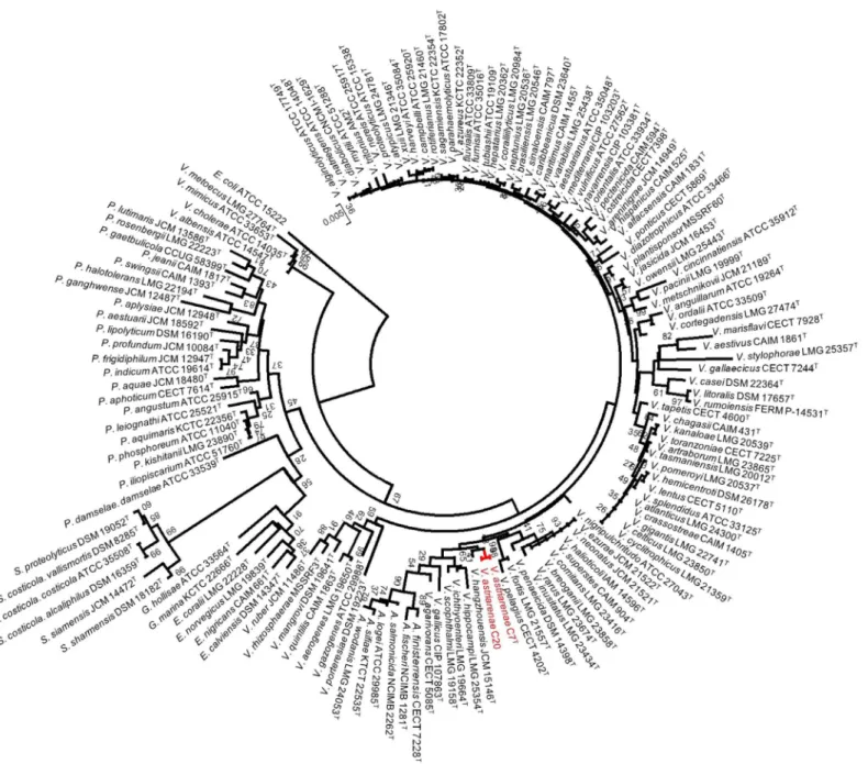
Fig 1. Phylogenetic tree on the basis of 16S rRNA gene sequences by neighbor-joining method.Bootstrap values are on 500 replicates. The topology of branch with the bootstrap value was also supported by maximum likelihood and maximum parsimony methods.
Phenotypic characterization and genome based phenotypes
All strains were cultured on ZoBell 2216E agar medium [36] and their main characteristics were determined as described previously [37–42]. Phenotypic features were obtained directly
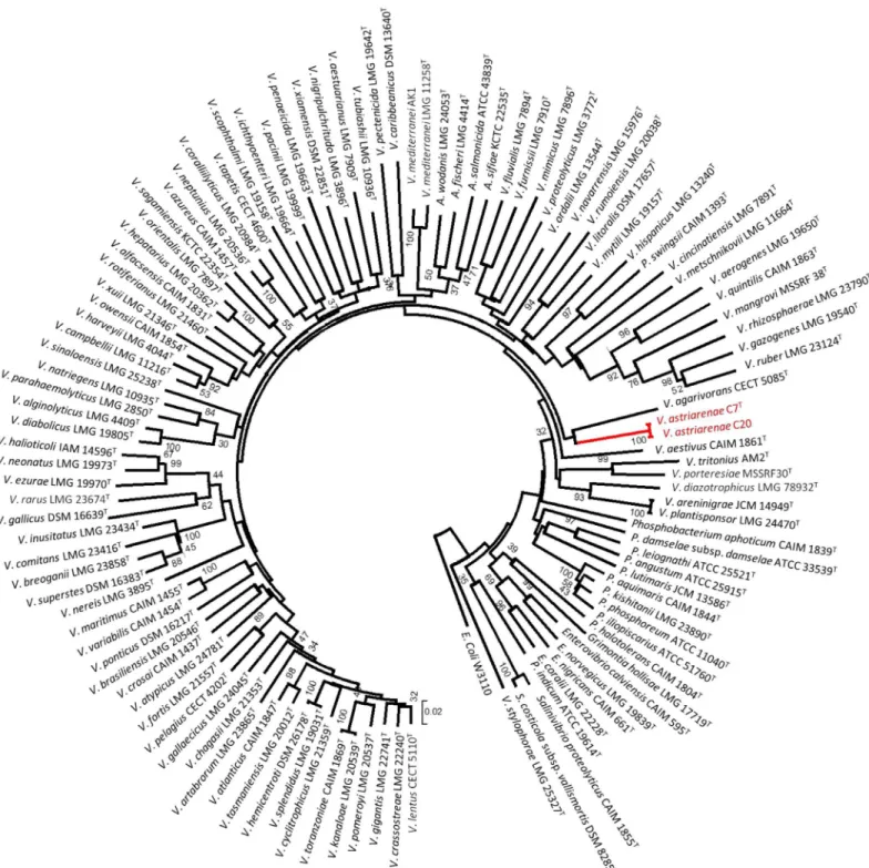
Fig 2. Phylogenetic tree on the basis ofpyrHgene sequences by neighbor-joining method.The tree were drawn by MEGA and the gene sequences were corresponded toE.colisequence at position 150 to 544. Bootstrap values are on 500 replicates. The topology of branch with the bootstrap value was also supported by maximum likelihood and maximum parsimony methods.
from the whole genome sequences by automatically searching and database comparisons of the genes that define the metabolic pathways of each diagnostic feature and their regulatory genes as described previously [5]. Briefly, genes coding the proteins responsible for each feature were detected using the RAST program and the KEGG metabolic database following subsequent identification using BLASTP algorithm [5].
Nucleotide sequence accession number
The genome data has been deposited at DDBJ/EMBL/GenBank under the accession numbers BBMQ01000001-336, BBMT01000001-45 and BBMS01000001-208 forVibrio astriarenaesp. nov. C7T(JCM 19233),V.maritimusC210 (JCM 19240) andV.variabilisC206 (JCM 19239), respectively.
The 16S rRNA gene sequences of C7Tand C20 performed by Sanger sequencing were deposited to GenBank under KP342514 and KP713778, respectively. Also, the housekeeping gene sequences for C20 andV.agarivoransCECT 5085Tperformed by the same method were deposited under the accession number shown inS4 Table.
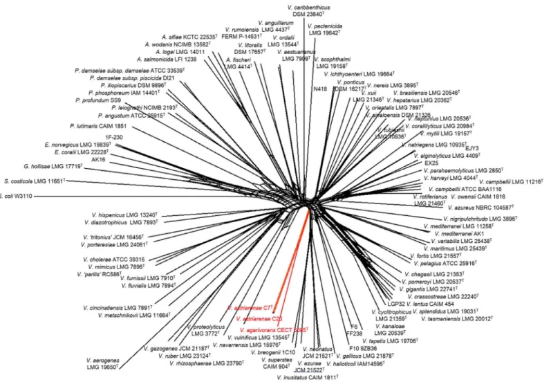
Fig 3. Concatenated split network tree based on eight gene loci.ThegapA,gyrB,ftsZ,mreB,pyrH,recA,rpoA, andtopAgene sequences of 100 taxa were concatenated including the representative of novel vibrios in the current study (Vibrio astriarenaesp. nov. C7Tand C20). Phylogenetic tree was generated using the SplitsTree4 program.Vibrio astriarenaesp. nov. C7Tand C20 formed a clade not associated any vibrio clades proposed previously.
Results and Discussion
Monophyletic premise guarantees common evolutionary lineages
Tindall et al. [43] in their article have listed the key elements and set out a guide on how a pro-karyote should be characterized for taxonomic purposes. The essence of the article is that the characterization of any new taxon should be as comprehensively as possible to place them within the existing hierarchical framework (Bacteriological Code, 1990 revision) and to provide a description of the taxa. In the current article, we described two potential new strains using both polyphasic and genomic approaches. Initially, the 16S rRNA gene sequences of strains C7Tand C20 were aligned by comparison to a database containing 138 aligned small-subunit rRNA gene sequences of typeVibrionaceae. The analyses by three different methods (NJ, ML and MP) showed no incongruence that they be included in the genusVibrio; more precisely in a subgroup close toV.hangzhouensisJCM 15146T(97.8% similarity) (Fig 1). The nucleotide sequences of the 16S rRNA gene of two strains ofV.astriarenaesp. nov. were identical.Fig 1
depicts the node of final phylogenetic tree (shown in red) obtained using the NJ method with high bootstrap value (98%) and supported by ML and MP analyses. The sequence similarities to type strains ofVibrionaceaehave further distinguished the novel strains fromV.agarivorans
by 97.3%. Furthermore, the similarities analyses toV.maritimusLMG 25439T,V.variabilis
LMG 25438TandV.brasiliensisLMG 20546Twere 98.8 to 99.0% which are in accordance to species delineation as proposed by Kita-Tsukamoto et al. [44]. The authors have drawn the cir-cumscription border of new species inVibrionaceaeat99.3% 16S rRNA similarity. On the
pyrHgene phylogeny,V.astriarenaesp. nov. C7Tand C20 formed a robust clade withV. agari-voransCECT 5085Tas the closest related species (Fig 2).
Note that separate 16S rRNA andpyrHgenes phylogenies ofV.astriarenaesp. nov. C7T and C20 are insufficient to demonstrate a monophyletic clade adjacent to the same reference type strain (close toV.hangzhouensisin 16S rRNA gene phylogeny, and toV.agarivorans
based onpyrHgene) on its own. This is supported by previous researches [33,45] which showed the individual gene analyses are known to have low interspecies resolution in Vibriona-ceae. The incongruence of the topologies on the basis of both genes may also indicate that C7T and C20 probably form a novel clade. The MLSA of the eight housekeeping genes (Fig 3) pro-vided a more robust inference of the evolutionary history of vibrios [33] verifying the mono-phyly of new candidates [6]. Previously [1], the singletonV.agarivoranswas not included in eight genes analysis due to the lack of some gene sequences. The fact that both novel strains andV.agarivoransare sharing some phenotypic characters (described below) and their close relationship inpyrHgene phylogeny (Fig 2) demands a complete eight genes ofV.agarivorans
to be included to our multilocus sequences dataset. Thus, inclusion of multilocus sequences of
V.agarivoransCECT 5085TandV.astriarenaesp. nov., strain C7Tand C20 into the pre-exist-ing clades data set [1] formed a branch that is strongly presumed to represent a new clade unre-lated to any others (Fig 3). Sawabe et al. [1] has also described the other clades in great detail. It is noted that the species within each clade shared>20% DDH,<5% G+C (mol%),>85%
MLSA sequence similarity and>89% AAI [1,33].
The concatenated network tree of 100 taxa ofVibrionaceaebased on eight gene loci has positioned the strain C7 and C20 closely toV.agarivoransCECT 5085Tsupported our earlier
similarity [1]. They also discovered similar incongruence in Mediterranei and Pectenicida clades. Single gene analysis is known to have different resolution according to the taxonomic groups due to different molecular clocks of the different genes [46]. Thompson et al. [46] in general suggested that the taxonomic resolution of 16S rRNA was restricted to genera differen-tiation amongVibrionaceaerather than the differentiation of species. This suggestion may partly explain the discrepancies. However, above all, the analyses presented herein have strongly proved the novelty ofV.astriarenaesp. nov. C7Tand C20. Today, the new clade can-didate may includeV.astriarenaesp. nov. C7Tand C20, andV.agarivorans, and the whole genome sequence ofV.agarivoransthat are currently in progress is expected to revealed more. The study will allows direct comparison of both species and describes the new clade more thor-oughly. For now, the name Agarivorans is proposed for the new clade.
Genomic coherence
—
demonstrates a stable taxonomic framework for
the novel strains
A group of novel bacterial species is defined as having>5% mol G+C difference of the genomic
DNA,<70% DDH similarity (both experimental andin silico),<96% AAI [47] and ANI [6,
43] against closely related species. The pre-determined cut-off values for each parameter is gen-erally in a good correlation to the boundary of 70% DDH similarity [43]. Such genomic coher-ence provides a stable taxonomic framework for species identification and is expected to acquire a certain degree of phenotypic consistency. The DNA G+C content of the novel strains are 46.4 and 46.1% for C7Tand C20 respectively. The mol percentages fall within the range of genus vibrio i.e. 46 to 52% [48] and support our initial phenotypic, andpyrHand 16S rRNA phylogeny data (Figs1and2). In spite of all the debates over the reliability of the parameters used and/or the circumscription threshold [2–3,6], our result is in agreement with Tindall et al. [43] as they pointed out that the DNA G+C content is still a useful parameter. Apparently, the HPLC-based DNA G+C content of C7Tand C20 has placed them into the genusVibrio.
DNA-DNA hybridization results showed that two strains ofV.astriarenaesp. nov. C7Tand C20 were conspecific when genomic DNA ofV.astriarenaesp. nov. C7Twas used as probe.
pyrHphylogeny data (Fig 2) had showed a close relation of both strains toV.agarivoransin support to their similar agarolytic characters. However, experimental DDH usingV. agarivor-ansCECT 5085Tas a probe showed only 17.3% and 25.7% DNA-DNA relatedness against C7T and C20, respectively. The DNA-DNA relatedness of strains C7Tand C20 were 8.4% and 7.1%, respectively, againstV.hangzhouensisJCM 15146Tas a probe. Previous 16S rRNA phylogeny data (Fig 1) also suggested a close relationship of novel bacteria to Mediterranei and Orientalis clades. Later, experimental DDH againstV.brasiliensisLMG 20546Tas a probe revealed only 9.7% and 12.1% relatedness for C7Tand C20, respectively.
Available draft genome sequence ofV.astriarenaesp. nov. C7T,V.maritimusC210,V. var-iabilisC206,V.brasiliensisLMG 20546T,V.mediterraneiAK1T,V.orientalisATCC 33934T,V.
sinaloensisDSM 21326 andV.tubiashiiATCC 19109Thas allowed simultaneousin silico anal-yses to provide a more rigid argument over their novelty.In silicoDDH (%) values (Formula 2, recommended) of C7TagainstV.maritimusC210,V.variabilisC206 andV.mediterranei
AK1Twere 31.6 ± 2.5%, 31.9 ± 2.5% and 30.5 ± 2.5%, respectively. Furthermore,in silicoDDH (%) values of C7TagainstV.brasiliensisLMG 20546T,V.orientalisATCC 33934T,V. sinaloen-sisDSM 21326 andV.tubiashiiATCC 19109T(representatives of Orientalis clade) were 31.4 ±2.5%, 30.9±2.5%, 31.4±2.5% and 32.0±2.5%, respectively. The values have further discerned C7Tfrom other of its immediate group.
C210,V.variabilisC206,V.mediterraneiAK1T,V.brasiliensisLMG 20546T,V.orientalis
ATCC 33934T,V.sinaloensisDSM 21326 andV.tubiashiiATCC 19109Twere 71.4, 71.8, 70.3, 73.4, 73.7, 73.5 and 73.5%, respectively. Similarly, C7Twas also found to have low average nucleotide identity (ANI) againstV.maritimusC210,V.variabilisC206,V.mediterranei
AK1T,V.brasiliensisLMG 20546T,V.orientalisATCC 33934T,V.sinaloensisDSM 21326 and
V.tubiashiiATCC 19109Tat 84.6, 84.8, 83.6, 84.7, 84.6, 84.7 and 84.8%, respectively. ANI is claimed to be the most acknowledged parameter used for microbial classification [6] and both AAI and ANI values described herein are below the threshold for species circumscription and we can consider that C7Tbelongs to a new species [32,47].
Phenotypic coherence
—
validates a common physiological, structural
and ecological characters among organisms of the same taxon
In microbial taxonomy classification, a new species within a genus can be distinguished by a set of specific phenotypic tests. The tests may include specific cultural characteristics, enzymes production and metabolism of specific organic compounds. Thus, the organisms of the same taxon do exhibit some degree of phenotypic coherence. Upon inoculation of seawater speci-mens collected from the coral reef off Iriomote-Ishigaki islands, two isolates with pronounced agarolytic activity were recovered from TCBS agar. The cells are rods and appear to be motile with polar flagella. Bacterial colonies are circular with the entire margin producing shallow cra-ters when cultured in ZoBell 2216E agar. These bacteria required salt for its growth, facultative anaerobes and were catalase and gelatinase positive. They produced lipase and DNase and fer-ment D-glucose, D-galactose, trehalose, D-mannitol and N-acetylglucosamine. Specific bio-chemical and physiological features ofV.astriarenaesp. nov. are shown inTable 1. It is noted that, the phenotypic profiles of bothV.astriarenaesp. nov. C7Tand C20 were almost identical except for some variables in oxidase reaction, nitrate reduction, acid production from D-glu-cose and assimilation of melibiose and lactose. The type strains were also compared toV. agari-voransCECT 5085Tand the profiles revealed some differences in carbon source use. Novel strains were capable of utilizing D-mannose, D-gluconate, trehalose and DL-malate as sole car-bon and energy sources whileV.agarivoransdid not (Table 1). Phenotypic and biochemical features of the reference strains includingV.hangzhouensisJCM 15146TandV.agarivorans
CECT 5085Tare also presented inS2 Tableas way of comparison to the novel strains [49–50]. The sole carbon source assimilation for both reference strains was tested in the basal medium of Baumann and Baumann using BiotypeR-100 strips (bioMerieux) containing 99 pure carbon sources including a negative control. The growth of the bacteria tested was recorded after incu-bation at selected temperature and time interval. The test allows direct comparison to our strains as it also requires the cells to grow and assimilate the carbon sources rather than a mere colour changes displayed by other identification kits. The results are depicted inTable 1. Other references were also included and presented inS2 Table, but be aware that the sole carbon assimilation tests forV.maritimusLMG 25439T,V.variabilisLMG 25438TandV.brasiliensis
LMG 20546Twere performed using the API ZYM, API 20E (bioMerieux) and Biolog GN2 met-abolic fingerprinting kits [48,51].
arginine decarboxylation, indole, acetoin production and fermentation of arabinose, sucrose, galactose, cellobiose, D-mannitol, trehalose, D-sorbitol, myo-inositol and D-mannose. A pro-totype vibriophenotyping program by Amaral et al. [5] allows an automated comparative anal-ysis of C7Twith 35 otherVibrionaceae. Some representatives of Mediterranei (V.mediterranei
andV.maritimus) and Orientalis clade (V.orientalisandV.tubiashii) were also included in the analyses. The results are correlated to experimental phenotypic characters in which 9 of 13 phenotypes tested were found to be negative and utilization of trehalose was found to be posi-tive in both methods (S3 Table). On the other hand, utilization of galactose, mannitol and cel-lobiose was found to be positive in laboratory methods but negative in thein silico
phenotyping.
Despite the potential ofin silicophenotyping, phenotypic identification in general, either by classic experiment orin silicostill suffers much lower resolution and has a more limited scope compared to genome-based identification [5]. Phenotypic observation of the sister species includingV.choleraeandV.harveyifor instance, reveals a relatively high similarity (>65%)
against its genomic counterpart with25%in silicoDDH indicating their individuality—dif-ferent species [5]. Regarding the data presented herein, threein vitrophenotypes were found to be incongruent in theirin silicotests. The incongruence between the two tests may result in either negativein vitrobut positivein silicoor vice versa. The former case has already been explained in Amaral et al. [5,52] in which the expression of genesin vitromay be arrested by some mutations and/or the absence of a global regulator responsible for the phenotype. How-ever, in the latter case (positivein vitro, negativein silico) observed in fermentation of galac-tose, mannitol and cellobiose herein, the possible explanation may include an irregular manner or alternative routes of the metabolism of these compounds as described in the assimilation of fructose inE.coli[52–53]. Also, low Pearson correlation of only 0.68 between phenotypic and genotypic similarity may in part explain the discrepancies [5].
Nevertheless, we propose a proper manual examination should be performed when using the automated phenotyping method particularly on every gene involved in the contradicted phenotype (the phenotypes that are different inin vitroandin silicoexperiment). The amino
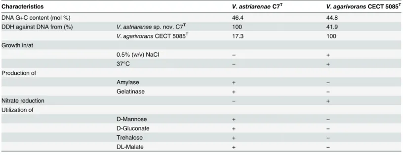
Table 1. Useful phenotypic and genotypic characteristics for distinguishingVibrio astriarenaesp. nov. andVibrio agarivorans.
Characteristics V.astriarenaeC7T V.agarivoransCECT 5085T
DNA G+C content (mol %) 46.4 44.8
DDH against DNA from (%) V.astriarenaesp. nov. C7T 100 41.9
V.agarivoransCECT 5085T 17.3 100 Growth in/at
0.5% (w/v) NaCl − +
37°C − +
Production of
Amylase + −
Gelatinase + −
Nitrate reduction − +
Utilization of
D-Mannose + −
D-Gluconate + −
Trehalose + −
DL-Malate + −
Data for utilization of organic compounds byV.agarivoransCECT 5085Twere obtained from Macián et al. (2004).
acid sequence of the genes can be used as queries for BLAST searches and re-verify their iden-tity. For instance, theoretically, a total of six genes should be present for the positive assimila-tion of galactose. Taking a close look at each gene involved, onlyα-D-galactose 1-phosphate permease was claimed not to be found inV.astriarenaesp. nov. C7Twith only 28.5% similar-ity. However, BLAST searches of the gene reveals up to 88% identity to symporter YagG of Vib-riospp. and with 96% query coverage, it shows 85% similarity to the putative permease of
Photobacterium profundum. The similarity is high and these proteins also belong to the sugar porter family of a major facilitator superfamily (MFS). Positive assimilation of galactosein vitroby C7Tmay be due to this similarity. Still, thorough investigation is needed before any other conclusion is made. In support to the prototype program developed forVibrionaceae[5], frequent usage of the program on manyVibriospecies will further validate and possibly draw its own circumscription value in determining a new species. Thus, based onin silico phenotyp-ing alone, strain C7Tis found to be closely related toV.alginolyticussupporting the above grouping method which grouped them together withVibriospecies. This experiment with combination of phylogenetic analyses (Figs1,2and3) showed that strains C7Tand C20 should be recognized not only as a new species but also as a new clade. The nameVibrio astriarenae
sp. nov. is proposed for strains C7Tand C20 indicating the islands from where they were originated.
Conclusion
In conclusion, all polyphasic taxonomic data combined with the genome based taxonomy now has confirmed the justification of our proposal ofV.astriarenaesp. nov. as a new species of vib-rio. BothpyrHand 16S rRNA gene sequence phylogenies, the most fundamental and strong selective measures, had grouped the strains into the genusVibrioand distinguished them from any knownVibriospp. Despite having similar agarolytic activity, the strains differed fromV.
agarivoransCECT 5085Tand its other closest relatives. Further genomic analyses including DNA G+C content, DNA-DNA hybridization (DDH), genomic sequencing, AAI, ANI andin silicoDDH of the type strain C7Tprovided a clear result that theV.astriarenaesp. nov. is the newest member ofVibrionaceae. Later, MLSA of eight housekeeping genes for both C7Tand C20 placed them into a new clade within the familyVibrionaceaetogether withV.agarivorans
CECT 5085T. The clade is named Agarivorans and further research and the sequencing of whole genome ofV.agarivoransCECT 5085Tand C20 is currently in progress. Comparative data on the basis of phenotypic characteristics also supports their novelty (Table 1andS2
Table) and simultaneously places them into the genusVibrio. Thirteen phenotypic features of
V.astriarenaesp. nov. C7Tcan be retrieved directly from its genome sequence using an auto-mated vibriophenotyping program
Description of
Vibrio astriarenae
sp. nov.
V.astriarenae. (as.tri.a.re'nae. L. n. astrum, star; L. n. arena, sand. N.L. gen. n. astriarenae, from ‘starry sand’. This refers to the isolation source of the sample site, Taketomi Island which is known for its starry sand due to the remains of foraminifera. The shells of these single-celled protists formed star shaped grains of sand along the beaches of Taketomi Island).
Gram-negative, facultatively anaerobic, motile with polar flagella rod isolated from seawater specimen collected from coral reef of Taketomi Island (24°20.5260' N; 124°05.6443' E). Cells are rod shaped, with rounded ends, and are 0.7 to 1.1μm in diameter and 2.4 to 3.1μm long
when the organism is grown in ZoBell 2216E agar. Polar flagella is observed when the organism is cultivated on solidified media and/or in liquid media. Colonies on ZoBell 2216E agar medium are non-pigmented, circular and smooth with entire edge. Sodium ion is essential for growth. Growth occurs at NaCl concentrations of 1.0 to 3.0% with optimum growth and appar-ent agarolytic activity at 3.0% NaCl. No growth is detected at 5.0% NaCl and beyond for both strains. Both strains grew at pH 5 to 10, optimally in pH 7.5. No growth was observed at pH
>10. The bacterium is a mesophilic chemoorganotroph which grows at temperatures between
20 and 30°C. No growth occurs at 15, 37 and 40°C. The type strain is negative for oxidase and positive for catalase, production of amylase, lipase, agarase, gelatinase and DNase; and assimi-lation ofD-glucose,D-xylose,D-mannose,D-galactose, trehalose, cellobiose, melibiose, lactose, D-gluconate,DL-malate,D-mannitol andN-acetylglucosamine. The bacterium is negative for gas production from glucose, acid production from glucose, nitrate reduction, acetoin, casei-nase and urease production, lysine decarboxylase, arginine dehydrolase, ornithine decarboxyl-ase, indole, luminescence, pigmentation; assimilation of sucrose,D-glucuronate, propionate, citrate, pyruvate,D-sorbitol,γ-aminobutyrate, putrescine,L-tyrosine andL-arabinose. The G+C content of the DNA from the type strain is 46.3 mol%. The strain C7Twas deposited in the Japan Collection of Microorganisms, Collection of Aquacultural Important Microorganisms, Mexico and BCCM/LMG Bacteria Collection, Belgium under respective accession numbers, JCM 19233T, CAIM 1900Tand LMG 28701T.
Supporting Information
S1 Table. List of reference strains used for the phylogenetic tree based on 16S rRNA gene sequence as shown inFig 1.
(DOCX)
S2 Table. Useful phenotypic characters for distinguishingVibrio astriarenaesp. nov. with
their closely related species. (DOCX)
S3 Table. Comparativein silicophenotypic characters ofV.astriarenaesp. nov. C7Twith
otherVibrionaceae. (DOCX)
S4 Table. Accession number of the reference strains used for the concatenated tree based on eight housekeeping gene sequences as shown inFig 3.
(DOCX)
S1 Dataset. The sequences of eight housekeeping gene ofVibrio astriarenaesp. nov. C7T
Acknowledgments
This work was supported by Genome Information Upgrading Program of National BioRe-source Project from the Ministry of Education, Culture, Sports, Science and Technology (MEXT) of Japan (MH and MO), Strategic Japanese-Brazilian Cooperative Program, Biomass and Bioenergy (TS and FLT), JSPS-CAPES bilateral cooperative program (TS and FLT), and Kaken (26660168) (TS). We are gratefully thank Mr. Tomioka for collecting seawater samples and special thanks go to Prof. Dr. Bernhard Schink from University of Konstanz, Germany for his advice on bacterial names. FLT and CT thank CAPES, CNPq, and FAPERJ for funding. PMM thanks CAPES for the PhD scholarship (4848-14-9 CAPES/JSPS) and MARA for the PhD education loan to NA.
Author Contributions
Conceived and designed the experiments: NA FG Tomoo Sawabe. Performed the experiments: NA FG AAKMR Kazumichi Sato Keisuke Sato SM PMM. Analyzed the data: NA FG AAKMR. Contributed reagents/materials/analysis tools: WS KO MH MO FLT CT GMAF BG Toko Sawabe Tomoo Sawabe. Wrote the paper: NA Tomoo Sawabe. Critical review, ideas and sug-gestion given during manuscript preparation: WS KO MH MO FLT CT GMAF BG Toko Sawabe Tomoo Sawabe.
References
1. Sawabe T, Ogura Y, Matsumura Y, Feng G, Amin AR, Mino S, et al. (2013) Updating theVibrioclades defined by multilocus sequence phylogeny: proposal of eight new clades, and the description ofVibrio tritonius sp. nov. Front Microbiol 4:1–14
2. Vandamme P, Peeters C. (2014) Time to revisit polyphasic taxonomy. Antonie Van Leeuwenhoek 106: 57–65. doi:10.1007/s10482-014-0148-xPMID:24633913
3. Thompson CC, Amaral GR, Campeão M, Edwards RA, Polz MF, Dutilh BE, et al. (2014) Microbial tax-onomy in the post-genomic era: Rebuilding from scratch. Arch Microbiol 197: 359–370. doi:10.1007/ s00203-014-1071-2PMID:25533848
4. Achtman M, Wagner M. (2008) Microbial diversity and the genetic nature of microbial species. Nat Rev Microbiol 6: 431–440. doi:10.1038/nrmicro1872PMID:18461076
5. Amaral GRS, Dias GM, Wellington-Oguri M, Chimetto L, Campeão ME, Thompson FL, et al. (2014) Genotype to phenotype: identification of diagnostic vibrio phenotypes using whole genome sequences. Int J Syst Evol Microbiol 64: 357–365. doi:10.1099/ijs.0.057927-0PMID:24505074
6. Rosselló-Móra R, Amann R (2015) Past and future species definitions for Bacteria and Archaea. Syst Appl Microbiol 38: 1–8
7. Gomez-Gil B (2003)Vibrio paciniisp.nov., from cultured aquatic organisms. Int J Syst Evol Microbiol 53: 1569–1573. PMID:13130050
8. List of prokaryotic names with standing in nomenclature. Available:www.bacterio.net. Accessed on: 16 May 2015.
9. Thompson FL, Gevers D, Thompson CC, Dawyndt P, Hoste B, Munn CB, et al. (2005) Phylogeny and molecular identification of vibrios on the basis of multilocus sequence analysis. Appl Environ Microbiol 71: 5107–5115. PMID:16151093
10. Boucher Y, Stokes HW. (2006) The roles of lateral gene transfer and vertical descent in vibrio evolution. In: Thompson FL, Austin B, Swings J, editors. The biology of vibrios. ASM Press: Washington, DC. pp. 84–94.
11. Thompson JR, Polz MF. (2006) Dynamics of vibrio population and their role in environmental nutrient cycling. In: Thompson FL, Austin B, Swings J, editors. The biology of vibrios. ASM Press: Washington, DC. pp. 190–203.
12. Macián MC, Ludwig W, Schleifer KH, Pujalte MJ, Garay E. (2001)Vibrio agarivoranssp.nov., a novel agarolytic marine bacterium. Int J Syst Evol Microbiol 51: 2031–2036. PMID:11760944
14. Chenl LC, Craigiel JS, Xie ZK. (1994) Protoplast production fromPorphyra linearisusing a simplified agarase procedure capable of commercial application. J Appl Phycol 6: 35–39.
15. Dipakkore S, Reddy CRK, Jha B. (2005) Production and seeding of protoplasts ofPorphyra okhaensis
(Bangiales, Rhodophyta) in laboratory culture. J Appl Phycol 17: 331–337.
16. Gupta V, Trivedi N, Kumar M, Reddy CRK, Jha B. (2013) Purification and characterization of exo-β-agarase from an endophytic marine bacterium and its catalytic potential in bioconversion of red algal cell wall polysaccharides into galactans. Biomass Bioenergy 49: 290–298.
17. Matsumura Y, Sato K, Al-Saari N, Nakagawa S, Sawabe T. (2014) Enhanced hydrogen production by a newly described heterotrophic marine bacterium,Vibrio tritoniusstrain AM2, using seaweed as the feedstock. Int J Hydrogen Energy 39: 7270–7277.
18. Marmur J. (1961). A procedure for the isolation of deoxyribonucleic acid frcom microorganisms. J. Mol. Biol. 3:208–218.
19. Tamaoka J, Komagata K. (1984) Determination of DNA base composition by reversed-phase high-per-formance liquid chromatography. FEMS Microbiol Lett 25:125–128.
20. Ezaki T, Hashimoto Y, Takeuchi N, Yamamoto H, Liu S-L, Miura H, et al. (1988) Simple genetic method to identify viridans group streptococci by colorimetric dot hybridization and fluorometric hybridization in microdilution wells. J Clin Microbiol 26: 1708–1713. PMID:3183018
21. Meinkoth J, Wahl G. (1984) Hybridization of nucleic acids immobilized on solid supports. Anal Biochem 138: 267–284. PMID:6204550
22. Ezaki T, Hashimoto Y, Yabuuchi E. (1989) Fluorometric deoxyribonucleic acid-deoxyribonucleic acid hybridization in microdilution wells as an alternative to membrane filter hybridization in which radioiso-topes are used to determine genetic relatedness among bacterial strains. Int J Syst Bacteriol 39:224–
229.
23. Sawabe T, Sugimura I, Ohtsuka M, Nakano K, Tajima K, Ezura Y, et al. (1998)Vibrio halioticolisp. nov., a non-motile alginolytic marine bacterium isolated from the gut of abaloneHaliotis discus hannai. Int J Syst Bacteriol 48: 573–580. PMID:9731299
24. Larkin MA, Blackshields G, Brown NP, Chenna R, McGettigan PA, McWilliam H, et al. (2007) Clustal W and Clustal X version 2.0. Bioinform 23: 2947–2948.
25. Tamura K, Stecher G, Peterson D, Filipski A, Kumar S. (2013) MEGA6: Molecular Evolutionary Genet-ics Analysis version 6.0. Mol Biol Evol 30: 2725–2729. doi:10.1093/molbev/mst197PMID:24132122
26. Saitou N, Nei M. (1987) The neighbor-joining method: a new method for reconstructing phylogenetic trees. Mol Biol Evol 4: 406–425. PMID:3447015
27. Aziz RK, Bartels D, Best A, DeJongh M, Disz T, Edwards RA, et al. (2008) The RAST Server: rapid annotations using subsystems technology. BMC Genomics 9:75. doi:10.1186/1471-2164-9-75PMID:
18261238
28. Meier-Kolthoff JP, Auch AF, Klenk H-P, Göker M. (2013) Genome sequence-based species delimita-tion with confidence intervals and improved distance funcdelimita-tions. BMC Bioinformatics 14:60. doi:10. 1186/1471-2105-14-60PMID:23432962
29. Camacho C, Coulouris G, Avagyan V, Ma N, Papadopoulos J, Bealer K, et al. (2009) BLAST+: architec-ture and applications. BMC Bioinformatics 10: 421. doi:10.1186/1471-2105-10-421PMID:20003500
30. Nelder JA, Wedderburn RWM. (1972) Generalized Linear Models. J R Stat Soc Ser A 135: 370–384.
31. Konstantinidis KT, Tiedje JM. (2005) Towards a genome-based taxonomy for prokaryotes. J Bacteriol 18: 6258–6264.
32. Thompson CC, Chimetto L, Edwards RA, Swings J, Stackebrandt E, Thompson FL. (2013) Microbial genomic taxonomy. BMC Genomics 14: 913. doi:10.1186/1471-2164-14-913PMID:24365132
33. Sawabe T, Kita-Tsukamoto K, Thompson FL. (2007) Inferring the evolutionary history of vibrios by means of multilocus sequence analysis. J Bacteriol 189: 7932–7936. PMID:17704223
34. Bandelt H-J, Dress AWM. (1992) Split decomposition: a new and useful approach to phylogenetic anal-ysis of distance data. Mol Phylogenet Evol 1: 242–252. PMID:1342941
35. Huson DH, Bryant D. (2005) Application of phylogenetic networks in evolutionary studies. Mol Biol Evol 23: 254–267. PMID:16221896
36. Oppenheimer CH, ZoBell CE. (1952) The growth and viability of sixty-three species of marine bacteria as influenced by hydrostatic pressure. J Mar Res 11:10–18.
37. Baumann P, Schubert RHW. (1984)Vibrionaceae. In Krieg NR, Holt JG, editors. Bergey's manual of systematic bacteriology Vol. 1. Williams and Wilkins: Baltimore. pp. 516–550.
39. Holt JG, Krieg NR, Sneath PHA, Staley JT, Williams ST. (1994) Facultatively anaerobic gram-negative rods. Subgroup 2: Family Vibrionaceae. In Bergey's manual of determinative microbiology, 9th edition. Williams and Wilkins: Baltimore. pp.190–253.
40. Leifson E. (1963) Determination of carbohydrate metabolism of marine bacteria. J Bacteriol 82:33–36.
41. Ostle AG, Holt JG. (1982) Nile blue A as fluorescent stain for poly-beta-hydroxybutyrate. Appl Environ Microbiol 44:238–241. PMID:6181737
42. West M, Burdash NM, Freimuth F. (1977) Simplified silver plating stain for flagella. J Clin Microbiol 6:414–419. PMID:72075
43. Tindall BJ, Rosselló-Móra R, Busse H-J, Ludwig W, Kämpfer P. (2010) Notes on the characterization of prokaryote strains for taxonomic purposes. Int J Syst Evol Microbiol 60: 249–266. doi:10.1099/ijs.0. 016949-0PMID:19700448
44. Kita-Tsukamoto K, Oyaizu H, Nanba K, Simidu U. (1993) Phylogenetic relationships of marine bacteria, mainly members of the familyVibrionaceae, determined on the basis of 16S rRNA sequences. Int J Syst Bacteriol 43: 8–19. PMID:8427811
45. Pascual J, Macián MC, Arahal DR, Garay E, Pujalte MJ. (2010) Multilocus sequence analysis of the central clade of the genusVibrioby using the 16S rRNA,recA,pyrH,rpoD,gyrB,rctBandtoxRgenes. Int J Syst Evol Microbiol 60: 154–165. doi:10.1099/ijs.0.010702-0PMID:19648344
46. Thompson CC, Vicente ACP, Souza RC, Vasconcelos ATR, Vesth T, Alves N, et al. (2009) Genomic taxonomy of Vibrios. BMC Evol Biol 9: 258. doi:10.1186/1471-2148-9-258PMID:19860885
47. Barton LL. (2005) Structural and functional relationships in prokaryotes. Springer Science: USA. pp. 3–43.
48. Chimetto LA, Cleenwerck I, Moreira APB, Brocchi M, Willems A, De Vos P, et al. (2011)Vibrio variabilis
sp. nov. andVibrio maritimussp. nov., isolated fromPalythoa caribaeorum. Int J Syst Evol Microbiol 61: 3009–3015. doi:10.1099/ijs.0.026997-0PMID:21296931
49. Xu X-W, Wu Y-H, Wang C-S, Oren A, Wu M. (2009)Vibrio hangzhouensissp. nov., isolated from sedi-ment of the East China Sea. Int J Syst Evol Microbiol 59: 2099–2103. doi:10.1099/ijs.0.008698-0
PMID:19605706
50. Macián MC, Garay E, Grimont P a D, Pujalte MJ. (2004)Vibrio ponticussp. nov., a neighbour ofV. flu-vialis—V.furnissiiclade, isolated from gilthead sea bream, mussels and seawater. Syst Appl Microbiol
27: 535–540. PMID:15490554
51. Thompson FL. (2003)Vibrio neptuniussp. nov.,Vibrio brasiliensissp. nov. andVibrio xuiisp. nov., iso-lated from the marine aquaculture environment (bivalves, fish, rotifers and shrimps). Int J Syst Evol Microbiol 53: 245–252. PMID:12656180
52. Amaral GRS, Campeão ME, Swings J, Thompson FL, Thompson CC. (2015) Finding diagnostic pheno-typic features ofPhotobacteriumin the genome sequences. Antonie Van Leeuwenhoek 107: 1351–
1358. doi:10.1007/s10482-015-0414-6PMID:25724129