Expression and Reduces M1T1 Group A Streptococcal
Systemic Virulence
Andrew Hollands1,4, Ramy K. Aziz2, Rita Kansal3, Malak Kotb3,4, Victor Nizet5,6, Mark J. Walker1* 1School of Biological Sciences, University of Wollongong, Wollongong, New South Wales, Australia,2Department of Microbiology and Immunology, Faculty of Pharmacy, Cairo University, Cairo, Egypt,3The VA Hospital, Memphis, Tennessee, United States of America,4The Department of Molecular Genetics, Biochemistry and Microbiology, The University of Cincinnati, College of Medicine, Cincinnati, Ohio, United States of America,5Department of Pediatrics, University of California San Diego, La Jolla, California, United States of America,6Skaggs School of Pharmacy and Pharmaceutical Sciences, University of California San Diego, La Jolla, California, United States of America
Abstract
Epidemiological studies of group A streptococcus (GAS) have noted an inverse relationship between SpeB expression and invasive disease. However, the role of SpeB in the course of infection is still unclear. In this study we utilize a SpeB-negative M1T1 clinical isolate, 5628, with a naturally occurring mutation in the gene encoding the regulator RopB, to elucidate the role of RopB and SpeB in systemic virulence. Allelic exchange mutagenesis was used to replace the mutatedropBallele in 5628 with the intact allele from the well characterized isolate 5448. The inverse allelic exchange was also performed to replace the intactropB in 5448 with the mutated allele from 5628. An intactropB was found to be essential for SpeB expression. While theropBmutation was shown to have no effect on hemolysis of RBC’s, extracellular DNase activity or survival in the presence of neutrophils, strains with the mutatedropBallele were less virulent in murine systemic models of infection. An isogenic SpeB knockout strain containing an intact RopB showed similarly reduced virulence. Microarray analysis found genes of the SpeB operon to be the primary target of RopB regulation. These data show that an intact RopB and efficient SpeB production are necessary for systemic infection with GAS.
Citation:Hollands A, Aziz RK, Kansal R, Kotb M, Nizet V, et al. (2008) A Naturally Occurring Mutation inropBSuppresses SpeB Expression and Reduces M1T1 Group A Streptococcal Systemic Virulence. PLoS ONE 3(12): e4102. doi:10.1371/journal.pone.0004102
Editor:Adam J. Ratner, Columbia University, United States of America
ReceivedSeptember 10, 2008;AcceptedNovember 21, 2008;PublishedDecember 31, 2008
Copyright:ß2008 Hollands et al. This is an open-access article distributed under the terms of the Creative Commons Attribution License, which permits unrestricted use, distribution, and reproduction in any medium, provided the original author and source are credited.
Funding:This work is funded by the NIH (grant AI077780 to VN and AI04198 to MK), National Health and Medical Research Council (grant number 459103 to MW, VN and MK), Veteran’s Affairs Merit Award (MK) and the Department of Education, Science and Training (International Linkages Grant CG110095 to MW, VN and MK). Andrew Hollands is the recipient of an Australian Postgraduate Award. Funding agencies played no role in the study and in the decision to submit the manuscript or its preparation.
Competing Interests:The authors have declared that no competing interests exist.
* E-mail: [email protected]
Introduction
Streptococcus pyogenes (group A streptococcus; GAS) is a Gram-positive, human-specific pathogen responsible for over 500,000 deaths each year [1]. Severe invasive GAS infections such as necrotizing fasciitis account for approximately 30% of these deaths, and the incidence of such acute conditions has been on the rise since the mid 1980’s [2]. This resurgence has been paralleled by the emergence of a globally disseminated GAS cone belonging to serotype M1T1 [3–5]. While the M1T1 GAS has become the most common cause of streptococcal pharyngitis, this clone is also overrepresented in cases of severe invasive disease [6,7].
Studies of M1T1 clinical isolates from invasive disease cases have revealed an inverse relationship between expression of the extracellular cysteine protease SpeB and clinical severity [8]. The existence of a SpeB-negative invasive phenotype has been hypothesized that results from mutations in the regulator covR/ S [9]. SpeB is a secreted cysteine protease initially expressed as 40 kDa zymogen which is then converted to the 28 kDa active form by autocatalytic processing [10]. SpeB is known to cleave numerous host proteins including components of the extracellular matrix, cytokine precursors, immunoglobulins and antimicrobial
peptides [11–13], which could interfere with host immune functions. However, SpeB has also been shown to cleave a range of GAS proteins such as the fibrinogen-binding M1 protein [14,15], various superantigens [16,17], the secreted plasminogen activator streptokinase [18] as well as the DNase Sda1 [17], and thus possibly interfere with the proven virulence functions of these bacterial factors. The precise role(s) of SpeB throughout the course of infection are undoubtedly complex, and not surprisingly, different studies using different in vivo animal models have produced seemingly contradictory results [19–21].
In this study we examined the effect of a natural mutation in the gene encoding the regulator RopB (also known as Rgg [22]) identified in a SpeB-negative GAS clinical isolate. RopB is a GAS transcriptional regulator that has been shown to be essential for expression of SpeB and binds directly to the promoter region of
Subsequent investigations into the effect of RopB on virulence have yielded differing results. A study utilizing a zebrafish intramuscular infection model with serotype M5 GAS showed that inactivation of RopB resulted in decreased virulence [30], whereas a study utilizing a murine intraperitoneal infection model with serotype M49 GAS showed that inactivation of RopB resulted in increased virulence [27].
While such global differences in virulence effects could in part result from the differing animal models used, it may also reflect strain-specific variation in the RopB regulon. For example, separate studies have shown ropB mutation to have either no effect on hemolysis and DNase activity or, alternatively, to increase expression of hemolysin and DNase-encoding genes and the associated phenotypic activities [23,26]. This strain-specific variation is highlighted in a recent work by Dmitriev et al.[29] that shows inter- and intra-serotypic variation in the transcriptome ofropBmutant GAS, with only members of the SpeB operon being commonly regulated in all strains tested.
It is in the light of the current uncertainty surrounding RopB and its role in virulence that we sought to investigate the role of this transcriptional regulator in the serotype M1T1 GAS background that is the leading agent of severe human infection. This analysis begins with a naturally-occuring mutation in ropB
identified in one such strain.
Materials and Methods
Bacterial strains, media and growth conditions
The M1T1 GAS clinical isolates, 5448 and 5628, used in this study have been previously described [31]. Both 5448 and 5628 are speA-positive M1T1 strains that were isolated from patients with STSS that were recruited through an ongoing population-based surveillance for invasive GAS infections in Ontario, Canada. Both strains were determined to be derived from the same clone as detailed elsewhere [3]. GAS strains were grown in Todd-Hewitt broth containing 1% yeast extract (THY), or on Todd-Hewitt agar plates (THA). Escherichia coli were grown in Luria-Bertani broth (LB) or on Luria-Bertani agar (LA). For antibiotic selection, erythromycin (Erm) was used at 5mg/ml for GAS and 500mg/ml forE. coli.
Allelic exchange mutagenesiswas performed essentially as previously described [32]. The ropB allele plus upstream and downstream flanking regions was amplified from 5448 or 5628 using the primers RopB-F-BamHI (59-CAG GAT CCC TCA TTT CAG TTG ACA AGA AAC-39) and RopB-R-XbaI (59-CGC TCT AGA TAC CAA AAG GCT AGA CCT CTG-39). The PCR products and temperature sensitive vector pHY304 were ligated using T4 ligase to create the plasmids pHY5448RopB and pHY5628RopB. The plasmid pHY5628RopB was transformed into GAS strain 5448 and the plasmid pHY5448RopB was transformed into GAS strain 5628, and Ermrtransformants were grown at the permissive temperature for plasmid replication (30uC). Single-crossover chromosomal insertions were selected by shifting to the nonpermissive temperature (37uC) while maintaining Erm selection. Single crossover colonies were then grown in the absence of antibiotic selection at 30uC and Ermscolonies were then screened for the presence of the appropriateropBallele using DNA sequence analysis. Confirmed allelic exchange mutants were designated 5448R2 (5448 containing the mutantropBallele from 5628) and 5628R+(5628 containing the wildtype (WT)ropBallele from 5448).
SpeB activity assays
Cysteine protease activity assays were performed as described by Collinet al.[33]. GAS strains were grown overnight at 37uC.
Cultures were then diluted 1:50 and grown for 17 h at 37uC. The cultures were centrifuged at 32006g, and the supernatants sterile-filtered through 0.2mm syringe-driven filters (Whatman). 200ml of filtered supernatant was mixed with 200ml of activation buffer (1 mM EDTA, 20 mM DTT in 0.1 M sodium acetate buffer, pH 5.0) and incubated for 30 min at 40uC. 400ml of 2% (w/v) azocasein in activation buffer was then added and incubated for a further 1 h at 40uC. Trichloro-acetic acid was then added to a final concentration of 15% (w/v) and thoroughly mixed. The mixture was then centrifuged at 15,0006g for 5 min and the OD366of the supernatant was then measured to indicate cleavage
of the azocasein by SpeB.
SpeB Western blot
For Western blot analysis, bacterial cultures were grown to late stationary phase (17 h) and pelleted by centrifugation at 3,2006g
for 10 min. The supernatants were sterile-filtered through 0.22mm syringe-driven filters (Millipore). Supernatants were diluted 1:8 and 10ml of each sample was run on a 10% Bis-Tris Gel with MOPS running buffer (Invitrogen). Proteins were then transferred to a Nitrocellulose membrane (Invitrogen) by use of a Trans-Blot SD semi-dry transfer cell (BioRad) for 1 h at 20 V. The presence of SpeB in culture supernatants was detected using primary rabbit anti-SpeB diluted 1:1,000 for 1.5 h. Following subsequent washing, the membrane was incubated with a secondary goat anti-rabbit-HRP conjugate diluted 1:10,000 for 1 h. SuperSignal West Pico Chemiluminescent Substrate (Thermo Scientific) was used to expose autoradiography film as per the manufacturer’s instructions.
Hemolytic activity assay
Fresh, heparinized human blood from healthy volunteers was washed twice with sterile PBS and resuspended to a final concentration of 2% (v/v) in PBS. Bacterial cultures were grown to mid-log phase (OD600= 0.4), pelleted by centrifugation at
3,2006g and the supernatants sterile-filtered through 0.22mm syringe-driven filters (Millipore). 100ml of blood solution was mixed with 100ml of serially diluted supernatant. The plates were incubated for 1 h at 37uC, then 1 h at 4uC. Following centrifugation at 1,5006gfor 10 min, 100ml was transferred into a fresh 96-well flat bottom plate and the absorbance at 405 nm was recorded.
DNase activity assays
DNase assays were performed as previously described [34,35]. Briefly, GAS strains were grown to mid-log phase (OD600= 0.4),
and the supernatants collected by centrifugation at 32006g. 2.5ml of bacterial supernatant was added to 7.5ml of calf thymus DNA (1mg/ml) and 40ml of DNase buffer (3 mM MgCl2, 3 mM
CaCl2, 300 mM Tris; pH 7.4). The reaction was incubated for
5 min at 37uC and then stopped by the addition of 12.5ml 0.33 M EDTA (pH 7.3). The relative DNA degradation was then compared by gel electrophoresis on a 1% agarose gel.
Neutrophil killing assays
Human neutrophils were isolated from venous blood of healthy volunteers using the PolyMorphPrep kit as per the manufacturer’s instructions (Axis-Shield, Norway). In 96-well plates, we mixed 26105 neutrophils and 26104 colony forming units (cfu) of
logarithmic phase GAS in RPMI containing 2% heat-inactivated autologous plasma in a total volume of 200ml. The plates were centrifuged at 5006gfor 5 min and incubated at 37uC in 5% CO2
incubated overnight at 37uC and cfu enumerated. Control wells containing bacteria but no neutrophils were used to determine survival. Percentage survival was calculated as [cfu/ml experi-mental well]/[cfu/ml control well]6100%.
In vivoSpeB switching studies
GAS strain 5628R+was grown to mid-log phase (OD600= 0.4),
pelleted by centrifugation at 3,2006gand washed twice with sterile PBS. Bacteria were then resuspended in PBS at a final concentration of 16109cfu/ml. 100ml of bacterial suspension (16108cfu) was injected subcutaneously into the flank of 10 week
old C57BL6/J mice. Three days post-infection, bacteria were recovered from the lesion and screened for SpeB status using the skim-milk agar method as previously described [36].
Tissue cage implantation andin vivobacterial growth
To obtain in vivo-derived RNA for microarray analysis, a previously described subcutaneous murine Teflon chamber model was used [31]. Approximately 108cfu of bacteria were injected in sterile Teflon chambers that had been surgically inserted under the skin of age-matched female BALB/c mice. After 24 h, bacteria were collected, tested for purity and phenotypic homogeneity on blood agar plates and Columbia-casein agar plates, respectively. Only pure isolates with homogeneous protease activity (either positive or negative) were further selected for RNA extraction. Selected isolates recovered from Teflon chambers were centri-fuged, and their pellets were washed twice in PBS, sheared with beads (QBiogene), then processed for RNA extraction according to the RNEasey protocol (Qiagen).
Expression microarrays
For transcriptome analysis, we used oligomer-based microarrays printed in the Molecular Resource Center at the University of Tennessee Health Science Center. Each array consists of duplicates of 2,328 oligomers (70-mers) that represent all ORFs in M1 GAS, strain SF370 (GenBank accession#NC_002737) in addition to oligomers representing ORFs from prophages in strains MGAS8232 (GenBank accession# NC_003485) and MGAS315 (GenBank accession#NC_004070). The arrays also contained positive and negative controls of streptococcal ribosomal DNA and alien DNA (Stratagene), respectively.
cDNA preparation and microarray hybridization
Bacterial RNA was treated with DNase Turbo (Ambion) for 1 h to remove any genomic DNA contamination, then converted it to dendrimer-labeled cDNA using the 3DNA Array 900TM kits (Genisphere, http://www.genisphere.com/array_detection_900. html) following the manufacturer’s protocol. Equal amounts of dendrimer-labeled cDNA from different pairs of isolates were mixed, applied to the glass microarrays, and incubated for 16 h. After this first hybridization, the arrays were washed, labeled with a mixture of Alexa Fluor 546 and Alexa Fluor 647 (Genisphere), incubated for 3 h, washed again, then scanned bv GenePix 4000B scanner (Axon Instruments, Inc.) We followed a cyclic design that allowed us to compare every condition to each other at least twice, and guaranteed dye swapping to eliminate effects of non-specific binding.
Analysis of microarray data
To analyze the microarray data, GenePixPro 4.0 software (Axon Instruments, Inc.) was used for image processing, fluorescent normalization and spot finding, then GeneSpring GX 7.3.1 (Agilent Technologies) was used for normalization, statistical analysis, clustering analysis and gene-list generation. All
primary microarray data were submitted to the NCBI Gene Expression Omnibus (GEO) in accordance with MIAME standards (GEO accession#GSE13656).
Murine systemic infection models
GAS strains were grown to mid-log phase (OD600= 0.4),
pelleted by centrifugation at 3,2006g and washed twice with
sterile PBS. For intravenous challenge, bacteria were then resuspended in PBS at a final concentration of 16109cfu/ml. 200ml of bacterial suspension (26108cfu) was injected into the
lateral tail vein of 10 week old C57BL6/J mice. For intraperitoneal infection, the bacteria were resuspended at a concentration of 2.56108 cfu/ml in PBS containing 5% (w/v) mucin. 200ml of bacterial suspension (56107cfu) was injected into the peritoneal
cavity of C57BL6/J mice. For both experiments, the mice were monitored for 10 days and deaths recorded every 24 h.
Results
Sequence analysis of clinical isolate 5628 reveals intact speBandcovR/Sbut mutation in ropB
M1T1 GAS clinical isolate 5628 was found on screening to lack SpeB activity by azocasein assay. DNAsequencing was performed using primers listed in Table 1. No mutations were found in the strain 5628 in thespeBgene nor in thecovR/Slocus, which has been previously linked to loss of SpeB expression [37,38]. Further sequencing was performed on the previously described regulators
luxS,rofA,ropAandropB[24,39–41]. This analysis revealed a point mutation inropB, leading to truncation of RopB at amino acid 170 of the 280 amino acid protein (Fig. 1A). The truncation of RopB suggested that this may be the cause of lack of SpeB expression in this strain.
Repair of the 5628ropB allele restores SpeB expression and activity
Western blot detected SpeB in overnight culture supernatants of 5628R+, containing the intactropBallele, but not in the clinical isolate 5628 nor the isogenic mutant 5628RDSpeB, containing the intact
ropBallele but lackingspeB(Fig. 1B). This expression correlated with a restoration of extracellular protease activity as detected by azocasein degradation assay (Fig. 1C). Protease activity was abrogated by the addition of the cysteine protease inhibitor E64. These data demonstrated that theropBpoint mutation was indeed responsible for the lack of SpeB expression in the clinical isolate 5628.
RopBmutation does not affect bacterial growth, hemolysis, DNase activity or resistance to neutrophil killing
GAS strain 5628 and its isogenic mutants 5628R+ and 5628RDSpeB were grown in THB, and the OD600was measured
over time. No significant difference was found among the growth rates of the three bacterial strains (Fig. 2A). Hemolysis assays were performed to determine the effect ofropBmutation on the cytolytic ability of GAS. No significant difference was seen in hemolytic activity among strains containing the WT or mutatedropB allele (Fig. 2B). Extracellular DNase activity of mid-log phase bacteria and GAS resistance to neutrophil killing were also unaffected by mutation inropB(Fig. 2C and 2D).
SpeB-positive bacteria revert to a SpeB-negative phenotype on subcutaneous infection
recovered from the lesion and screened for SpeB status using the skim-milk agar method. While the inoculum was 100% SpeB-positive, bacterial populations recovered from the lesions of individual mice were found to be 2%, 4%, 6%, 8%, and 24% SpeB-negative. Five representative colonies were picked, and the
covR/S locus was sequenced in each of them. All colonies sequenced were found to have substitution mutations in covR/S
resulting in truncation of CovS. This finding reveals that when SpeB-negative colonies are selected for in vivo, this selection is predominantly a phenomenon associated with CovR/S
inactiva-Table 1.Primers used for sequencing theropBlocus (includingspeB), thecovR/Slocus,luxS,rofAandropA.
Primer name Sequence (59–39)
RopB-F1 GACTGTTCGTTAGAAAGCCA
RopB-F2 CTCCTGATACGATGATAA
RopB-F3 AAAGTTTTCTTTCAAGGC
RopB-F4 CTTTGATTTGTTCGACAT
RopB-F5 TACCATGAATGGTAATAG
RopB-F6 TATCTCACTACCATTTTGC
RopB-F7 TGAGTTTCTCTTTATTAG
RopB-F8 GAACGGTGTTGTGTGTCT
RopB-R9 AGTCACCCATTGATAAAG
RopB-R10 AGGCGGCTTCAACGGTTA
RopB-R11 AGACTACACTTACACACT
RopB-R12 ATCCAAAAATCAGCAGCTATC
RopB-R13 TTAACAAAATGAGAACGG
RopB-R14 TGATAGTCGCTTATGATA
RopB-R15 CATATTGACAAACATCCGAATCG
RopB-R16 GCTGTTGAGATAAACTAC
RopB-R17 CTAGACCTCTGCTCACTAG
CovRS-F1 GCTATTCCGGTACAGGTGT
CovRS-F2 GTCAATGGTCGTGAAGGGT
CovRS-F3 GATGTCTATATTCGTTATCTCC
CovRS-F4 GATGATTTTTACCACAGATAAC
CovRS-F5 GCATATTGGTCTCTTACAAC
CovRS-F6 GCAAATTGTAGATGGGTATCA
CovRS-R7 GCGGAAAATAGCACGAATAC
CovRS-R8 AGGCAATCAGTGTAAAGGCA
CovRS-R9 CTTGTGCCAAATAACTCAACA
CovRS-R10 ATCAAAAGCCTGCTCAAATGA
CovRS-R11 CTTTCATGTCATCCATCATTG
CovRS-R12 TTGCTCTCGTGTGCCATCT
LuxS-F1 GCAGCTCTATTGCACCTAT
LuxS-F2 AAGAAGTTATCGTCGAAA
LuxS-F3 AATCCTACTGACCTATTT
LuxS-R4 TAGTGGCAACACGGTGAA
LuxS-R5 TGAAAACCTGTTCGACAG
LuxS-R6 ATAATGGCAATGGTTAC
LuxS-R7 GTACCTTACAATCAAGATGTT
RofA-F1 TCTTGAGCTAATGCAACCGT
RofA-F2 GAATCCGTTAGGAGATGA
RofA-F3 GTTTCGATAATATCATGG
RofA-F4 AAAGGATGTGTAAATTGG
RofA-F5 ACAAGGTTTCCAAATAAG
RofA-R6 AAGCAATTAACATAAGCG
RofA-R7 TCTGCAACATTTTATTCC
RofA-R8 GGCATTAAAGTTTATGAC
RofA-R9 TAGGAAGAGAGGTCCCTT
RofA-R10 GAACTTGAATCTGGATTTATTG
RopA-F1 TCTTGTCCTGCAAATACGTC
RopA-F2 TCTTCTTGAGTTGTACCA
RopA-F3 CTCAACACCATCAACTGA
Primer name Sequence (59–39)
RopA-F4 GATTTGTAGCTTTGTTTTC
RopA-R5 TTAAGGAACAAAACGTACAAG
RopA-R6 ATGTTGACACACTTGAAG
RopA-R7 TGTTGTGTCAATGGAAAA
RopA-R8 ATGCCATAGTCATCCGTT
RopA-R9 AATCCTTTCTTTGATAGTTTATC
doi:10.1371/journal.pone.0004102.t001 Table 1.cont.
Figure 1. Mutation inropBresults in truncation of the RopB protein and abrogation of SpeB expression. (A) Schematic representation of the RopB protein expressed by GAS strains 5448 and 5628. In 5628, white region represent homology to 5448, shaded region indicates unique protein sequence in 5628. (B) Western blot for SpeB using overnight culture supernatants. (C) SpeB activity assay using azocasein substrate.
tion. TheropB mutation of strain 5628 was not recapitulated on passage in the mouse subcutaneous infection model.
SpeB is the principal target of RopB regulationin vivo
Microarray analysis was performed onin vivo-derived RNA from the well characterized GAS strain 5448 and its derivative 5448R2, containing the mutatedropB allele from 5628. Fourty-seven genes were found to be down-regulated and 52 genes were found to be up-regulated in 5448R2greater than 2-fold (P,0.05). The most strongly down-regulated genes in 5448R2are members of the SpeB operon, as would be expected from the lack of SpeB expression and activity in 5628 (Fig. 3). In addition to the SpeB operon, genes of the streptolysin S operon were also found to be
strongly down-regulated in theropBmutant strain. Apart from the gene encoding the superantigen SmeZ, there was an absence of virulence-related genes found to be strongly up-regulated in the strain 5448R2, which expressed the truncated RopB.
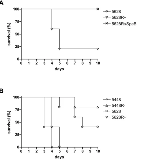
RopB is required for virulence in systemic infection
To determine the effect of ropB mutation on virulence, C57BL6/J mice were subjected to intraperitoneal infection with mid-logarithmic phase bacteria. GAS strain 5628R+expressing a full length RopB showed increased virulence compared to the strain 5628 with a truncated RopB (Figure 4A). Strain 5628RDSpeB containing a full-length RopB but lacking SpeB also showed reduced virulence, suggesting that the reduced
virulence of 5628 may primarily result from the lack of SpeB expression in this strain. Virulence was also examined in an intravenous model of systemic infection. The well characterized strain 5448 and its isogenic mutant 5448R2, containing theropB
allele from 5628, were further included to examine the effect of this allelic variation in a well-characterized virulent strain of GAS. The two strains containing a truncated RopB (5628 and 5448R2) demonstrated similarly reduced virulence compared to the two strains containing a full-length RopB (5628R+and 5448) (Fig. 4B), confirming that inactivation of RopB results in decreased virulence in systemic infection.
Discussion
The cumulative contribution of the secreted cysteine protease SpeB to the pathogenesis of invasive GAS infection is at present unclear, and studies in various GAS serotypes and animal models have produced varying results [19–21]. In the globally-dissemi-nated clonal M1T1 serotype associated with epidemic invasive GAS infection, an inverse relationship has been found between SpeB activity and clinical disease severity [8]. Recently, we and others have found a connection between inactivation of SpeB through mutation in the two-component regulatory system covR/ S and development of invasive disease in the murine model [37,38]. In this study, we have investigated a second mechanism of inactivation of SpeB, namely through truncation of the regulator RopB. We have shown that in M1T1 GAS, when thecovR/Slocus is intact and DNase Sda1 activity is unaffected, ropB point mutation results in reduced virulencein vivo, despite inactivation of SpeB activity.
The SpeB-negative, serotype M1T1 clinical isolate 5628 was used to investigate the role of RopB in virulence. RopB has previously been shown to be necessary for SpeB production [23,24] as well as regulation of other GAS virulence factors [25– 29]. The M1T1 strain 5628 contains a point mutation in theropB
gene that results in a truncation of the last 110 amino acids of the 280 amino acid protein. Restoration of full-length RopB in
5628R+resulted in gaining SpeB expression, but did not result in changes in hemolysis, DNase activity or resistance to killing by neutrophils.RopBmutation in this particular strain does not result in the kind of global phenotypic change found with covR/S
mutation where differential expression was observed of multiple genes encoding virulence determinants, includingsic,ska,slo,speA,
speJ, scpC and the hyaluronic acid synthesis operon [37] which were all unaffected in theropBmutant examined in this work.
Recently, we have demonstrated that WT, SpeB-positive bacteria undergo a phase-shift to a SpeB-negative phenotype after subcutaneous infection of mice [17]. This phase switch was observed to be the result of mutations in the two-component regulatory system covR/S [38]. CovR/S is a global regulator, whose inactivation is linked to an invasive phenotype and involves up-regulation of many genes encoding virulence factors such as streptodornase, streptokinase, streptolysin O, streptococcal inhib-itor of complement and the hyaluronic acid capsule synthesis operon [37]. Of the down-regulated genes in CovR/S mutant strains, SpeB would appear to be one of the most important due to its ability to degrade many host and self-proteins. It is the loss of SpeB expression in these mutant bacteria that is hypothesized to allow the invasive spread of GAS by sparing from SpeB degradation self-proteins involved in plasminogen accumulation and activation [42]. In this study we found that SpeB-positive bacteria with a restored RopB reverted to a SpeB-negative phenotype on subcutaneous infection. Furthermore, the switch to a SpeB-negative phenotype appeared to occur exclusively through mutations incovR/S. This result suggests thatcovR/Smutations are the predominant method of phenotypic switching in GAS and that
ropBmutation is not readily selected forin vivo. This leads to the conclusion thatropBmutation in 5628 does not represent a similar mechanism to covR/S mutation and may (a) be an incidental occurrence or (b) arise secondary to different selection pressures than those associated with the shift to invasive infection.
Microarray analysis was conducted usingin vivo-derived RNA, since the transcriptome of bacteria grownin vitromay differ greatly
Figure 3. Microarray analysis emphasizes the down-regulation of the SpeB operon.The figure shows the top 10 genes up- and down-regulated in theropBmutant bacteria compared to WT when the bacteria are inoculatedin vivo(P,0.05). The values plotted represent log ratios (log mutant/WT), and genes co-clustered in operons are highlighted (black bars, SpeB operon; white bars, SLS operon).
from the transcriptome found during the course of infection. Implantation of tissue cages allows for bacteria to be grown in an
in vivo environment for 24 hours prior to recovery and RNA extraction. The microarray data showed members of the SpeB operon to be the most strongly down-regulated genes following inactivation of RopB. This finding supported thein vitroandin vivo
data that suggested SpeB is the main target of RopB regulation in this strain.
Virulence studies utilizing systemic models of infection showed that a full-length RopB was required for virulence. The clinical isolate 5628 expressing a truncated RopB, as well as the allelic exchange mutant 5448R2 also expressing the truncated RopB from 5628, showed reduced virulence compared to strains containing a full-length RopB. These data illustrate the necessity for an intact RopB for full virulencein vivo. In addition, the strain 5628RDSpeB expressing an intact RopB but lacking SpeB showed similarly reduced virulence to the strain with a truncated RopB. While the down-regulation of the streptolysin S operon may contribute to the reduced virulence, this finding implies that the loss of SpeB in this strain is the main cause of the lack of virulence exhibited. This finding leads to the conclusion that inactivation of SpeB alone is not sufficient to initiate invasive disease. Moreover,
these data suggest that SpeB loss, only in the context of mutation of thecovR/Sregulatory circuit, promotes invasion. Of note, the additional virulence factors streptokinase and M1 protein, are both up-regulated incovR/Smutant M1T1 strains [37], but not in the
ropBmutant under investigation in this study. Streptokinase and M1 protein are believed to play a role in the accumulation of host plasmin at the GAS cell surface, a process which is thought to accentuate invasive disease [5,42,43].
Together these data provide evidence of RopB’s role in virulence. In M1T1 GAS, an intact RopB and efficient SpeB production are necessary for systemic infection.
Acknowledgments
The authors wish to thank William L. Taylor (University of Tennessee, Molecular Resource Center) for his help and guidance in hybridizing and scanning microarrays.
Author Contributions
Conceived and designed the experiments: AH VN MJW. Performed the experiments: AH RKA RK. Analyzed the data: AH RKA RK MK VN MJW. Contributed reagents/materials/analysis tools: RKA RK. Wrote the paper: AH MK VN MJW.
Figure 4. RopB and SpeB-negative bacteria show reduced virulence in systemic infection models.(A) Intraperitoneal infection of C57BL6/J mice with 56107cfu of GAS strains with 5% mucin. (B) Intravenous challenge of C57BL6/J mice with 26108cfu of GAS strains.
References
1. Carapetis JR, Steer AC, Mulholland EK, Weber M (2005) The global burden of group A streptococcal diseases. Lancet Infect Dis 5: 685–694.
2. Kaplan EL (1991) The resurgence of group A streptococcal infections and their sequelae. Eur J Clin Microbiol Infect Dis 10: 55–57.
3. Chatellier S, Ihendyane N, Kansal RG, Khambaty F, Basma H, et al. (2000) Genetic relatedness and superantigen expression in group A streptococcus serotype M1 isolates from patients with severe and nonsevere invasive diseases. Infect Immun 68: 3523–3534.
4. Tart AH, Walker MJ, Musser JM (2007) New understanding of the group A Streptococcus pathogenesis cycle. Trends Microbiol 15: 318–325.
5. Walker MJ, McArthur JD, McKay F, Ranson M (2005) Is plasminogen deployed as aStreptococcus pyogenesvirulence factor? Trends Microbiol 13: 308–313. 6. Demers B, Simor AE, Vellend H, Schlievert PM, Byrne S, et al. (1993) Severe
invasive group A streptococcal infections in Ontario, Canada: 1987–1991. Clin Infect Dis 16: 792–800; discussion 801–792.
7. Cleary PP, Kaplan EL, Handley JP, Wlazlo A, Kim MH, et al. (1992) Clonal basis for resurgence of seriousStreptococcus pyogenesdisease in the 1980s. Lancet 339: 518–521.
8. Kansal RG, McGeer A, Low DE, Norrby-Teglund A, Kotb M (2000) Inverse relation between disease severity and expression of the streptococcal cysteine protease, SpeB, among clonal M1T1 isolates recovered from invasive group A streptococcal infection cases. Infect Immun 68: 6362–6369.
9. Sumby P, Barbian KD, Gardner DJ, Whitney AR, Welty DM, et al. (2005) Extracellular deoxyribonuclease made by group A Streptococcus assists pathogenesis by enhancing evasion of the innate immune response. Proc Natl Acad Sci U S A 102: 1679–1684.
10. Musser JM, Stockbauer K, Kapur V, Rudgers GW (1996) Substitution of cysteine 192 in a highly conservedStreptococcus pyogenesextracellular cysteine protease (interleukin 1beta convertase) alters proteolytic activity and ablates zymogen processing. Infect Immun 64: 1913–1917.
11. Cunningham MW (2000) Pathogenesis of group A streptococcal infections. Clin Microbiol Rev 13: 470–511.
12. Hynes W (2004) Virulence factors of the group A streptococci and genes that regulate their expression. Front Biosci 9: 3399–3433.
13. Nyberg P, Rasmussen M, Von Pawel-Rammingen U, Bjorck L (2004) SpeB modulates fibronectin-dependent internalization of Streptococcus pyogenes by efficient proteolysis of cell-wall-anchored protein F1. Microbiology 150: 1559–1569.
14. Raeder R, Woischnik M, Podbielski A, Boyle MD (1998) A secreted streptococcal cysteine protease can cleave a surface-expressed M1 protein and alter the immunoglobulin binding properties. Res Microbiol 149: 539–548. 15. Ringdahl U, Svensson HG, Kotarsky H, Gustafsson M, Weineisen M, et al.
(2000) A role for the fibrinogen-binding regions of streptococcal M proteins in phagocytosis resistance. Mol Microbiol 37: 1318–1326.
16. Kansal RG, Nizet V, Jeng A, Chuang WJ, Kotb M (2003) Selective modulation of superantigen-induced responses by streptococcal cysteine protease. J Infect Dis 187: 398–407.
17. Aziz RK, Pabst MJ, Jeng A, Kansal R, Low DE, et al. (2004) Invasive M1T1 group A Streptococcus undergoes a phase-shift in vivo to prevent proteolytic degradation of multiple virulence factors by SpeB. Mol Microbiol 51: 123–134. 18. Rezcallah MS, Boyle MD, Sledjeski DD (2004) Mouse skin passage of Streptococcus pyogenesresults in increased streptokinase expression and activity. Microbiology 150: 365–371.
19. Svensson MD, Scaramuzzino DA, Sjobring U, Olsen A, Frank C, et al. (2000) Role for a secreted cysteine proteinase in the establishment of host tissue tropism by group A streptococci. Mol Microbiol 38: 242–253.
20. Ashbaugh CD, Wessels MR (2001) Absence of a cysteine protease effect on bacterial virulence in two murine models of human invasive group A streptococcal infection. Infect Immun 69: 6683–6688.
21. Lukomski S, Burns EH Jr, Wyde PR, Podbielski A, Rurangirwa J, et al. (1998) Genetic inactivation of an extracellular cysteine protease (SpeB) expressed by Streptococcus pyogenesdecreases resistance to phagocytosis and dissemination to organs. Infect Immun 66: 771–776.
22. Chaussee MS, Somerville GA, Reitzer L, Musser JM (2003) Rgg coordinates virulence factor synthesis and metabolism inStreptococcus pyogenes. J Bacteriol 185: 6016–6024.
23. Chaussee MS, Ajdic D, Ferretti JJ (1999) Thergggene ofStreptococcus pyogenes NZ131 positively influences extracellular SPE B production. Infect Immun 67: 1715–1722.
24. Neely MN, Lyon WR, Runft DL, Caparon M (2003) Role of RopB in growth phase expression of the SpeB cysteine protease ofStreptococcus pyogenes. J Bacteriol 185: 5166–5174.
25. Chaussee MA, Callegari EA, Chaussee MS (2004) Rgg regulates growth phase-dependent expression of proteins associated with secondary metabolism and stress inStreptococcus pyogenes. J Bacteriol 186: 7091–7099.
26. Dmitriev AV, McDowell EJ, Kappeler KV, Chaussee MA, Rieck LD, et al. (2006) The Rgg regulator of Streptococcus pyogenes influences utilization of nonglucose carbohydrates, prophage induction, and expression of the NAD-glycohydrolase virulence operon. J Bacteriol 188: 7230–7241.
27. Pulliainen AT, Hytonen J, Haataja S, Finne J (2008) Deficiency of the Rgg regulator promotes H2O2resistance, AhpCF-mediated H2O2decomposition, and virulence inStreptococcus pyogenes. J Bacteriol 190: 3225–3235.
28. Chaussee MS, Sylva GL, Sturdevant DE, Smoot LM, Graham MR, et al. (2002) Rgg influences the expression of multiple regulatory loci to coregulate virulence factor expression inStreptococcus pyogenes. Infect Immun 70: 762–770. 29. Dmitriev AV, McDowell EJ, Chaussee MS (2008) Inter- and intraserotypic
variation in theStreptococcus pyogenesRgg regulon. FEMS Microbiol Lett 284: 43–51.
30. Neely MN, Pfeifer JD, Caparon M (2002) Streptococcus-zebrafish model of bacterial pathogenesis. Infect Immun 70: 3904–3914.
31. Kazmi SU, Kansal R, Aziz RK, Hooshdaran M, Norrby-Teglund A, et al. (2001) Reciprocal, Temporal Expression of SpeA and SpeB by Invasive M1T1 Group A Streptococcal Isolatesin vivo. pp 4988–4995.
32. Jeng A, Sakota V, Li Z, Datta V, Beall B, et al. (2003) Molecular Genetic Analysis of a Group A Streptococcus Operon Encoding Serum Opacity Factor and a Novel Fibronectin-Binding Protein, SfbX. pp 1208–1217.
33. Collin M, Olsen A (2000) Generation of a mature streptococcal cysteine proteinase is dependent on cell wall-anchored M1 protein. Mol Microbiol 36: 1306–1318.
34. Buchanan JT, Simpson AJ, Aziz RK, Liu GY, Kristian SA, et al. (2006) DNase expression allows the pathogen group A Streptococcus to escape killing in neutrophil extracellular traps. Curr Biol 16: 396–400.
35. Aziz RK, Ismail SA, Park HW, Kotb M (2004) Post-proteomic identification of a novel phage-encoded streptodornase, Sda1, in invasive M1T1 Streptococcus pyogenes. Mol Microbiol 54: 184–197.
36. Ashbaugh CD, Warren HB, Carey VJ, Wessels MR (1998) Molecular analysis of the role of the group A streptococcal cysteine protease, hyaluronic acid capsule, and M protein in a murine model of human invasive soft-tissue infection. J Clin Invest 102: 550–560.
37. Sumby P, Whitney AR, Graviss EA, DeLeo FR, Musser JM (2006) Genome-wide analysis of group A streptococci reveals a mutation that modulates global phenotype and disease specificity. PLoS Pathog 2: e5.
38. Walker MJ, Hollands A, Sanderson-Smith ML, Cole JN, Kirk JK, et al. (2007) DNase Sda1 provides selection pressure for a switch to invasive group A streptococcal infection. Nat Med 13: 981–985.
39. Beckert S, Kreikemeyer B, Podbielski A (2001) Group A streptococcalrofAgene is involved in the control of several virulence genes and eukaryotic cell attachment and internalization. Infect Immun 69: 534–537.
40. Marouni MJ, Sela S (2003) The luxSgene of Streptococcus pyogenes regulates expression of genes that affect internalization by epithelial cells. Infect Immun 71: 5633–5639.
41. Lyon WR, Caparon MG (2003) Trigger factor-mediated prolyl isomerization influences maturation of theStreptococcus pyogenescysteine protease. J Bacteriol 185: 3661–3667.
42. Cole JN, McArthur JD, McKay FC, Sanderson-Smith ML, Cork AJ, et al. (2006) Trigger for group A streptococcal M1T1 invasive disease. Faseb J 20: 1745–1747.