http://www.uem.br/acta ISSN printed: 1806-2636 ISSN on-line: 1807-8672
Doi: 10.4025/actascianimsci.v38i3.31910
Aquatic microbiota diversity in the culture of Nile tilapia
(Oreochromis niloticus) using bioflocs or periphyton: virulence factors
and biofilm formation
Jéssica Lucinda Saldanha da Silva1*, Davi de Holanda Cavalcante2, Fátima Cristiane Teles de Carvalho1, Regine Helena Silva dos Fernandes Vieira1, Marcelo Vinícius do Carmo e Sá2 and Oscarina Viana de Sousa1
1
Laboratório de Microbiologia Ambiental e do Pescado, Instituto de Ciências do Mar, Universidade Federal do Ceará, Avenida Abolição, 3207, 60165-081, Fortaleza, Ceará, Brazil. 2
Departamento de Engenharia de Pesca, Centro de Ciência Agrárias, Universidade Federal do Ceará, Fortaleza, Ceará, Brazil. *Author for correspondence. E-mail: [email protected]
ABSTRACT. The following research isolated and identified the main bacterial groups present in the culture of juvenile Nile tilapia in the presence of bioflocs and/or periphyton. The strains were also tested for the production of exoenzymes, indicative of potential virulence factors, and ability to form biofilm. The water samples were taken from tilapia cultured in the presence of bioflocs (T1), in the presence of bioflocs and periphyton (T2), from traditional culture (T3) and from culture in the presence of periphyton (T4). In the growth and selection of the bacterial groups, pour plate method was used, along with the following media: Plate Count Agar (PCA - DIFCO), Aero Pseudo Selective Agar (GSP - Himedia) and Nutrient Agar (AN - Merck). 46 strains were isolated in the following distribution: T1 (n = 12); T2 (n = 10); T3 (n = 14) and T4 (n = 10). Among the isolates, the most frequent genera were: Pseudomonas spp.,
Aeromonas spp., Staphylococcus spp., Bacillus spp., Mycobacterium spp., Micrococcus spp., and Corybacterium spp. Bacterial isolates in treatments T1 and T3 tested positive for five virulence profiles each, while those isolated from T2 and T4 for two and three virulence profiles, respectively. Treatments in bioflocs and periphyton (T2) or only periphyton (T4) yielded bacteria of less pathogenic potentials. In relation to the fish growth, T1 and T4 resulted in a higher final weight.
Keywords: fish farming, microbial aggregates, pathogenicity.
Diversidade da microbiota aquática em cultivo de tilápia do Nilo (
Oreochromis niloticus
)
utilizando bioflocos ou perifiton: fatores de virulência e formação de biofilme
RESUMO. A presente pesquisa isolou e identificou os principais grupos bacterianos presentes no cultivo de juvenis de tilápia do Nilo na presença de bioflocos e/ou perifíton. Verificou-se a produção de exoenzimas como indicadoras de potenciais fatores de virulência e a capacidade de formação de biofilme. As amostras de água foram retiradas do cultivo das tilápias na presença de bioflocos (T1), cultivo na presença de bioflocos e perifíton (T2), cultivo tradicional (T3) e cultivo na presença de perifíton (T4). Para o crescimento e seleção dos grupos bacterianos foi utilizada a técnica da semeadura em profundidade com os seguintes meios de cultura: Ágar para Contagem em Placas (PCA – DIFCO), Aero Pseudo Seletive Ágar (GSP – Himedia) e Ágar Nutriente (AN – Merck). Foram isoladas 46 cepas, distribuídas da seguinte forma: T1 (n = 12); T2 (n = 10); T3 (n = 14) e T4 (n = 10). Entre os isolados, os gêneros mais frequentes foram:
Pseudomonas spp., Aeromonas spp., Staphylococcus spp., Bacillus spp., Mycobacterium spp., Micrococcus spp., e
Corybacterium spp. Os isolados bacterianos dos tratamentos T1 e T3 expressaram cinco perfis de virulência enquanto aqueles isolados dos tratamentos T2 e T4 apresentaram dois e três perfis de virulência, respectivamente. Os tratamentos com presença de bioflocos e perifíton (T2) ou somente perifíton (T4) apresentaram bactérias com menor potencial patogênico. Em relação ao crescimento dos peixes, o T1 e T4 possibilitaram maior ganho em peso final.
Palavras-chave: piscicultura, agregados microbianos, patogenicidade.
Introduction
Aquaculture is the food industry sector that presents the highest growth rate (Food and Agriculture Organization [FAO], 2014),
organic matter from the feed and toxic nitrogen compounds (Avnimelech, 2006). Different culture systems create an artificial environment which are able to directly influence microorganisms, increasing the selection, adaptation and growth of specific bacterial communities in regular aquatic microbiota (Jiravanichpaisal, Miyazaki, & Limsuwan, 1994; Aguirre-Guzmán, Ruíz, & Ascencio, 2004). Moreover, a number of diseases might affect tilapia production, generating high mortality rates and, consequently, economic losses. Among the main pathogenic bacteria in the culture of tilapia are
Flavobacterium columnaris, Aeromonas sp., Vibrio sp.,
Streptococcus iniae, S. agalactiae, Edwardisiella tarda, Francisella sp. (Kubtiza, 2008; Soto, Hawake, Fernandez, & Morales, 2009).
Aiming to minimize problems generated by aquaculture, new technologies have been employed to improve feeding, water quality and effluents, securing a healthy cultivation environment and helping in the development of an environmentally sustainable and correct activity (Crab, Avnimelech, Defoirdt, Bossier, & Verstraete, 2007).
The use of microbial communities is one of the alternatives that have been used in aquaculture. These communities can grow attached to substrates such as periphyton or in suspension as flakes (bioflocs). They are formed by diverse microorganisms, such as bacteria, fungi, microalgae and protozoa, aside from detritus and organic matter (Azim, Milstein, Wahab, & Verdegam, 2003; Azim, Beveridge, Van Dam, & Verdegem, 2005; Emerenciano et al., 2007). Many researches have shown the efficiency of these two systems in aquaculture (Zhang, Lin, Wang, & Xu, 2010; Sakr, Shalaby, Wassef, El-Sayed, & Moneim, 2015) and the positive results are attributed to the presence of microorganisms, specially bacteria, and the metabolites they produce (Avnimelech, 1999; Crab, Lambert, Defoirdt, Bossier, & Verstraete, 2010).
The microorganisms benefit acquaculture systems with numerous functions, such as nutrient cycling, reduction in the amount of toxic nitrogen compounds (Keshavanath et al., 2002; Asaduzzaman et al., 2009; Audelo-Naranjo, Martínez-Córdova, Voltolina, & Gómez-Jiménez, 2011) and extra source of organic foodstuff (Avnimelech, 2007; Uddin, Azim, Wahab, & Verdegem, 2009), compensating for a decrease in feeding levels.
Some of the negative factors that might be caused by the microorganisms are the development of diseases, toxin production, excessive consumption of dissolved oxygen and excretion of nitrogen metabolites (Martínez-Cordova, Emerenciano,
Miranda-Baeza, & Martínez-Porchas, 2015). For this reason, the main components of these microbial aggregates, as well as their diversity and dynamic, should be further studied. Aquatic microorganisms not only influence the environment, but are also known for bearing relations to the physiological state of fish and diseases (Al-Harbi & Uddin, 2005). The pathogenicity of microorganism is linked to virulence factors and the formation of biofilms, both regulated by the quorum sensing process (Zhao, Chen, Quan, & Fan, 2015). Bacterial enzymes expression is important and has many functions in the culture system of tilapia, such as organic matter processing, adaptation to different conditions and environments (Jose, Giridhar, Anas, Bharathi, & Nair, 2011) and is also a parameter for potential pathogenicity factors (Costa, Amorim, Araújo, & Vieira, 2013).
The comprehension of these biological systems, the main components of the microbial aggregates, their diversity, dynamics, and possible adaptation or mechanisms processes are fundamental steps in the mastery of technologies that favor beneficial microbial groups. The present research aimed to isolate, identify and subject the bacterial strains to tests for potential virulence factors and biofilm formation, in order to characterize the environmental strategies of the isolates present in the culture of juvenile Nile tilapia in four different treatments.
Material and methods
Experimental system
The culture of juvenile Nile tilapia, weighting 0.99 ± 0.04 g, was conducted in the premises of an aquaculture laboratory in the Fishing Engineering Department of the Federal University of Ceará. The fish were kept in polyethylene tanks with 250 L of usable volume, in a density of 18 fish per tank (72 fish m-3) and fed with commercial feed in pellets
(40% BP- Brute Protein; 0.8 – 1.2 mm), four times a day (8, 11, 14 and 16 hour) for 10 weeks.
substrates for fixation. The carbon source used to keep the C:N relation in 15:1 was molasses powder, according to Schryver and Verstraete (2009).
The dissolved oxygen was monitored weekly (following Apha, 2005), whereas pH was measured daily with a pH-meter. In the evaluation of growth and survival rate of the fish, biometric tests were conducted every two weeks.
Water samples processing
In the growth and selection of bacterial groups, pour-plate method was used along with the following media: Plate Count Agar (PCA - DIFCO), Aero Pseudo Selective Agar (GSP - Himedia) and Nutrient Agar (AN - Merck). Water samples of 1.0 L were collected from the tanks and serial dilutions were made in saline solution at 0.85% (10-1 to 10-5). Parts of the diluted sample
(1.0 mL) were distributed in Petri dishes and covered with 15.0 mL of the culture media specific to each bacterial group (pour plate technique) and left in an incubator for 48 hours at 35ºC (Downes & Ito, 2001). After that, five colonies were isolated for each culture media used.
After purification, the bacterial cultures were subjected to Gram staining characterization (Tortora, Funke, & Case, 2005) and biochemical tests according to Bergey’s (2014).
Tests for the detection of biofilm formation
Ability to form biofilms was determined through phenotypic test in plates containing Congo red agar (CRA) according to Freeman, Falkiner, and Keane (1989) and Abdallah, Chaieb, Zmantar, and Kallel (2009) with adaptations, and through glass addhesion test (GAT) tubes, with modifications (Christensen, Simpson, Bisno, & Beachey, 1982).
Detection of potential virulence factors
All bacterial isolates from the Nile tilapia culture water were subjected to the tests of virulence factor expression, with the help of the following exoenzymes: gelatinase (GEL), elastase (Elas), caseinase (CAS), lipase (LIP), phospholipase (Phos) and haemolytic activity (β-hemolysis in sheep blood) (β-hem), following the methodologies of Rust, Messing, and Iglewski (1994), Rodrigues, Ribeiro, Alves, and Hofer (1993) and Furniss and Donovan (1979).
Results and discussion
Water quality variables and zootechnical performance
Concentration of dissolved oxygen has not varied among the treatments tested, in average they
remained within the range of 5.9 ± 0.8 mg L-1.
Values for pH were, in average, 7.02 ± 0.18, 7.03 ± 0.17, 8.10 ± 0.60, 8.13 ± 0.61 for T1, T2, T3 and T4, respectively. Adjustment of C: N relation had influence in the decrease of pH values, while the presence of periphyton bore no such influence. Values for oxygen and pH are considered acceptable for the culture of Nile tilapia. Survival rate of the fish was higher than 80% and their final weight was, in average, 7.79 ± 0.82, 28.14 ± 2.7, 24.35 ± 1.04, 26.04 ± 2.07 for T1, T2, T3 and T4, respectively. For further details on the culture system, variables in water quality and zootechnical performance, refer to Cavalcante, Lima, Rebouças, and Sá (2016). It is noticeable that the presence of bioflocs (T1 and T2) and of periphyton (T4) contributed to a higher final weight in comparison to T3. Their presence in the culture system yields extra natural feed for the cultured organisms, increasing their weight and productivity (Avnimelech, 2007; Uddin et al., 2009).
Isolation and identification
From the water samples collected in each treatment, 46 strains were isolated, distributed in the following way: T1 (n = 12); T2 (n = 10); T3 (n = 14) and T4 (n = 10).
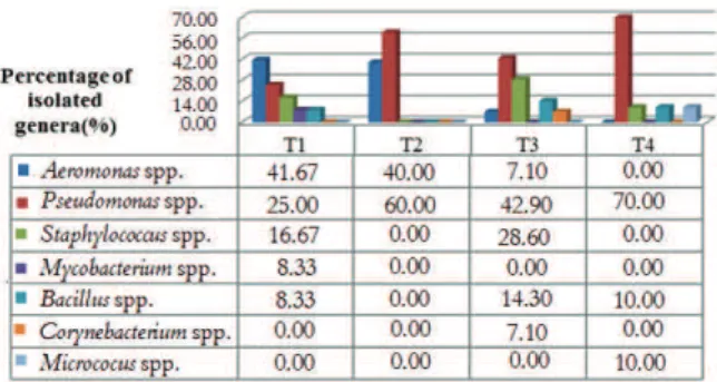
In Figure 1, identification and percentage of isolated bacterial strains for each treatment is shown. In Treatment T1 (biofloc technology-BFT), genus
Aeromonas spp. represented more than 41%, followed by 25% of Pseudomonas spp., 16.67% of
Staphylococcus spp., and 8.33% of Mycobacterium spp. and Bacillus spp., respectively. Likewise, works of Monroy-Dosta et al. (2013) on the culture of tilapia with bioflocs during 14 weeks, found the genera
Aeromonas, Vibrio, Pseudomonas, Bacillus, Micrococcus
among others.
Figure 1. Identification and frequency of bacterial genera isolated in the four treatments employed in the culture of juvenile Nile tilapia (O. niloticus) with or without periphyton and bioflocs.
would be a higher diversity of bacterial genera, as the C: N relation was adjusted. Moreover, an artificial substrate was added to the tank, offering better environmental conditions for the development of microorganisms, as the bacteria do not live as isolated colonies in nature, but typically form biofilms, characterized by a structured and functional microbial community (Tortora, Funke, & Case, 2012).
In treatment T3, genus Pseudomonas spp. was the most prominent (42.9%), followed by Staphylococcus
spp. (28.6%) and Bacillus spp. (14.3%), while the genera Corynebacterium spp. and Aeromonas spp. presented the lowest isolation percentage (7.1%). Differently from the present results, Newaj-Fyzul, Mutani, Ramsubhag, and Adesiyun (2008) found the following proportions of isolates, in decreasing order: Bacillus spp. (80%), Staphylococcus spp.,
Alcaligenes spp. and Aeromonas spp. (60%), and
Pseudomonas spp. (53%). The prevalence of other organisms may be attributed to seasonal variations and different culture procedures, such as feeding rates, which contribute to the increase or decrease of substrates for the establishment of certain bacterial groups in spite of others.
In treatment T4, the most observed genus was
Pseudomonas spp. (70%) followed by Staphylococcus
spp., Bacillus spp. and Micrococcus spp., (10% each). Corroborating with the results, Shilta, Chadha, Pandey, and Sawant (2016), in a study on the culture of Etroplus suratensis (a type of cichlid) in the presence of periphytic biofilm, reported the dominance of the genera Bacillus, Pseudomonas and
Micrococus in the water. Heterotrophic bacteria such as Pseudomonas spp. and Bacillus spp., are known for their ability to degrade organic matter (Monroy-Dosta et al., 2013). Thus, the presence of an inductor substrate for periphyton had a purifying effect in the culture water, as it offered a larger surface for the adhesion of heterotrophic microbiota.
It is possible to suggest that the culture systems employed in the present research influenced the diversity and proportion of bacterial genera isolated, which are differently adapted to each environment tested.
Among all treatments, the most frequently isolated genera, in decreasing order, are Pseudomonas
spp., Aeromonas spp., Staphylococcus spp., Bacillus spp.,
Mycobacterium spp., Micrococcus spp., and
Corybacterium spp..
Members of the Pseudomonas spp. are frequently found in soil, continental and oceanic
waters (Raaijmakers, Weller, & Thomashow, 1997). They belong to the heterotrophic microbiota in water, helping on the mineralization of nutrients. In addition to that, they produce bioactive compounds that are used as pathogen inhibitors in shrimp farming (Vijayan et al., 2006; Rattanachuay, Kantachote, Tantirungkij, Nitoda, & Kanzaki, 2010).
The Aeromonas genus is widely known as the etiological agent in fish infections, causing septicemia and hemorrhagic ulcers, generating significant mortality rates in freshwater as well as in marine species, harming the economy of aquaculture (Hu, Wang, Pan, Lu, & Liu, 2012; Beaz-Hidalgo, & Figueras, 2013). Therefore, presence of these microorganisms in tilapia culture waters should be of concern, calling for the use of procedures which favor beneficial bacterial groups to grow in the culture environment, decreasing or even eliminating Aeromonas spp..
Staphylococcus spp., Mycobacterium spp., Micrococcus
spp. and Corynebacterium spp. genera belong to the regular human microbiota (Kaushal, Gupta, & Van Hoek, 2016), or might be associated to it, hence it is believed that the presence of these groups are the consequence of human activity on the environment, which directly contribute to the dissemination of these microorganisms. Some bacterial species in these genera are opportunistic pathogens both to humans and fish (Austin & Austin, 2007; Santos et al., 2007).
Many species of the Bacillus genus are used as probiotics in aquaculture, in the strengthening of the immunological systems (Wang, Tian, Yao, & Li, 2008), stimulation of digestive enzymes, zootechnical improvement of the cultured animals (Standen et al., 2013; Jatobá & Mouriño, 2015), production of antibacterial compounds (Panigrahi & Azad, 2007), and degradation of organic matter. Therefore, it is considered a beneficial genus for the culture.
Once the main bacterial genera are identified in each treatment, adjustments in the water or feed can be made, in order to favor the main groups of interest in improving the environment, aside from preventing diseases from spreading.
Virulence factors and biofilm formation
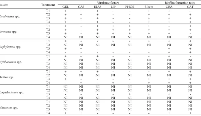
water, inanition, oxydative stress, etc (Stewart, 2002). Hence, bacteria possess adaptive mechanisms, biochemical or genetic (Meer Van Der, 2006), including new metabolic routes and pathogenic expression, for example. With that in mind, it can be noticed that the bacterial isolated from each treatment showed differences in the expression of potential virulence factors and biofilm formation, as can be observed in Table 1.
In treatments T1 (presence of biofloc) and T3 (control) the isolated genera displayed a higher pathogenic potential, as they produced up to five from the six virulence factors tested (Table 1). Aside that, it was possible to identify four and five virulence profiles, respectively, as can be seen on Table 1, and a positive result in at least one test for biofilm formation.
Potentially pathogenic bacteria were isolated and identified in the treatment with biofloc technology - BFT (T1). However, no case of disease was observed during the whole culture period. Some authors relate that the employment of biofloc may protect the cultivated organisms against the infection of pathogenic bacteria, because of the ‘natural probiotic’ effect acting against, for example, Vibrio
spp. and ectoparasites. This effect is triggered by large groups of microorganism, mainly by bacteria that are considered to be on the first trophic level in
the system (Crab et al., 2010; Emerenciano, Gaxiola, & Cuzon, 2013).
It is worth noting that in the control treatment (T3) no case of disease was observed, and it was necessary to conduct in vivo tests to ascertain the beneficial potential of bioflocs for fish. It is known that the presence of potentially pathogenic microorganisms only preferably causes damage when there is some unbalance in one or more of the following three elements: environment, host and pathogen. Thus, the culture is considered stable for the whole experimental period. In the culture of tilapia in the presence of both periphyton and bioflocs (T2), only two genera were isolated (Pseudomonas spp. and Aeromonas spp.). The
Pseudomonas spp. group produced gelatinase, caseinase, elastase and hemolysin with β-hemolytic activity. Aeromonas spp., in the other hand, tested positive for the other enzymes, excepting caseinase, a total of five from six analyzed factors. Both groups were positive for biofilm formation. In this treatment only two virulence profiles were observed. In the case of environmental unbalance or the use of inadequate procedures in the culture, diseases might appear, causing high mortality rates and economic losses, since these genera are known for possessing various opportunistic pathogenic species (Peixoto, Sá, Gordiano, & Costa, 2012).
Table 1. Analysis of potential virulence factors and tests for the detection in the formation of biofilm of bacterial isolates for each treatment employed in the cultivation of juvenile Nile tilapia.
Isolates Treatment Virulence factors Biofilm formation tests
GEL CAS ELAS LIP PHOS β-hem CRA GAT
Pseudomonas spp.
T1 + + + - - + + -
T2 + + + - - + + +
T3 + + + - - + + +
T4 + + + - - + + +
Aeromonas spp.
T1 + - + + + + - +
T2 + - + + + + + +
T3 + - + + + + + -
T4 NI NI NI NI NI NI NI NI
Staphylococcus spp.
T1 + - - - - + + +
T2 NI NI NI NI NI NI NI NI
T3 + + - - - - + +
T4 - - - - + +
Mycobacterium spp.
T1 + + + - - - + -
T2 NI NI NI NI NI NI NI NI
T3 NI NI NI NI NI NI NI NI
T4 NI NI NI NI NI NI NI NI
Bacillus spp.
T1 + + + - - - + +
T2 NI NI NI NI NI NI NI NI
T3 + - - - - + + +
T4 - + - + - + + +
Corynebacterium spp.
T1 NI NI NI NI NI NI NI NI
T2 NI NI NI NI NI NI NI NI
T3 - - - - + + + -
T4 NI NI NI NI NI NI NI NI
Micrococcus spp.
T1 NI NI NI NI NI NI NI NI
T2 NI NI NI NI NI NI NI NI
T3 NI NI NI NI NI NI NI NI
T4 + + - - - - + +
The strains isolated from Treatment 4 presented up to four of the six virulence factors, while also being positive for biofilm formation (Table 1). Moreover, only three virulence profiles were attested (Table 2). This treatment contributed to a more balanced culture environment. Characterization of bacterial strains exoenzymes, plus biofilm formation were used as an expression of virulence potential factor in this study. With these parameters in mind, it can be argued that this treatment resulted in the isolated genera expressing less pathogenicity, seeing that they were positive with less exoenzymes. It is proved that the presence of periphytic biofilm in cultivation environments reduces the occurrence of pathogens (Thompson, Abreu, & Wasielesky, 2002) and helps in the fortification of the fish immune system, offering higher resistance to diseases (Zhang et al., 2010; Anand et al., 2014; Kumar et al., 2015).
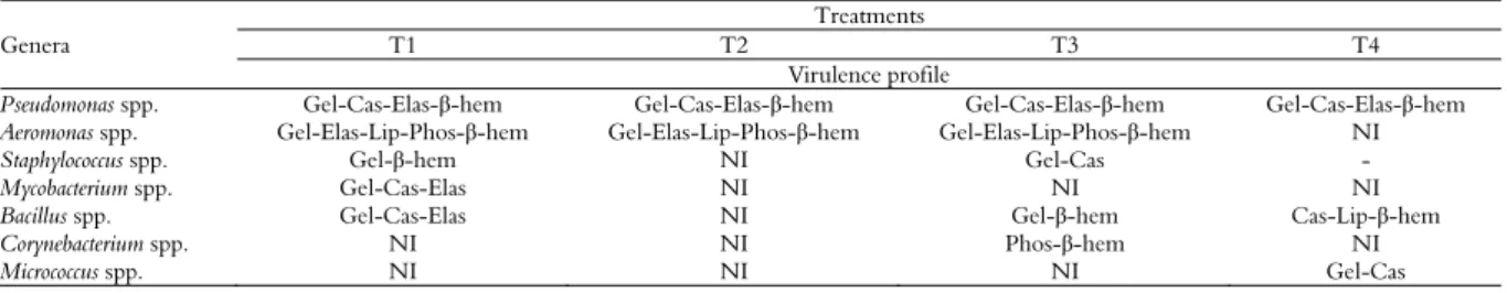
Observing virulence profiles in Table 2, one can identify and establish the following order of potentially pathogenic strains: Aeromonas>
Pseudomonas> Bacillus> Staphylococcus> Mycobacterium, Corynebacterium e Micrococcus.
Pseudomonas spp. and Aeromonas spp. did not alter the virulence profile, remaining the same in all treatments: Gel-Cas-Elas-β-hem and Gel-Elas-Lip-Fosf-β-hem, respectively (Table 2).
Sung and Hong (1997) isolated Aeromonas and
Pseudomonas from freshwater shrimp Macrobrachium rosembergii and identified that these bacteria were able to produce four and five extracellular products, respectively. The virulence of Aeromonas is considered multifactorial, as it is practically impossible to establish a hierarchy in the classification of its virulence factors according to their part in the disease process, and the following factors have been identified as virulence indicators: hemolysins, proteases, lipases, enterotoxins, gelatinases, elastases, among others (Rabaan, Gryllos, Tomas, & Shaw, 2001; Nam & John, 2007; Peixoto et al., 2012; Yadav, Verma, Pradhan, Dobriyal, & Sood, 2014).
Bacillus spp. and Staphylococcus spp. group exhibited three different virulence profiles, and were also positive for biofilm formation, being the third
group in pathogenic potential. Many studies in literature have used Bacillus species as probiotics (Cruz, Ibáñez, Hermosillo, & Saad, 2012; Albuquerque, Marengoni, & Boscolo, 2013), however, in this study, the isolated strains showed an enzymatic profile that probably conditioned them to the non-expression of their beneficial effects on the cultured organisms. Staphylococcus spp. possess various virulence mechanisms (Otto, 2012), and some of its species cause diseases in fish (Carvalho, Belém-Costa, & Porto, 2015). Although the genera
Mycobacterium spp., Corynebacterium spp. and
Micrococcus spp. isolated in this research have presented only one virulence profile, they are considered important pathogens of fish (Austin & Austin, 2007).
All genera identified were positive to at least one of the biofilm formation tests, independent of the treatment from which they were isolated. This biofilm formation factor, associated with the remaining exoenzyme production tests, presents a risk for cultivation, as biofilm formation is also considered an important factor in the pathogenicity of microorganisms, and can be used as an advantage in resisting environmental factors, aside from avoiding cellular and chemical defenses of the hosts immune system (Wakimoto et al., 2004; Dong, Fan, Wang, Shi, & Zhang, 2013).
Conclusion
Treatments T1 and T3 presented a higher total of isolates, which expressed five virulence profiles. While genera from treatments T2 and T4 had two and three virulence profiles, respectively. The genus
Aeromonas, considered one of the groups most pathogenic to fish, was not isolated in T4. The fish cultivated with bioflocs (T1) and periphyton (T4) presented a higher final weight.
The employment of growth inductor substrate for the periphyton and the manipulation of the C: N relation is a good alternative to aquaculture, since it is possible to increase the diversity of less pathogenic bacteria.
Table 2. Profile of potential virulence factors in bacterial isolates for each treatment tested in the culture of juvenile Nile tilapia (O. niloticus).
Genera
Treatments
T1 T2 T3 T4
Virulence profile
Pseudomonas spp. Gel-Cas-Elas-β-hem Gel-Cas-Elas-β-hem Gel-Cas-Elas-β-hem Gel-Cas-Elas-β-hem
Aeromonas spp. Gel-Elas-Lip-Phos-β-hem Gel-Elas-Lip-Phos-β-hem Gel-Elas-Lip-Phos-β-hem NI
Staphylococcus spp. Gel-β-hem NI Gel-Cas -
Mycobacterium spp. Gel-Cas-Elas NI NI NI
Bacillus spp. Gel-Cas-Elas NI Gel-β-hem Cas-Lip-β-hem
Corynebacterium spp. NI NI Phos-β-hem NI
Referências
Abdallah, F. B., Chaieb, K., Zmantar, T., & Kallel, H. (2009). Adherence assays and slime production of
Vibrio alginolyticus and Vibrio parahaemolyticus. Amina Bakhrouf Brazilian Journal of Microbiology, 40(2), 394-398. Aguirre-Guzmán, G., Ruíz, H. M., & Ascencio, F. (2004). A review of extracellular virulence product of Vibrio species important in diseases of cultivated shrimp.
Aquaculture Research, 35(2), 1395-1404.
Albuquerque, D. M., Marengoni, N. G., & Boscolo, W. R. (2013). Probióticos em dietas para tilápia do Nilo durante a reversão sexual. Ciência Rural, Santa Maria, 43(8), 1503-1508.
Al-Harbi, A. H., & Uddin, N. (2005). Bacterial diversity of tilapia (Oreochromis niloticus) cultured in brackish water in Saudi Arabia. Aquaculture, 250(3), 566-572. Anand, P. S. S., Kohli, M. P. S., Dam Roy, S., Sundaray, J.
K., Kumar, S., Sinha, A., … Sukham, M. K. (2014). Effect of dietary supplementation of periphyton on growth, immune response and metabolic enzyme activities in Penaeus monodon. Aquaculture Research, 46(9), 2277-2288.
Apha. (2005). Standard methods for the examination of water and waste water (21st ed.). Washington, D.C.: American Public Health Association.
Asaduzzaman, M., Wahab, M. A., Verdegem, M. C. J., Benerjee, S., Akter, T., Hasan, M. M., & Azim, M. E. (2009). Effects of addition of tilapia Oreochromis niloticus
and substrates for periphyton developments on pond ecology and production in C/N-controlled freshwater prawn Macrobrachium rosenbergii farming systems.
Aquaculture, 287(3), 371-380.
Audelo-Naranjo, J. M., Martínez-Córdova, L. R., Voltolina, D., & Gómez-Jiménez, S. (2011). Water quality, production parameters and nutritional condition of Litopenaeus vannamei (Boone, 1931) grown intensively in zero water exchange mesocosms with artificial substrates. Aquaculture Research, 42(9), 1371-1377.
Austin, B., & Austin, A. (2007). Bacterial fish pathogens disease of farmed and wild fish. Ellis Horwood: Chichester, 196-224.
Avnimelech, Y. (1999). Carbon and nitrogen ratio as a control element in Aquaculture systems. Aquaculture, 176(3-4), 227-235.
Avnimelech, Y. (2006). Bio-filters: The need for a new comprehensive approach. Aquacultural Engineering, 34, 172-178.
Avnimelech, Y. (2007). Feeding with microbial flocs by tilapia in minimal discharge bio-flocs technology ponds. Aquaculture, 264(3), 140-147.
Azim, M. E., Beveridge, M. C. M., Van Dam, A. A., & Verdegem, M. C. J. (2005). Periphyton and aquatic production: an introduction. In M. E. Azim, M. C. J. Verdegem, A. A. Van Dam, & M. C. M. Beveridge (Eds.), Periphyton - ecology, exploitation and management
(p. 1-14). Cambridge, UK: CABI Publishing.
Azim, M. E., Milstein, A., Wahab, M. A., & Verdegam, M. C. J. (2003). Periphyton–water quality relationships in fertilized fishponds with artificial substrates.
Aquaculture, 228(1), 169-187.
Beaz-Hidalgo, R., & Figueras, R. M. J. (2013). Aeromonas
spp. whole genomes and virulence factors implicated in fish disease. Journal of Fish Diseases, 36(4), 371-388. Bergey’s. (2014). Manual of determinative bacteriology (2nd
ed., v. 2, part B). New York: Springer.
Brasil. (2014). Produção da pecuária municipal, 2013. Brasília, DF: IBGE.
Carvalho, E., Belém-Costa, A., & Porto, J. I. R. (2015). Identificação bioquímica de bactérias patogênicas isoladas de peixes ornamentais no estado do Amazonas.
Revista Brasileira de Saúde e Produção Animal, 16(1), 170-178.
Cavalcante, D. H., Lima, F. R. S., Rebouças, V. T., & Sá, M. V. C. (2016). Association between periphyton and bioflocs systems in intensive culture of juvenile Nile tilapia. Acta Scientiarum. Animal Sciences, 38(2), 119-125. Christensen, G. D., Simpson, W. A., Bisno, A. L., &
Beachey, E. H. (1982). Adherence of slime-producing strains of Staphylococcus epidermidis to smooth surfaces. Infectious and Immunity, 37(1), 318-326.
Costa, R. A., Amorim, L. M. M. C., Araújo, R. L., & Vieira, R. H. S. F. (2013). Multiple enzymatic profiles of Vibrio parahaemolyticus strains isolated from oysters.
Revista Argentina de Microbiologia, 45(4), 267-270. Crab, R., Avnimelech, Y., Defoirdt, T., Bossier, P., &
Verstraete, W. (2007). Nitrogen removal techniques in aquaculture for a sustainable production. Aquaculture, 270(1), 1-14.
Crab, R., Lambert, A., Defoirdt, T., Bossier, P., & Verstraete, W. (2010). The application of bioflocs technology to protect brine shrimp (Artemia franciscana) from pathogenic Vibrio harveyi. Journal of Applied Microbiology, 109(5), 1643-1649.
Cruz, P. M., Ibáñez, A. L., Hermosillo, O. A. M., & Saad, H. C. R. (2012). Use of probiotics in aquaculture.
International Scholarly Research Network, 2012, Article ID 916845, 1-13.
Dong, X., Fan, X., Wang, B., Shi, X., & Zhang, X.-H. (2013). Invasin of Edwardsiella tarda is essential for its haemolytic activity, biofilm formation and virulence towards fish. Journal of Applied Microbiology, 115(1), 12-19.
Downes, M. P., & Ito, K. (2001). Compendium of methods for the microbiological examination of foods (4th ed.).
Washington, DC:APHA.
Emerenciano, M. G., Wasielesky, W. J., Soares, R. B., Ballester, E. C., Izeppi, E. M., & Cavalli, R. O. (2007). Crescimento e sobrevivência do camarão-rosa (Farfantepenaeus paulensis) na fase de berçário em meio heterotrófico. Acta Scientiarum. Biological Sciences, 29(1), 1-7.
Matovic (Ed.), Biomass now – cultivation and utilization
(p. 301-328). Rijeka, Croatia: In Tech.
Food and Agricultural Organization [FAO]. (2014). The State of world fisheries and aquaculture - SOFIA. Roma: FAO.
Furniss, A. L., & Donovan, T. J. (1979). The Víbrio.
[Monograph Series, 58]. London: Public Health Laboratory Sevice.
Freeman, D. J., Falkiner, F. R., & Keane, C. T. (1989). New method for detecting slime production by coagulase negative staphylococci. Journal of Clinical Pathology, 42(8), 872-874.
Hu, M., Wang, N., Pan, Z. H., Lu, C. P., & Liu, Y. J. (2012). Identity and virulence properties of Aeromonas
isolates from diseased fish, healthy controls and water environment in China. Letters in Applied Microbiology, 55(3), 224-233.
Jatobá, A., & Mouriño, J. L. P. (2015). Lactobacillus plantarum effect on intestinal tract of Oreochromis niloticus fingerlings. Ciência Animal Brasileira, 16(1), 45-53.
Jiravanichpaisal, P., Miyazaki, T., Limsuwan, C. (1994). Histopathology, Biochemistry, and Pathogenicity of
Vibrio harveyi Infecting Black Tiger Prawn Penaeus monodon. Journal of Aquatic Animal Health, 6(1), 27-35. Jose, J., Giridhar, R., Anas, A., Bharathi, P. A. L., & Nair,
S. (2011). Heavy metal pollution exerts reduction/adaptation in the diversity and enzyme expression profile of heterotrophic bacteria in Cochin estuary, India. Environmental Pollution, 159(10), 2775-2780.
Kaushal, A., Gupta, K., & Van Hoek, M. L. (2016). Characterization of Cimex lectularius (bedbug) defensin peptide and its antimicrobial activity against human skin microflora. Biochemical and Biophysical Research Communications, 470(4), 955-960.
Keshavanath, P., Gangadhar, B., Ramesh, T. J., Van Dam, A. A., Beveridge, M. C. M., & Verdegem, M. C. J. (2002). The effect of periphyton and supplemental feeding on the production of the indigenous carps Tor khudree and Labeo fimbriatus. Aquaculture, 213(3), 207-218.
Kubtiza, F. (2008). Sanidade aquícola. Panorama da aquicultura, 18(107), 28-37.
Kumar, S., Anand, P. S. S., Ravichandran, P., Panigrahi, A., Dayal, J. S., Ananda, R. A., … Ponniah, A. G. (2015). Effect of periphyton on microbial dynamics, immune responses and growth performance in black tiger shrimp Penaeus monodon Fabricius, 1798. Indian Journal Fiseries, 62(3), 67-74.
Martínez-Cordova, L. R., Emerenciano, M., Miranda-Baeza, A., & Martínez-Porchas, M. (2015). Microbial-based systems for aquaculture of fish and shrimp: an updated review. Reviews in Aquaculture, 7(2), 131-148. Meer Van Der, J. R. (2006). Environmental pollution
promotes selection of microbial degradation pathways.
Frontiers in Ecology and the Environment, 4(1), 35-42.
Monroy-Dosta, M., Del, C., Lara-Andrade, R., Castro-Mejía, J., Castro-Castro-Mejía, G., & Coelho-Emerenciano, M. G. (2013). Composición y abundancia de comunidades microbianas asociadas al biofloc en un cultivo de tilapia. Revista de Biología Marina y Oceanografía, 48(3), 511-520.
Nam, I. Y., & John, K. (2007). Rapid detection of virulence of Aeromonas isolated from a trout by hexaplex-PCR. Journal of Microbiology, 45(4), 297-304. Newaj-Fyzul, A., Mutani, A., Ramsubhag, A., & Adesiyun,
A. (2008). Prevalence of bacterial pathogens and their anti-microbial resistance in tilapia and their pond water in Trinidad. Adesiyun Zoonoses and Public Health, 55(4), 206-213.
Otto, M. (2012). MRSA virulence and spread. Cellular Microbiology, 14(10), 1513-1521.
Panigrahi, A., & Azad, I. S. (2007). Microbial intervention for better fish health in aquaculture: the Indian scenario. Fish Physiology Biochemistry, 33(4), 429-440. Peixoto, L. J. S., Sá, M. C. A., Gordiano, L. A., & Costa,
M. M. (2012). Aeromonas spp.: Fatores de virulência e perfis de resistência a antimicrobianos e metais pesados.
Arquivos do Instituto Biológico, 79(3), 453-461.
Raaijmakers, J., Weller, D. M., & Thomashow, L. S. (1997). Frequency of antibiotic-producing Pseudomonas
spp. in natural environments. Applied Environmental Microbiology, 63(3), 881-887.
Rabaan, A. A., Gryllos, I., Tomas, J. M., & Shaw, J. G. (2001). Motility and the polar flagellum are required for Aeromonas caviae adherence to HEp-2 cells. Infection and Immunity, 69(7), 4257-4267.
Rattanachuay, P., Kantachote, D., Tantirungkij, M., Nitoda, T., & Kanzaki, H. (2010). Inhibition of shrimp pathogenic vibrios by extracellular compounds from a proteolytic bacterium Pseudomonas sp. W3. Electronic Journal of Biotechnology, 13(1), 1-11.
Rodrigues, D. P., Ribeiro, R. V., Alves, R. M., & Hofer, E. (1993). Evaluation of virulence factors in environmental isolates of Vibrio species. Memórias do Instituto Oswaldo Cruz, 88(4), 589-592.
Rust, L., Messing, C. R., & Iglewski, B. H. (1994). Elastase assays. Methods Enzymol, 235(1), 554-562. Sakr, E. M., Shalaby, S. M., Wassef, E. A., El-Sayed, A. F.
M., & Moneim, A. I. A. (2015). Evaluation of Periphyton as a food source for Nile tilapia (O. niloticus) juveniles fed reduced protein levels in cages.
Journal of Applied Aquaculture, 27(1), 50-60.
Santos, A. L., Santos, D. O., Freitas, C. C., Ferreira, B. L. A., Afonso, I. F., Rodrigues, C. R., & Castro, C. H. (2007). Staphylococcus aureus: visitando uma cepa de importância hospitalar. Jornal Brasileiro de Patologia e Medicina Laboratorial, 43(6), 413-423.
Schryver, P., Verstraete, W. (2009). Nitrogen removal from aquaculture pond water by heterotrophic nitrogen assimilation in lab-scale sequencing batch reactors. Bioresource Technology, 100(3), 1162-1167. Shilta, M. T., Chadha, N. K., Pandey, P. K., & Sawant, P.
growth of Etroplus suratensis (Bloch, 1790). Aquaculture International, 24(2) 661-674.
Standen, B. T., Rawling, M. D., Davies, S. J., Castex, M., Foey, A., Gioacchini, G., … Merrifield, D. L. (2013). Probiotic Pediococcus acidilactici modulates both localised intestinal and peripheral-immunity in tilapia (O. niloticus). Fish & Shellfish Immunology, 35(4), 1097-1104. Stewart, P. S. (2002). Mechanism of antibiotic resistance
in bacterial biofilms. International Journal of Medical Microbiology, 292(2), 107-113.
Soto, E., Hawke, J. P., Fernandez, D., & Morales, J. A. (2009). Francisella sp., an emerging pathogen of tilapia,
Oreochromis niloticus (L.), in Costa Rica. Journal of Fish Diseases, 32(8), 713-722.
Sung, H. H., & Hong, T. Y. (1997). The gram-negative bacterial flora in Hepatopancreas of giant freshwater prawn (Macrobrachium rosenbergii): antibiotic sensitivities and production of extracellular products. Journal of Fisheries Society Taiwan, 24(3), 211-223.
Thompson, F. L., Abreu, P. C., & Wasielesky, W. (2002). Importance of biofilm for water quality and nourishment in intensive shrimp culture. Aquaculture, 203(3), 263-278.
Tortora, G. J., Funke, B. R., & Case, C. L. (2005).
Microbiologia (8a ed.). Porto Alegre, RS: Artmed. Tortora, G. J., Funke, B. R., Case, C. L. (2012). Microbiologia
(10a ed., cap. 3, 69-70). Porto Alegre, RS: Artmed. Uddin, M. S., Azim, M. E., Wahab, M. A., & Verdegem,
M. C. J. (2009). Effects of substrate addition and supplemental feeding on plankton composition and production in tilapia (O. niloticus) and freshwater prawn (Macrobrachium rosenbergii) polyculture. Aquaculture, 297(1-4), 99-105.
Vijayan, K. K., Singh, I. S. B., Jayaprakash, N. S., Alavandi, S. V., Pai, S. S., Preetha, R., … Santiago, T. C. (2006).
A brackishwater isolate of Pseudomonas PS-102, a potential antagonistic bacterium against pathogenic vibrios in penaeid and non-penaeid rearing systems.
Aquaculture, 251(2-4), 192-200.
Wakimoto, N., Nishi, J., Sheikh, J., Nataro, J. P., Sarantuya, J., Shita, M., ... Tokuda, K. (2004). Quantitative biofilm assay using a microtiter plate to screen for enteroaggregative Escherichia coli. The American Journal of Tropical Medicine and Hygiene, 71(5), 687-690.
Wang, Y. B., Tian, Z. Q., Yao, J. T., & Li, W. F. (2008). Effect of probiotics, Enteroccus faecium, on tilapia (O. niloticus) growth performance and immune response.
Aquaculture, 277(3-4), 203-207.
Yadav, S., Verma, D. K., Pradhan, P. K., Dobriyal, A. K., & Sood, N. (2014). Phenotypic and genotypic identification of Aeromonas species from aquatic environment. International Journal of Aquatic Science, 5
(1), 13-20.
Zhang, B., Lin, W., Wang, Y., & Xu, R. (2010). Effects of artificial substrates on growth, spatial distribution and non-specific immunity factors of Litopenaeus vannamei
in the intensive culture. Turkish Journal of Fisheries and Aquatic Sicences, 10(4), 491-497.
Zhao, J., Chen, M., Quan, C. S., & Fan, S. D. (2015). Mechanisms of quorum sensing and strategies for quorum sensing disruption in aquaculture pathogens.
Journal of Fish Diseases, 38(9), 771-786.
Received on May 13, 2016. Accepted on June 1, 2016.