O R I G I N A L A R T I C L E
Histopathology findings in common marmosets (
Callithrix jacchus
Linnaeus, 1758) with chronic weight loss associated with bile tract
obstruction by infestation with
Platynosomum
(Loos, 1907)
Maria Bernardete Cordeiro SousaÆ
Adriano Castro Lea˜oÆJose´ Fla´vio Vidal CoutinhoÆ Ana Maria de Oliveira Ramos
Received: 16 January 2008 / Accepted: 11 September 2008 / Published online: 8 October 2008
ÓJapan Monkey Centre and Springer 2008
Abstract Chronic weight loss in marmosets is often associated with wasting marmoset syndrome (WMS), an important disease that occurs in callitrichid colonies around the world. Even though its etiology is very difficult to determine, particular variables, such as weight loss, diarrhea and alopecia, associated or not with infestation in the pancreatic ducts with Trichospirura leptossoma (Nematoda: Thelazioidea), seem to be linked with the syndrome. This study investigated the histopathology of the lungs, duodenum, liver, gallbladder, extrahepatic bile ducts and pancreatic ducts of six common marmosets (Callithrix jacchus) suffering from severe non-diarrheic weight loss. Three individuals died naturally and the other three were euthanized. Microscopic findings showed the presence of adult flukes (Platynosomum) in the liver. These flukes, which provoke common infection in cats, were also observed inside the gallbladder as well as in the intra and extrahepatic bile ducts in common marmosets. Portal fibrosis was observed in two animals, which developed chronic fibrosing hepatopathy (biliary pattern, grade 3). The disease progresses without diarrhea and without pan-creatic lesions or infestation. With the progression, the animals presented with ascending cholangitis, cholestasis and portal fibrosis, sometimes culminating in secondary biliary cirrhosis. Therefore, this infirmity, associated with
chronic weight loss in common marmosets, could be another etiological factor linked with WMS.
Keywords Common marmosetWeight loss
PlatynosomumChronic biliary obstruction
Wasting marmoset syndrome
Introduction
The common marmoset (Callithrix jacchus) is a small Brazilian New World primate that has been used exten-sively in biomedical research. The continued use of C. jacchus for research and studies in the biomedical field encompasses areas such as behavior, neuroscience, toxi-cology, drug development and reproductive biology (Tardif et al. 2003; Mansfield 2003). It is imperative that these captive animals be provided with adequate breeding con-ditions and preventive veterinary care, thereby keeping the animals healthy and thriving (Ludlage and Mansfield
2003).
Many studies point out a range of characteristics that make C. jacchus a good choice for experimental proce-dures. Some of the most cited are: small size, female reproductive profile of four offspring per year (Hearn
1978), similarities between marmoset and human repro-ductive physiology, a 28-day ovarian cycle (Hearn 1983) and copulation with gestating or non-estrous females (Dixson and Lunn 1987). On the other hand, in captivity these primates may present with wasting marmoset syn-drome (WMS), characterized by the clinical manifestation of rapid weight loss, with skeletal and muscular atrophy more prominently seen in the lower extremities (Ialeggio and Baker 1995). The latter study confirmed that animals affected by WMS were identified in 60% of C. jacchus
M. B. C. Sousa (&)A. C. Lea˜oJ. F. V. Coutinho
Departamento de Fisiologia,
Universidade Federal do Rio Grande, Caixa Postal, 1511, Natal, RN 59078-970, Brazil
e-mail: [email protected]
A. M. de Oliveira Ramos
State Service for Ascertaining Death in the State of Rio Grande do Norte, Natal, Brazil
breeding institutions. According to Tardif et al. (1984) and Logan and Khan (1996), the occurrence of WMS increases both the susceptibility to, and incidence of, parasitic infections. In our common marmoset colony, around 10% of the animals showed weight loss that evolved to WMS; antiparasitic agents are regularly administrated to prevent infestation. Melo and Martins (1986) describe the pinworm Primasubulura jacchi as the most frequent nematode encountered in a colony ofC. penicillata. In C. geoffroyi specimens confiscated from the illegal wildlife trade, Melo (2004) found other helminthes besidesP. jacchi, including Platynosomum amazonensis (family Dicrocoeliidae). Pre-vious infestation inC. jacchusbyP. amazonensiswas also reported in captive animals in Oak Ridge, Tennessee. Although these parasites are spread throughout the world, they are more frequently found in tropical countries. They are small flukes found in the bile ducts and gallbladder of cats and their intermediate hosts include the snail Sub-lima octona, while lizards and toads could also be paratenic hosts (Xavier et al.2007).
In this study, we describe hepatic and biliary histopa-thological findings in C. jacchus due to infestation by Platynosomumin animals clinically diagnosed with WMS.
Methods
Animals belonging to the common marmoset colony at the Primatology Center (IBAMA register 1/24/92/0039-0) of the University Federal do Rio Grande do Norte (UFRN), Natal, Brazil are housed in outdoor cages exposed to nat-ural lighting, temperature, and humidity conditions. The experiment took place between September 2003 and October 2004, during which time the temperature was around 28.2°C and the humidity between 66 and 90%.
The animals are kept in family groups, same sex dyads or isolated, in cages made of brick walls and a galvanized wire door with a cement or sand floor. Water was provided
ad libitum and the animals were fed with a protein mash and fresh fruits or vegetables, twice daily, in the morning and afternoon. This diet was supplemented three times a week with boiled chicken or fish and raisins and cereals. A number of episodes of lizards, bees, butterflies being captured and eaten by the monkeys were recorded. As the colony is located within the University Campus in the urban area of the city, cats were occasionally seen around the colony perimeter. To prevent parasitic infestation, antihelminthic (ivermectin 1%, 200lg/kg, subcutaneously; albendazole 4%, 25 mg/kg, orally), antigiardiasis and an-tiamebiasis (tinidasol 10%, 50 mg/kg, orally) agents were administered every 6 months.
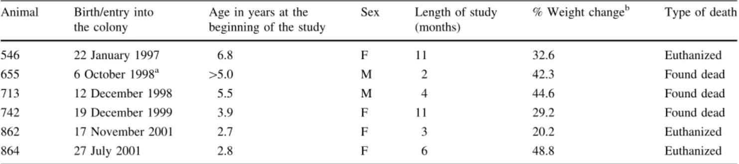
Six adult marmosets (four females and two males) were used in this study. This experimental group comprised animals considered to be affected by WMS, whose initial diagnosis was made based on monthly weight evolution. Animals that lost more than 10% of their body weight were isolated in a new cage and subjected to weekly weighing in addition to having their muscles and fur examined. The monitoring period varied from 2 to 11 months from the beginning of the expression of clinical signs such as weight loss, alopecia and reduction in muscle mass as determined by palpation. None of the experimental animals had chronic diarrhea. The mean age of these animals was 4.3±1.77 years, an age within the range associated with highest WMS incidence (Table1).
During the monitoring, three deaths were recorded in animals that also showed jaundice, extreme muscle mass loss and severe alopecia in the body and tail. The three remaining animals were sacrificed using sodium thiopental (Abbott, Sa˜o Paulo, SP, Brazil, 40 mg/kg, i.p.) to achieve deep anesthesia. After both natural death and sacrifice, the animals immediately underwent necropsy. The necropsies were performed in the conventional manner, by total evisceration and fixing organs in 10% formalin. The organs were routinely processed and stained using hematoxylin– eosin (H&E) and Masson’s trichrome techniques.
Table 1 Animal identification, physiological and monitored data
Results
All six animals showed decreasing mean weights over the monitoring period. Weight measured before death showed that the animals lost between 20.8 and 48.8% of the final weight measured before weight loss was detected (Table1).
In all six necropsied animals, dilatation of the intrahe-patic biliary pathways and the presence of blackish material in the lumen were identified. There was a slight fibrous thickening of the gallbladder wall. Two of the livers examined had increased consistency and greenish-brown coloration, along with an irregular surface showing small nodules (Fig.1a). The number of fluke specimens in the biliary tree was not quantified, as the viscera were already fixed in formalin, prior to dissection of the biliary pathways and total fluke count. The necropsy revealed tiny black foreign bodies, resembling flat fusiform flukes, in the
gallbladder and main bile ducts of all six animals. Only one animal had slightly dilated pancreatic ducts. The remaining viscera were macroscopically normal.
We identified innumerable sections of adult flukes in the biliary tree, along with portal and periportal lymphocytic inflammatory infiltrate in all six animals. Microscopic alterations associated with infestation of the biliary path-ways by adult flukes were also seen in all six marmosets. Several flat and fusiform adult flukes, measuring on aver-age 30 mm912 mm, occupied the lumen of the gallbladder and of the large bile ducts. The flukes had ovaries and testes and ovoid eggs with a thick yellowish shell and poorly defined opercula. The eggs measured 30– 40lm920–25lm, characteristics that, along with their habitat and morphology, allow their positive identification as Platynosomum(Fig.1b).
The liver of three animals showed clear and significant bilirubinostasis and cholestasis (Fig.1c), and one of these
Fig. 1 aMacroscopic section of liver showing fine nodulation in the parenchyma and dilation of the intra and extrahepatic biliary tree; presence of dark viscous material in the lumen of the biliary pathways and fibrous thickening of the gallbladder wall.bGallbladder (mucous on the right) containing adult fluke specimens (H&E9100).cBile lake and intense cholestasis (H&E9400).
dPolymorphonuclear neutrophils in the lumen of a dilated bile duct (ascending cholangitis) (H&E9400).e
Portal fibrosis with septum formation and the presence of nodules in the parenchyma, characterizing a biliary pattern of chronic hepatopathy, grade 3 (Masson’s trichrome and H&E
revealed the presence of polymorphonuclear neutrophils in portal areas and in the lumen of interlobular bile ducts (ascending cholangitis) (Fig.1d). Two animals with hel-minthic infestation had chronic biliary obstruction (bilirubinostasis and cholestasis), intense ductal neofor-mation and expansive fibrosis of the portal spaces with the focal presence of nodules in the hepatic parenchyma (Fig.1e, f).
Slight atrophy of the duodenal mucosa was observed in two animals, with a reduction in the villos/crypt ratio (2:1, normal: 3:1), without superficial enterocyte lesion and with a slight increase in lymphocytes and plasmocytes in the lamina propria. Sclerosis of renal glomeruli was detected in one of the animals with chronic fibrosing hepatopathy (biliary pattern, grade 3).
Discussion
The mean weight of the experimental animals was lower than that described for healthy captive animals of the colony (Arau´jo, 2000; A.C. Lea˜o, A.D. Do´ria-Neto and M.B.C. Sousa, unpublished data): mean weight of adult captive males/females=374.63 g±45.03). In this case the physiopathological mechanisms underlying the weight loss and the other signs of WMS in common marmosets seem to be a consequence of cholestasis resulting from defective canalicular secretion of bile or obstruction of bile flow. As has been observed for humans (Hoffman 2002) and experimental models using mice (Goergiev et al.
2008), the decrease, or even absence, of bile acid in the small intestinal lumen, provokes lipid maldigestion and fat-soluble vitamin malabsorption, accompanied by severe nutritional deficits leading to reduction in muscle mass and anorexia (Arau´jo et al.2002).
Considering the extensive literature on how WMS affects primates, this disease seems to have a multifactorial etiology. The clinical signs, which include chronic diar-rhea, alopecia and low weight, are linked to a high mortality rate. According to Logan and Khan (1996), ani-mals with WMS are more susceptible to a higher incidence of parasitical infections. In this study, microscopic
altera-The anatomopathological aspects of the necropsies performed on captiveC. jacchusat the UFRN Primatology Center show that the deaths were due to chronic hepatobil-iary compromise. These results show a significant relationship between WMS andPlatynosomumparasitism of the gallbladder and intra and extrahepatic biliary tree. The course of the disease can include portal lymphocytic inflammation, ascending cholangitis, and biliary flux obstruction with intense hepatic bilirubinostasis. It may also evolve into portal fibrosis extending into the parenchyma, progressing to chronic fibrosing hepatopathy (biliary pat-tern) and chirrosis. Different from other WMS deaths, generally associated with diarrhea and Trichospirura lep-tostomapancreatic duct infestation, the chronic weight loss in the present study was not related to diarrhea, but rather was associated withPlatynosomumparasitic infestation.
Since the main and intermediate hosts were present in the colony when most of the infested animals were living in cages with sand floors, we introduced procedures to pre-vent new cases of platynosomiasis. The floors were cemented, an additional antihelminthic agent was pre-scribed (praziquantel, 60 mg/kg, orally), the vegetation was pruned more frequently and a campaign to capture stray cats was instigated by the University.
Acknowledgments We are grateful to Edno´lia Camara, Antoˆnio B. da Silva and Geniberto C. dos Santos for assisting with the care of the animals and to Dr. Eveline Pipolo for reviewing the parasitological findings. We are also grateful to three anonymous referees, whose suggestions significantly improved this manuscript. During this pro-ject M.B.C.S. was supported by CNPq grants (524409/96, 470601/ 2003-5 and 308280/2006-7). The maintenance and use of the exper-imental animals followed the Brazilian Society of Neuroscience and Behavior guidelines, as well as the recommendations of the Society for Neuroscience (USA).
References
Arau´jo A, Arruda MF, Alencar AI, Albuquerque F, Nascimento MC, Yamamoto ME (2000) Body weight of wild and captive marmosets. Int J Primatol 21:317–324
Ialeggio DM, Baker AJ (1995) Results of a preliminary survey into wasting marmoset syndrome in callitrichid collections. In: Proceedings of the first annual conference of the nutrition advisory group of the American Zoo and Aquarium Association, pp 148–158
Kingston N, Cosgrove GE (1967) Two new species ofPlatynosomum
(Trematoda: Dicrocoeliidae) from South American monkeys. Proc Helminth Soc Washington 34:147–151
Logan AC, Khan KNM (1996) Clinical pathologic changes in two marmosets with Wasting Syndrome. Lab Anim Pathol 24:707– 709
Ludlage E, Mansfield K (2003) Clinical care and diseases of the common marmoset (Callitrix jacchus). Comp Med 53:369–382 Mansfield K (2003) Marmoset models commonly used in biomedical
research. Comp Med 53:383–392
Melo AL (2004) Helminth parasites of Callithrix geoffroyi. Lab Primate Newsl 43:7–9
Melo AL, Martins WA (1986) Sobre o parasitismo porPrimasubulura jacchi emCallithrix penicillata(in Portuguese). In: Melo MT
(ed) A Primatologia no Brasil, vol 2. Sociedade Brasileira de Primatologia, Brası´lia, pp 483–487
Tantalean M, Gozalo A, Montoya E (1990) Notes on some helminth parasites from Peruvian monkeys. Lab Primate Newsl 29:6–8 Tardif SD, Richter CB, Carson RL (1984) Effects of sibling-rearing
experience on future reproductive success in two species of Callitrichidae. Am J Primatol 6:377–380
Tardif SD, Smucny DA, Abbott DH, Mansfield K, Schultz-Darken N, Yamamoto ME (2003) Reproduction in captive common mar-mosets (Callithrix jacchus). Comp Med 53:364–368
Warren KS, Swan RA, Hobbs RP, Eva H, Kuhn M, Jonathan L (1998)
Platynosomum fastosumin orangutans ex-captive in Indonesia. J Wildl Dis 3:644–646
Xavier FG, Morato GS, Dario AR, Maiorka PC, Spinosa HS (2007) Cystic liver disease related to high Platynosomum fastosum