Rev. Col. Bras. Cir. 2016; 43(5): 401-403
DOI: 10.1590/0100-69912016005006
Right hepatic artery aneurysm
Aneurisma de artéria hepática direita
Astrid del PilAr ArdilA BernAl1; PAulo loures, tCBC- rJ2; JuAn CristóBAl osPinA CAlle1; BeAtriz CunhA2; JuAn CAmilo CórdoBA1.
INTRODUCTION
T
he first reported case of hepatic artery aneurysm (HAA) is attributed to James Wilson, an anatomist in the year 1809, and the first successful repair was credited to the German surgeon Dr. Hans Kehr in the year 19031.This is a potentially fatal disease when pre-senting rupture. The rupture rate is controversial and varies between 20% and 80%, clearly determined by the inability to detect asymptomatic aneurysms. The aneurysms of the hepatic artery and its branches are unusual vascular lesions, corresponding to approxi-mately 21% to 44% of all visceral aneurysms1.
CASE REPORT
A female, 89-year-old patient was hospitalized with a longstanding abdominal pain which had worsened in the days prior to hospitalization, associated with severe anemia and one episode of hematemesis studied with upper endoscopy (UE), which showed no bleeding, whether active or recent. The abdominal computed tomography (CT) showed dilatation of bile ducts, aerobilia, as well as gallbladder with thickened walls. The suggested diagnosis was acute complicated cholecystitis. She was admitted and medical treatment started.
Three days after admission, the patient persisted with anemia and need for blood transfusion. A new UE showed hemobilia and new abdominal CT without contrast showed: dilatation of intra and extrahepatic bile ducts, especially in the right lobe, with heterogeneous content; intra-pancreatic bile duct of 14 mm in diameter; gallbladder with thickened
1 - Carlos Chagas Medical Postgraduate Institute, Rio de Janeiro, RJ, Brazil. 2 - Carlos Chagas State Hospital, Rio de Janeiro, RJ, Brazil.
Case Report
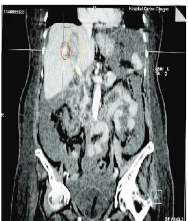
Figure 1. Abdominal CT scan with contrast showing right hepatic ar-terial malformation (smaller circle) and intra-parenchymal hematoma (larger circle).
A B S T R A C T
We report a case of an aneurysm of the right hepatic artery and its multidisciplinary management by general surgery, endoscopy and radiology services. Being a case of extremely low incidence, it is important to show its diagnostic and therapeutic approach.
402
Rev. Col. Bras. Cir. 2016; 43(5): 401-403
Bernal
Right hepatic artery aneurysm
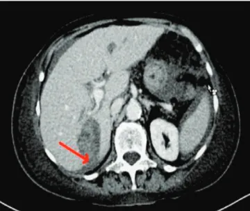
walls and heterogeneous content, with interposed gas focus; free fluid in the peritoneal cavity, of heterogeneous density in the parieto-colic gutters, suggesting hemoperitoneum; and questionable intrahepatic vascular ectasia of the right lobe. The complete CT study, this time with venous contrast, showed hemoperitoneum and a hyperdense formation relative to the hepatic parenchyma in segments VII and VIII measuring approximately 2.32x5.44 cm, suggestive of malformation of the right hepatic artery (Figures 1 and 2). When compared to the previous CT, the hepatic intra-parenchymal hematoma had increased, with rupture of hepatic capsule and extravasation into the peritoneal cavity (Figure 3).
The fall in hematocrit continued, for which she again received transfusion in the same day. Given the impossibility of performing liver angiography and instability of the patient, the emergency service staff decided to perform an exploratory laparotomy. The inventory of the cavity revealed massive hemoperito-neum, hepatomegaly, gallbladder with thickened walls containing multiple calculi, common bile duct with sig-nificant dilation and a ruptured hematoma in the he-patic segment VII, with active bleeding.
We evacuated the hemoperitoneum, com-pressed the hematoma, and dissected the hepatic hilum with identification of the common hepatic artery bifurca-tion and ligabifurca-tion of the right hepatic artery. We
proceed-ed to cholecystectomy with exploration of the bile duct, from where we removed a large calculus and a large clot.
DISCUSSION
The HAA is clinically important because of its high mortality rate (25% to 70%) when rupture occurs. The most common incidence is between the fifth and sixth decades of life and the most common location is extrahepatic. Hepatic aneurysms compromise the common hepatic artery 70% to 80% of cases. Multiple etiologies have been described including atherosclerosis, abdominal trauma, surgical procedures, degenerative diseases, infections, collagen vascular disease, and congenital anomalies.
Multiple complementary tests are available for diagnosis, such as abdominal ultrasound, CT, angio-CT, MRI, endoscopy and angiography. The latter is not only a diagnostic tool, but also a therapeutic modality of choice in splanchnic aneurysms through embolization. It can also provide evidence of collateral circulation, determine the size and shape of the aneurysm, uncover arterioportal fistulas and provide accurate anatomical information necessary for embolization or surgery.
In general, the location of the aneurysm elects its therapeutic approach: Intrahepatic HAA – prefer-ably treated by selective embolization or partial liver resection; Extrahepatic HAA – can be treated by per-Figure 2. CT: section showing abundant hemoperitoneum. Figure 3. CT: hematoma rupture and extravasation into the peritoneal
403
Rev. Col. Bras. Cir. 2016; 43(5): 401-403
Bernal
Right hepatic artery aneurysm
cutaneous obliteration in patients with high operative risk, but the ideal treatment is resection and arterial reconstruction. When the HAA is located in the com-mon hepatic artery, one may use embolization or an-eurysm ligation without arterial reconstruction, but
when located in the proper hepatic artery, it requires vascular reconstruction to avoid hepatic ischemia sec-ondary to interruption of the collateral circulation re-flowing through the gastroduodenal and right gastric arteries1-5.
R E S U M O
Relatamos um caso de aneurisma da artéria hepática direita conduzido de forma multidisciplinar pelos Serviços de Cirurgia Geral, Endos-copia e Radiologia. Em se tratando de caso de incidência baixíssima, é importante mostrar o enfoque diagnóstico e terapêutico usado em seu manejo.
Descritores: Aneurisma. Vísceras. Artéria Hepática. Hemobilia.
REFERENCES
1. Merrell SW, Schneider PD. Hemobilia, evolution of current diagnosis and treatment. West J Med. 1991;155(6):621-5.
2. Jaunoo SS, Tang TY, Uzoigwe C, Walsh SR, Gaunt ME. Hepatic artery aneurysm repair: a case report. J Med Case Reports. 2009;3:18.
3. Morisse D, Musante C, Heredia P. Aneurisma de arteria hepática.Rev Argent Cardiol. 2011;79(3):255.
4. Parolim MB, Lopes RW. Doenças vasculares do fígado. In: Kalil AN, Coelho JUC, Strauss E. Fígado e vias bili-ares: clínica e cirurgia. Rio de Janeiro: Revinter, 2001. 5. Garcia RG, Kamya CA, Ishikawa WY, Menezes MR,
Cerri GG. Aneurismas arteriais esplâncnicos – ex-periência do Serviço de Radiologia de Emergência – Departamento de Radiologia da Faculdade de Me-dicina da USP. Disponível em: http://www.hcnet. usp.br/inrad/departamento/radiops.htm
Received in: 11/02/2016
Accepted for publication: 01/05/2016 Conflict of interest: none.
Source of funding: none.
Mailing address:
Astrid del Pilar Ardila Bernal