Volume 2013, Article ID 156734,11pages http://dx.doi.org/10.1155/2013/156734
Review Article
Genetic Structure and Expression of
the Surface Glycoprotein GP82, the Main Adhesin of
Trypanosoma cruzi
Metacyclic Trypomastigotes
Paulo Roberto Ceridorio Correa, Esteban Mauricio Cordero, Luciana Girotto Gentil,
Ethel Bayer-Santos, and José Franco da Silveira
Departamento de Microbiologia, Imunologia e Parasitologia, Escola Paulista de Medicina, Universidade Federal de S˜ao Paulo, Rua Botucatu 862, 04023-062 S˜ao Paulo, SP, Brazil
Correspondence should be addressed to Jos´e Franco da Silveira; [email protected]
Received 7 December 2012; Accepted 30 December 2012
Academic Editors: S. Amaral Gonc¸alves da Silva, P. M. L. Dutra, P. Grellier, and J. Martins de Santana
Copyright © 2013 Paulo Roberto Ceridorio Correa et al. his is an open access article distributed under the Creative Commons Attribution License, which permits unrestricted use, distribution, and reproduction in any medium, provided the original work is properly cited.
T. cruziimproves the likelihood of invading or adapting to the host through its capacity to present a large repertoire of surface molecules. he metacyclic stage-speciic surface glycoprotein GP82 has been implicated in host cell invasion. GP82 is encoded by multiple genes from the trans-sialidase superfamily. GP82 shows a modular organization, with some variation of N-terminal region lanking a conserved central core where the binding sites to the mammalian cell and gastric mucin are located. he function of GP82 as adhesin in host cell invasion process could expose the protein to an intense conservative and selective pressure. GP82 is
a GPI-anchored surface protein, synthesized as a 70 kDa precursor devoid ofN-linked sugars. GPI-minus variants accumulate in
the ER indicating that GPI anchor acts as a forward transport signal for progressing along the secretory pathway as suggested for
T. cruzimucins. It has been demonstrated that the expression of GP82 is constitutive and may be regulated at post-transcriptional level, for instance, at translational level and/or mRNA stabilization. GP82 mRNAs are mobilized to polysomes and consequently translated, but only in metacyclic trypomastigotes. Analysis of transgenic parasites indicates that the mechanism regulating GP82
expression involves multiple elements in the 3�UTR.
1. Introduction
Trypanosoma cruziis a protozoan parasite that causes Chagas disease, also called American trypanosomiasis, a debilitating and incurable disease afecting millions of people in Latin America. he life cycle of T. cruzi has multiple develop-mental stages: two in the invertebrate vector (triatomine hematophagous insects) and two in the vertebrate hosts. he infective forms are the trypomastigote stages found in the bloodstream of mammalian hosts and the metacyclic trypomastigotes present in the digestive tract of triatomines. Metacyclic trypomastigotes when eliminated in the feces of the triatomine can initiate the infection of mammalian hosts by invading a variety of cell types. hey express a stage-speciic surface glycoprotein of 82 kDa (GP82) involved in host cell invasion that has no counterpart in bloodstream
trypomastigotes [1–4]. GP82 is the major cell adhesion molecule of metacyclic forms that induces the activation of Ca2+ signaling cascades, leading to host cell cytoskeleton rearrangement and recruitment of lysosomes at the site of parasite entry, events required for the formation of the parasitophorous vacuole, and parasite internalization [1,2,5–
8]. In this paper, we will mainly focus on the genetic structure of GP82 family and regulation of its expression by post-transcriptional control mechanisms.
2. Structure of
GP82
Gene
1 10 20 30 40 50 60 70 80 90
100 110 120 130 140 150 160 170 180
190 200 210 220 230 240 250 260 270
280 290 300 310 320 330 340 350 360
370 380 390 400 410 420 430 440 450 460
470 P4 480 490 500
P3
P3
P8
P7 510 520 530 540 550
560 570 580 590 600 610 620 630 640
650 660 670 680 690 700 710 720 731
5.4G6 C03 R31 JI8
5.4G6 CO3 R31 JI8
5.4G6 C03 R31 JI8
5.4G CO3 R31 JI8
5.4G6 C03 R31 JI8
5.4G6 C03 R31 JI8
5.4G6 C03 R31 JI8
5.4G6 C03 R31 JIB
II III
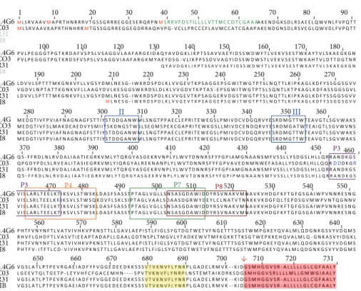
Figure 1: Alignment of amino acid sequences of some representatives of the GP82 family. Sequences are encoded by cDNA clones isolated fromT. cruzimetacyclic trypomastigotes: 5.4G6 (ABR19835); CO3 (ABO28970); R31 (AF128843); J18 (AAA21303). GenBank accession numbers are in parentheses. Potential initiator methionines (M) and a predicted N-amino terminal signal peptide are indicated in red and green, respectively. he Asp boxes (bacterial sialidase motifs) are boxed and indicated by Roman numerals II and III. he epitope for Mab3F6 (P3), mammalian cell binding sites (P4 and P8), and gastric-mucin binding site (P7) are boxed and indicated by diferent colors. Note the overlapping between P3 and P4 sites, and P7 and P8 sites. he subterminal VTV motif, characteristic of the TS superfamily, and the potential GPI-anchor sequence are shaded in yellow and magenta, respectively. he arrow denotes the cleavage site for GPI anchor addition.
heat inactivatedT. cruzi metacyclic forms [3, 4]. Since the determination of the irst GP82 gene sequence in 1994 [9] many other sequences have become available [10–14], including those fromT. cruzi genome sequencing projects [15–17]. he original analysis by Araya et al., 1994, showed the presence of two highly conserved Asp box domains (SxDxGxTW), previously described in bacterial sialidases, and a subterminal (VTVxNVFLYNR) motif (Figure 1) that are characteristics of the trans-sialidase (TS) superfamily of
T. cruzi[18]. For this reason GP82 was classiied in the TS superfamily [9,18].
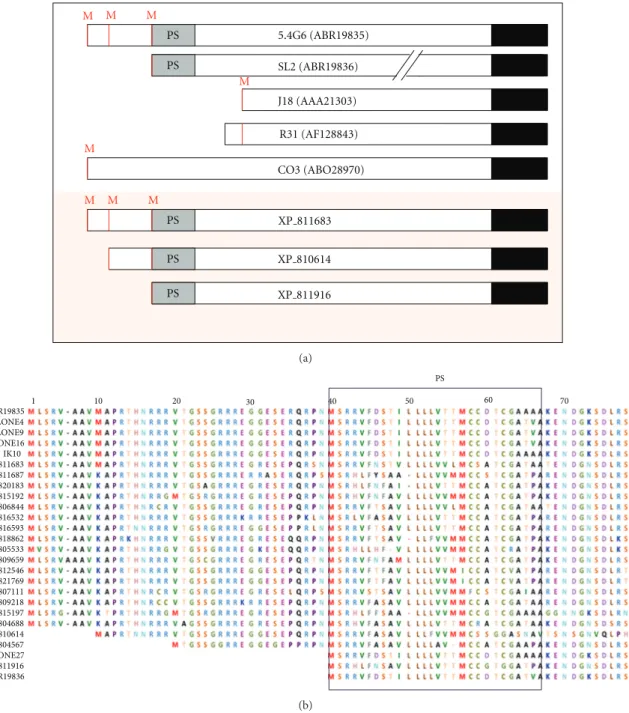
Figure 2 shows the comparison of ive GP82 sequence variants isolated in our laboratory by cDNA cloning and three genomic sequences of clone CL Brener (T. cruzigenome project). Although all variants code for a glycosylphos-phatidylinositol (GPI) anchor addition signal sequence at the carboxy-terminal (C-terminal), several of them do not have a signal peptide sequence at the amino-terminal (N-terminal), suggesting that they are not translocated into the endoplasmic reticulum (ER) and do not receive the GPI anchor.
Among the sequences annotated as GP82, the cDNA clone 5.4G6 (GenBank: EF154827) represents a complete transcript including the 5� and 3� untranslated regions (UTRs) (Figures1and2) [12]. his clone encodes a protein of
726 amino acids (GenBank: ABR19835) that reacts with the Mab3F6 (Figure 1) [12]. he open reading frame (ORF) has three ATG initiation codons in the same reading frame, but only the third codon is inserted within the Kozak sequence context (Figure 2) [12]. It has been proposed that the Kozak sequence (consensus: GCCRCCaugG, R designates a purine base and aug the initiation codon), located upstream of the initiation codon, facilitates the mRNA translation [19]. Fur-thermore, clone 5.4G6 encodes an N-terminal signal peptide of 27 amino acids located just ater the third methionine at position 39 and a signal sequence for cleavage/addition of GPI anchor at the C-terminal (Figures1and 2). Taking into account that the third ATG initiation codon follows the consensus Kozak sequence and the encoded protein has a predicted signal peptide, it is presumed that the translation initiates at the third methionine (Figure 2). he presence of 2-3 initiation codons in the same reading frame is relatively common among T. cruzi surface proteins, and ater the second or the third methionine there is a sequence encoding a cryptic signal peptide as described in many genes as GP85 [20], Tc85 [21], ASP-2 [22], CRP-10 [23], GP90 [24], and SAP [25].
PS PS
M M M
PS
PS
M
M
M M
PS M
5.4G6 (ABR19835)
SL2 (ABR19836)
J18 (AAA21303)
XP 811683
XP 810614 CO3 (ABO28970)
R31 (AF128843)
XP 811916
(a)
ABR19835 CLONE4 CLONE9 CLONE16 IK10 XP811683 XP811687 XP820183 XP815192 XP806844 XP816532 XP816593 XP818862 XP805533 XP809659 XP812546 XP821769 XP807111 XP809218 XP815197 XP804688 XP810614 XP804567 CLONE27 XP811916 ABR19836
1 10 20 30 40 50 60 70
PS
(b)
Figure 2: he modular architecture of GP82 family. (a) Structure of GP82 core proteins deduced from cDNA and genomic sequences.
Sequences from cDNA clones are listed in the legend ofFigure 1. SL (ABR19836) is a truncated cDNA sequence obtained by RT-PCR. he
slashes indicate that sequence is interrupted. For ease of viewing, the putative C-terminal was drawn in the same line. GenBank accession
numbers are in parentheses. he genomic sequences (GeneBank: XP 811683, XP 810614, XP 811916) were from theT. cruzigenome sequencing
project (clone CL Brener). Potential initiator methionines (M), predicted N-amino terminal signal peptide (SP), and potential GPI-anchor sequence are indicated in red, gray, and black, respectively. Not drawn to scale. (b) Alignment of GP82 sequences showing the variation of N-terminal region. Potential initiator methionines (M) are indicated in red and the predicted N-amino terminal signal peptide (PS) is boxed.
cDNA sequences: ABR19835, clones 4, 9, 16, IK10 and 27, ABR19835, ABR19836; genomic sequences were extracted from theT. cruzigenome
sequencing project (clone CL Brener) and are indicated by the preix XP .
the clone C03 (Figure 1) which is located at the lagellum of metacyclic forms of the CL strain [13]. Such GP82 pro-tein displaying lagellar localization is not involved in the invasion of mammalian cells by metacyclic forms [13]. Data obtained with monospeciic anti-GP82 antibodies support
the hypothesis that GP82 proteins that have no N-terminal signal peptide are located intracellularly and are not involved in host cell invasion [13].
V) at the N-terminal region, which acts as a signal peptide addressing the nascent protein into the ER. Analysis of the N-terminal region of the CRP-10 from TS superfamily indicates that this sequence functions as signal peptide [23]. Based on the analysis of GP82 variants characterized to date, we could suggest that metacyclic trypomastigotes simultaneously express diferent variants of GP82, and their cellular localization is determined by the N-terminal signal peptide. However, as the N-terminal of native GP82 has not been determined, we cannot rule out the possibility that the translation starts in diferent ATG codons.
he epitope recognized by Mab3F6 and the site of adhe-sion of GP82 to the host cells were identiied by incubating recombinant proteins and synthetic peptides in mammalian
in vitroinvasion assays. Regions of GP82 gene coding for the C-terminal, central, and N-terminal domains were subcloned into plasmid pGEX and expressed inE. coli. he reactivity with Mab3F6 and the ability of each recombinant protein to inhibit cell invasion were tested [6], both the Mab3F6 (P3) binding site and the host cell adhesion sites (P4 and P8) were identiied in the central domain of GP82 (Figure 1) as will be discussed in the topic below.
3. Organization of
GP82
Gene Family
T. cruzi genome comprises more than 50% of repetitive sequences including several multigene families that encode surface proteins. Among them, the most abundant is the TS superfamily [17]. he genome sequencing of clone CL Brener [17] conirmed the complexity of the TS superfamily by identiication of 1,430 sequences, including 737 genes and 693 pseudogenes. hese sequences have been annotated as trans-sialidase in theT. cruzigenome project with no mention to which group or family they could be included.
According to sequence identity, molecular weight, and function, members of TS superfamily were classiied into four groups or families [18,26–29]. Members of a family or group exhibit≥60% similarity among each other, whereas similarity among members of diferent families or groups may vary from 20 to 40%. Group I of the TS superfamily comprises proteins with enzymatic activity; that is, they are enzymes (trans-sialidase) able to transfer sialic acid from a donor to the mucins present atT. cruzisurface [18,26–29].
he members of group II were also called “trans-sialidase like” proteins because they have no enzymatic activity [18,
26–29]. hese proteins have complete or degenerate Asp box motifs (SxDxGxTW), the VTVxNVFLYNR motif char-acteristic of all TS members, and the signal sequence for cleavage/addition of GPI anchor at the C-terminal region [18, 26–29]. Group II comprises the surface glycoproteins GP85, Tc85, TSA-1, SA85, GP90, GP82, ASP-1, and ASP-2 which are involved in adhesion and invasion of mammalian cells [18, 20–22, 24, 26–30]. Several proteins of this group are also targets of the host immune system and may induce protective immunity in animal models [30]. he proteins of group III (CRP, FL160, CEA, and TESA) inhibit the classical and alternative pathways of complement activation and are recognized by sera from patients with Chagas disease [18,
26–29, 31, 32]. Group IV is composed of genes encoding trypomastigote surface antigens that have no deined biolog-ical function [18,26,27].
Recently, Freitas et al., 2011, [33] reported an extensive and detailed analysis of TS sequences of clone CL Brener that resulted in the redistribution of members in 8 diferent groups designated as TcSgroupI to TcSgroupVIII. he sequences analyzed in this study (� = 508) were categorized according to structure, function, presence of conserved motifs, chro-mosomal localization, expression proiling, and antigenic properties. TcSgroupI to TcSgroupIV (� = 176) correspond to groups I to IV described above. here is a good correlation with the classiication proposed previously [18, 26,27] and with the prior annotation made in our laboratory [16]. he new classiication proposed by Freitas et al., 2011, [33] could categorize 329 sequences that were included in the groups TcSgroupV–TcSgroupVIII.
To identify the repertoire ofGP82 genes in the genome of clone CL Brener (the reference clone ofT. cruziGenome Project), we carried out a BLASTP search using the GP82 encoded by clone 5.4G6 (GenBank: ABR19835) as query [10]. We identiied 19 complete sequences with>60% identity with the query which were considered as GP82 and distributed as follows: 2 proteins (GenBank: XP 811663 and XP 804688) with 70–81% identity and the remaining with 61–68% identity (seeFigure 2). Pseudogenes and truncated sequences were discarded from the analysis. Although GP82 are encoded from a relatively small number of genes, the repertoire is quite variable. his contrasts with other TS-like protein families which are composed of large sets of genes such as GP85, Tc85, GP90, and ASP [16,20,21,24,33].
he ability of genes to be robust to mutations at the codon level has been suggested as a key factor for understanding adaptation features. It has been proposed that genes relevant to host-parasite interactions will tend to exhibit high volatility or “anti-robust” patterns, which may be related to the parasite capacity of evading the host immune system [34]. We inves-tigated the potential capacity ofT. cruzisurface protein genes to maximize phenotypic variation, which may be seen as a key attribute to expand the repertoire of surface antigens [34]. he robustness of a parasite gene against mutations was addressed in terms of several genevolatilityanddiversityindicators. he potential impact of point-mutation errors on surface antigen genes based on the analysis of codon usage and its potential for generating diferent amino acid mutants were explored. hese data were consistent with the low rate of volatility calculated using the GP82 sequences deposited in GenBank [16].GP82genes have “low volatility” which means that the mutations are generally synonymous or lead replacing amino acids with others of the same polarity.GP82genes seem to be genetically “robust”; that is, they exhibit a tendency to neutralize the mutations encoding the same amino acid or an amino acid of the same polarity.
of GP82 genes suggests that they are “robust” or are not susceptible to mutation [16].
In the human protozoan parasites Trypanosoma brucei
and Plasmodium falciparum,the subtelomeric regions play an important role in generation of new variants of surface antigen genes and in the control of gene expression [35–
37]. We reported that T. cruzi subtelomeric regions are enriched in (pseudo)genes from the TS superfamily, DGF-1, and retrotransposon hot spot protein (RHS) families [38,39]. he abundance of surface protein genes in the subtelomeric regions suggests that these regions may have acted as a site for DNA recombination, expansion, and the generation of new variants of surface proteins. Moraes Barros et al. (2012) [39] demonstrated that all the groups of the TS superfamily are represented in the subtelomeric regions of clone CL Brener; most of the sequences (� = 83) are members of group II (GP82, GP85, TC85), which includes 22 complete genes. It is interesting to note that 7 out of 19GP82genes identiied in clone CL Brener are located at subtelomeric regions.
4. Synthesis and Processing of GP82
GP82 is attached to the outer parasite’s cell membrane by a GPI anchor [3, 40]. It is synthesized as a 70 kDa precursor devoid of N-linked sugars and when mature, it has an apparent molecular weight of 82 kDa. GP82 binds to the target cell in a dose-dependent and saturable fashion and reduces the infection of Vero cells by metacyclic forms of CL and Tulahuen strains by 90 to 97 and 50%, respectively [8].
he immunological screening of a metacyclic cDNA library with the Mab3F6 allowed the isolation of a 2,140 bp clone, named J18 (GenBank L14824), which encodes a protein of 516 amino acids containing the functional domains of GP82 [9]. Analysis of the deduced amino acid sequence showed the presence of three sialidase domains (two con-served and one slightly degenerated), a VTV motif, four putative N-glycosylation sites, and a GPI-anchor addition signal, which allows us to classify the GP82 in group II of TS superfamily [9].
Recombinant expression of clone J18 and a series of step-wise deletions enabled the identiication of the domain involved in the adhesion to the mammalian cells and indi-rectly, the region containing the epitope for Mab3F6 [6]. A central region spanning 132 amino acids was identiied as the responsible for the adhesin properties and ten overlapping peptides encompassing this central domain were synthesized to further characterize the region. he authors found two non-contiguous peptides with signiicant adhesive proper-ties, named P4 and P8; thus, they speculated about a putative conformational binding-domain in the native protein, in which these two peptides would be in close proximity [6].
To further address the adhesin activity of these peptides (P4 and P8) and to rule out any peptide’s solubility and conformational artifacts, both peptides were expressed in a non-adherent microorganism. he expression of GP82 cell binding peptides P4 or P8 in the fourth surface-exposed loop of the transmembrane protein LamB ofEscherichia coli
conferred the ability to this microorganism to adhere to the
surface of HeLa cells [41]. Between the two populations of bacteria, those carrying the P4 peptide were almost twice more eicient to adhere to HeLa cells than the population expressing the P8. In the same way, the expression of GP82 protein on the outer membrane of non-infective T. cruzi
epimastigotes enabled these non-adherent forms to attach to the surface of HeLa cells [42].
A more detailed analysis on the central domain of GP82 was performed by Manque et al., 2000, [43] using the same peptides described above by Santori et al., 1996, [6] and variants of GP82 lacking the regions corresponding to the peptides P4 and P8. his strategy allowed the authors to identify the peptide P3 as the epitope recognized by Mab3F6. As the peptide P3 has ten amino acids overlapping with the peptide P4 (cell binding site), this inding provided support for the inhibition of parasite’s invasion by Mab3F6, which is probably due to sterical hindrance. Additionally, the authors were able to conirm the GP82 conformational cell-binding domain hypothesis raised by Santori et al., 1996, [6] by means of the hybrid peptide P4/8 which contained 17 amino acids from P4 and 5 amino acids from P8 peptide. his peptide P4/8 was more eicient than P4 and P8 peptides to inhibit the binding of the recombinant GP82 to the HeLa cells [43].
Recently, it was demonstrated that GP82 binds speciically to gastric mucin in the oral infection [44,45]. he implication of GP82 in adhesion of metacyclic forms to the gastric mucin was irst described by Neira et al., 2003, [45] and further conirmed by Staquicini et al., 2010, [44] using a GP82 recombinant protein Del-4/8 lacking the central domain of the molecule. he GP82 binding to the gastric mucin may direct the adhesion and invasion of the stomach epithelium by the metacyclic forms. Tests of invasion inhibition of the gastric mucosa showed that peptide P7 (Figure 1), located in the central domain of the molecule, contains the binding site to the gastric mucin [44]. hein vitro inhibitory efect of peptide P7 was reproduciblein vivoin murine model [44].
Experimental evidence of the GP82 GPI-anchor was given by Cardoso De Almeida & Heise (1993) [40] through digestion with phosphatidylinositol-speciic phospholipase C (PI-PLC) and phase separation in Triton X-114. Araya et al., 1994, [9] predicted the putative GP82 GPI-anchor cleavage/addition site based on the sequence of the clone J18. Similarly, Ramirez et al., 1999, [14,46] analyzed in more detail the GPI-anchor signal of GP82 and otherT. cruziproteins and conducting homologous and heterologous expressions of GP82 inT. cruziepimastigotes and mammalian cell systems. Despite the absence of a typical signal peptide in the protein encoded by the J18 clone, the authors found that T. cruzi
indings indicated that the requirements for GPI-anchoring are diferent betweenT. cruziand mammalian cells [14,46].
In order to further dissect the requirements for GPI-anchoring between mammals and T. cruzi, a site-directed mutagenesis was performed in the GPI cleavage/addition signal [47]. he putative GPI-anchor acceptor domain deter-mined by Ramirez et al., 1999, [46] is formed by the amino acids aspartic (�), glycine (� + 1), and serine (�+ 2) (DGS) where the aspartic acid is linked to the GPI-anchor. A single mutation was introduced changing the aspartic acid to serine generating the sequence (SGS) which previously proved to be a feasible signal for GPI anchoring of T. bruceiVariant Surface Glycoprotein (VSG) in mammals [48]. An additional construct lacking the GPI-anchor was created and transfected either in mammalian cells orT. cruziepimastigotes [47].
Confocal analyses on transfected parasites showed that the point mutation had no detectable efect on the GPI-anchoring eiciency [47]. he deletion of the GPI-signal resulted in a protein that was not anchored but accumulated in the parasite cytoplasm instead [47]. hese indings were in agreement with those obtained in T. brucei by B¨ohme and Cross (2002) [49] where the parasite was able to anchor several mutated proteins but not those in which the GPI-anchor signal was deleted. On the other hand, the mam-malian cells failed to express all the transfected proteins on the cell surface, even the point mutation which proved to be functional for GPI anchoring in mammals [48].
Isolation of GPI-anchored proteins can be accomplished by digestion with enzymes that cleave speciically this struc-ture as glycosylphosphatidylinositol-speciic phospholipase C (GPI-PLC) or phosphatidylinositol-speciic phospholipase C (PI-PLC). his very simple approach can be hindered by the presence of acylation in position 6 of the inositol ring (sometimes the acylation occurs at position 5) due to steric hindrance.
Due to the presence of the GPI anchor, proteins carrying this modiication acquire an overall hydrophobic behavior. Based on this property, detergents can be used to con-centrate/enrich fractions in this particular kind of proteins by temperature-induced partition. Bordier 1981 [50] irst described the suitability of the detergent Triton X-114 (TX-114) to concentrate hydrophobic proteins due to its nearly physiological clouding point temperature. At temperatures above 23∘C a formerly homogenous solution containing the detergent TX-114 will split into two phases: an upper layer depleted of detergent (hydrophilic) and a lower phase enriched in detergent’s micelles (hydrophobic). Using this physical property, Cordero et al., 2009, [51] concentrated the GPI-anchored proteins of metacyclic and epimastigotes of T. cruzi ater several consecutive partitions in TX-114. Mass spectrometry analyses on those fractions detected several members of TS superfamily, among those, the surface glycoprotein GP82 had 22% of its sequence covered by tryptic peptides. Among those peptides, the Asp boxes, VTV, and cell binding site P8 were mapped [51]. Other important region mapped in this study was peptide P7, which was identiied as the binding site for the gastric mucin [44].
Several proteomics studies have been conducted in meta-cyclic forms of T. cruzi, but to the best of our knowledge,
there is only one additional report of GP82 elsewhere [52]. Recently, a quantitative proteomic study was performed in parasites undergoing metacyclogenesis [53]. Among the proteins identiied in this study, authors found 38 members of TS superfamily. Due to lack of a uniied/standardized annotation among the databases and the absence of the peptide sequences used in this study, it was not possible to determine the presence of GP82 among them. Some of those annotated TSs shared a high degree of identity with GP82 protein, but because of the missing peptide sequences it is impossible to assign an unambiguous classiication. he lack of a common nonredundant annotation represents an issue that must be taken in consideration with an urge to amend.
Recently, Cortez et al., 2012, [54] compiled all the biochemical, physicochemical, and functional information available on GP82 in order to create the most updated model of the protein structure. he authors based this model on the homology ofT. cruziGP82 (GenBank: L14824) withT. rangeli
sialidase (PDB 1N1T A), a close related molecule which had its crystallographic structure (inhibitor-bound) already solved. he sketched GP82 appears as two clearly diferent and separated domains (an amino-terminal�-propeller and a�-sandwich C-terminal domain) linked together by an� -helix. In this layout, P3, P4, P7, and P8 motifs have a variable degree of access to the solvent. he cell-binding peptide P4 encompasses 2/3 of the�-helix that bridges the protein together and is fully exposed. On the other hand, peptide P8 located in the carboxy-terminal domain, although exposed, has limited solvent accessibility. he partial exposure of the P8 motif complies with the experimental data and gives a topological explanation for the limited role of P8 in the GP82 binding to the cell [41]. As expected, the P3 motif containing the epitope for the 3F6 antibody was fully exposed and accessible to the solvent, reinforcing the Mab3F6 inhibitory efect by steric hindrance. he gastric mucin-binding motif P7 was poorly exposed, mostly due to its high hydrophobicity. he amino-terminal residues of this motif (P7) are completely buried, leaving just the C-terminal portion partially exposed, mainly because it overlaps with the P8 motif. In summary, this model seems to fulill the requirements for structural analysis and provides an appropriated support to the biological experimental data.
5. Post-Transcriptional Control Mechanisms
of GP82 Expression
In trypanosomes, post-translational control mechanisms play an important role in gene expression regulation due to unique features related to transcription, mRNA maturation, and stabilization over the parasite life cycle. T. cruzi genome is organized in large gene clusters separated by divergent strand-switch regions [55], and transcription of these clus-ters produces large primary transcripts that are processed by trans-splicing and polyadenylation to generate mature mRNAs [56]. hese processes are guided by pyrimidine-rich regions contained in the polycistronic transcripts [57,
(epimastigote, metacyclic trypomastigote, amastigote, and bloodstream trypomastigote) demonstrated that there was a signiicant change in relative protein abundance throughout life cycle [52]. Furthermore, microarray analysis showed that at least 50% ofT. cruzigenes are regulated during its life cycle [59]. GP82 is one of these diferentially regulated proteins, and the mechanisms regulating its stage-speciic expression began to be clariied.
Steady-state levels of GP82 transcripts from T. cruziG strain were determined by northern blot, dot-blot hybridiza-tion, and quantitative real-time PCR, demonstrating that there is a signiicant increase in GP82 mRNA levels in metacyclic forms when compared with the other three stages [9, 11, 60]. Northern blot analysis revealed a single band of 2.2 kb mRNA only in metacyclic forms [9]. Dot-blot hybridization showed that GP82 transcript levels were around 5.5-fold higher in metacyclic trypomastigotes than in other stages [60]. Similar results were obtained using quantitative real-time PCR (unpublished data). Moreover, expression analysis of other three GP82 gene subfamilies from Peru-2 strain, called groups A, B, and C, showed an increase in mRNA accumulation (4.7 to 9.3-fold) in metacyclic forms when compared to epimastigotes [11]. Additionally, GP82 protein stage-speciic expression was also showed by western blot using the Mab3F6 [3]. Even though GP82 mRNA and protein were barely detected in epimastigotes, nuclear run-on analysis demrun-onstrated that GP82 gene was transcribed in both epimastigote and metacyclic forms, conirming that transcript accumulation in metacyclic forms is not due to an increased transcription rate, but rather to some post-translational control [60].
Changes in GP82 mRNA stability were detected and thought to be responsible for diferences in its steady-state level. Parasites treated with actinomycin D had their GP82 transcript half-lives estimated to be about 6 h in metacyclic forms and 0.5 h in epimastigotes [60]. Cycloheximide treat-ment increased GP82 levels in epimastigotes, suggesting that a labile protein factor was responsible for destabilizing mRNA in these forms and prevent mRNA translation. In addition, GP82 mRNAs were only found associated with polysomes in metacyclic forms [60], indicating that transcript mobilization to polysomes might be involved in regulating GP82 expression, as was reported for anotherT. cruzigene [61].
here are at least three known factors that modu-late mRNA steady-state level: cis-acting elements, trans -acting factors, and the apparatus involved in mRNA turnover and degradation [62].Cis-acting elements are non-coding sequences that act from inside the same molecule (intramolecular action). Trans-acting factors are difuse molecules, usually proteins, that act from a diferent molecule to regulate a target mRNA (intermolecular action) [63]. he fate of transcripts is determined by the interaction of cis -acting sequences present in the 3�UTR with speciictrans -acting protein factors containing RNA-binding domains that subsequently recruit the protein machinery to destroy or stabilize mRNAs [64]. he involvement of GP82 3�UTR in mRNA stability was analyzed using a reporter green luo-rescent protein (GFP) fused upstream to the GP82 3�UTR.
Parasites transfected with an episomal plasmid carrying this construct had their GFP protein and mRNA levels analyzed, revealing that the 3�UTR was able to downregulate GFP in epimastigotes and upregulate it in metacyclic forms [65]. Similar mechanisms for controlling mRNA stability by 3�UTR sequences have also been described for other TS family members, such as the lagellum-associated surface protein FL-160 (TcS group III) [66], two genes coding for active trans-sialidase enzymes from TcS group I, described by Jager et al., 2008, [67], and another TS member [64]. here are pieces of evidence that stem-loop secondary structures formed in the 3�UTR might be responsible for the interaction with RNA-binding proteins [68]. Prediction of GP82 3�UTR secondary structure was performed in silico using mfold program [69], revealing the presence of stem-loop structures; however, the role of these structures was not analyzed so far. Regulatory cis-acting elements of variable sizes were identiied in the 3�UTR of some trypanosomatid genes (reviewed in [62,70]). In the case of GP82, four step-wise deletions were performed to search for regulatory elements in its 3�UTR. Results indicated that more than one region was responsible for changing GFP mRNA and protein levels in epimastigotes and metacyclic forms [65], suggesting that multiplecis-acting elements are present in GP82 3�UTR and might bind to distinct RNA-binding proteins (RBP). he irst
trans-acting factor identiied inT. cruziwas TcUBP1 (T. cruzi
uridine binding protein 1), which binds to AU-rich elements of the TcSMUG mRNA leading to its destabilization [71]. In addition to TcSMUG, other 39 transcripts were found bound to TcUBP1 by co-immunoprecipitation assays [68]. One commoncis-acting element was identiied in the 3�UTRs of the majority of these TcUBP1 target mRNAs. his cis -element was used to predict novel UBP1 target mRNAs and GP82 was one of them [68]. herefore, TcUBP1 could be one of thetrans-acting factors involved in GP82 mRNA stability. A schematic representation of the mechanism controlling
GP82gene expression is shown inFigure 3.
here are growing pieces of evidence suggesting the pres-ence of post-transcriptional operons in trypanosomes, medi-ated by the coordinmedi-ated interaction betweencis-elements and
trans-acting factors [68,72]. It was demonstrated that a group ofT. bruceistage-regulated proteins share a speciic sequence motif in the 3�UTR (reviewed in [72]). Also, two RBPs from
T. cruzi, TcUBP1, and TcUBP3, preferentially associate with a set of functionally related transcripts bearing the same RNA motif that is recognized by each protein [68]. hese post-transcriptional operons could explain how coordinately expression regulation is achieved in organisms where gene-speciic transcriptional control is absent.
6. Concluding Remarks and Perspectives
Epimastigote
N
mRNA destabilization
mRNA decay GP82
mRNA GP82
RBP GP82
RBP
5′
5′UTR CDS 3′UTR
5′ AAA 3′
AAA 3
′
(a)
Metacyclic
N
Translation in polysomes mRNA stabilization
GP82
mRNA GP82
RBP GP82
RBP
AAA 3
′
AAA 3
′ AAA 3′
5′UTR CDS 3′UTR
5′
5′
5′
(b)
Figure 3: Comparison of GP82 mRNA post-transcriptional control mechanisms in (a) epimastigotes and (b) metacyclic trypomastigotes.
In epimastigotes, GP82 mRNA interacts with possibly more than one RNA-binding protein (RBP), which binds to diferentcis-elements in
the 3�UTR region (small black rectangles), leading to mRNA destabilization and decay. Conversely, in the metacyclic trypomastigote stage, a
diferent set of RBPs interacts with thecis-elements present in the 3�UTR, promoting mRNA stabilization and translation in polysomes.
genes suggests that they are not susceptible to mutation. he many isoforms of GP82 and its multiple N-terminal variants suggest that some GP82 family members might display diferent cellular localizations and functions. he challenge is to ascertain the relationships betweenGP82gene sequences, protein isoforms, and its distinct or overlapping functions.
GP82 is a GPI-anchored surface protein, synthesized as a 70 kDa precursor devoid ofN-linked sugars and when mature has an apparent molecular weight of 82 kDa. GPI-minus vari-ants accumulate in the ER indicating that GPI anchor acts as a forward transport signal for progressing along the secretory pathway as suggested forT. cruzimucins [73]. Heterologous expression of GP82 into mammalian cells indicated that the requirements for GPI-anchoring are diferent between T. cruziand mammalian cells. hese diferences could be targets for the development of parasite-speciic therapeutic agents.
Several studies demonstrated that the transcription of GP82 is constitutive and may be regulated at post-transcriptional level, for instance, at translational level and/or mRNA stabilization. GP82 mRNAs are mobilized to polysomes and consequently translated, but only in metacyclic trypomastigotes. It has been suggested that the stabilizing mechanism acting in metacyclic trypomastigotes and the destabilizing mechanism in epimastigotes could be mediated by a cis-acting element present in the 3�UTR of transcripts. A series of step-wise deletions in the 3�UTR was created and results suggest that the mechanism regulating GP82 expression involves multiple elements in the 3�UTR. Interestingly, the 3�UTR of GP82 transcript promotes higher
expression of the green luorescent protein (GFP) reporter in metacyclic trypomastigotes than in epimastigotes.
In conclusion, while our knowledge of the structure and function of GP82 is large, there still remain many questions to be answered. Additional studies are carried out to analyze the expression, localization, and involvement in host cell invasion of each GP82 variant identiied to date.
Acknowledgments
his work was supported by grants from FAPESP and CNPq (Brazil) to J. F. da Silveira. P. R. C. Correa and E. Bayer-Santos are awarded a doctoral fellowship from FAPESP and CNPq, Brazil, respectively.
References
[1] N. Yoshida, K. M. Tyler, and M. S. Llewellyn, “Invasion mechanisms among emerging food-borne protozoan parasites,”
Trends in Parasitology, vol. 27, no. 10, pp. 459–466, 2011. [2] N. Yoshida, “Molecular basis of mammalian cell invasion by
Trypanosoma cruzi,”Anais da Academia Brasileira de Ciencias, vol. 78, no. 1, pp. 87–111, 2006.
[3] M. M. G. Teixeira and N. Yoshida, “Stage-speciic surface
antigens of metacyclic trypomastigotes ofTrypanosoma cruzi
identiied by monoclonal antibodies,”Molecular and
Biochemi-cal Parasitology, vol. 18, no. 3, pp. 271–282, 1986.
[4] N. Yoshida, “Surface antigens of metacyclic trypomastigotes of
[5] R. C. Ruiz, S. Favoreto, M. L. Dorta et al., “Infectivity of
Trypanosoma cruzistrains is associated with diferential
expres-sion of surface glycoproteins with diferential Ca2+signalling
activity,”Biochemical Journal, vol. 330, part 1, pp. 505–511, 1998.
[6] F. R. Santori, M. L. Dorta, L. Juliano et al., “Identiication
of a domain ofTrypanosoma cruzimetacyclic trypomastigote
surface molecule gp82 required for attachment and invasion of
mammalian cells,”Molecular and Biochemical Parasitology, vol.
78, no. 1-2, pp. 209–216, 1996.
[7] M. L. Dorta, A. T. Ferreira, M. E. M. Oshiro, and N. Yoshida,
“Ca2+ signal induced by Trypanosoma cruzi metacyclic
try-pomastigote surface molecules implicated in mammalian cell
invasion,”Molecular and Biochemical Parasitology, vol. 73, no.
1-2, pp. 285–289, 1995.
[8] M. I. Ramirez, R. De Cassia Ruiz, J. E. Araya, J. F. Da Silveira, and N. Yoshida, “Involvement of the stage-speciic
82-kilodalton adhesion molecule ofTrypanosoma cruzimetacyclic
trypomastigotes in host cell invasion,”Infection and Immunity,
vol. 61, no. 9, pp. 3636–3641, 1993.
[9] J. E. Araya, M. I. Cano, N. Yoshida, and J. F. Da Silveira, “Cloning and characterization of a gene for the stage-speciic 82-kDa
surface antigen of metacyclic trypomastigotes ofTrypanosoma
cruzi,”Molecular and Biochemical Parasitology, vol. 65, no. 1, pp. 161–169, 1994.
[10] P. R. C. Correa, Study of genetic variability of the surface
antigen GP82 of metacyclic trypomastigotes of Trypanosoma cruzi: identiication of transcripts repertoire and the binding-site to the mammalian cell [Ph.D. Doctoral hesis], Federal University of S˜ao Paulo (UNIFESP), S˜ao Paulo, Brazil, 2012. [11] D. Songthamwat, K. Kajihara, M. Kikuchi et al., “Structure
and expression of three gp82 gene subfamilies ofTrypanosoma
cruzi,”Parasitology International, vol. 56, no. 4, pp. 273–280, 2007.
[12] E. M. Cordero,In vitro and in vivo expression of genes encoding
for surface antigens GP82 and GP90 of metacyclic trypomastig-otes of Trypanosoma cruzi [Ph.D. Doctoral hesis], Federal University of S˜ao Paulo (UNIFESP), S˜ao Paulo, Brazil, 2012. [13] V. D. Atayde, M. Cortez, R. Souza, J. F. Da Silveira, and N.
Yoshida, “Expression and cellular localization of molecules of
the gp82 family inTrypanosoma cruzimetacyclic
trypomastig-otes,”Infection and Immunity, vol. 75, no. 7, pp. 3264–3270, 2007.
[14] M. I. Ramirez, S. B. Boscardin, S. W. Han et al., “Heterologous
expression of aTrypanosoma cruzisurface glycoprotein (gp82)
in mammalian cells indicates the existence of diferent signal
sequence requirements and processing,”Journal of Eukaryotic
Microbiology, vol. 46, no. 6, pp. 557–565, 1999.
[15] O. Franzen, E. Arner, M. Ferella et al., “he short non-coding
transcriptome of the protozoan parasiteTrypanosoma cruzi,”
PLoS Neglected Tropical Diseases, vol. 5, no. 8, Article ID e1283, 2011.
[16] F. Azuaje, J. L. Ramirez, and J. F. Da Silveira, “An exploration of the genetic robustness landscape of surface protein families
in the human protozoan parasite Trypanosoma cruzi,”IEEE
Transactions on Nanobioscience, vol. 6, no. 3, pp. 223–228, 2007. [17] N. M. El-Sayed, P. J. Myler, D. C. Bartholomeu et al., “he
genome sequence of Trypanosoma cruzi, etiologic agent of
chagas disease,”Science, vol. 309, no. 5733, pp. 409–435, 2005.
[18] W. Colli, “Trans-sialidase: a unique enzyme activity discovered
in the protozoanTrypanosoma cruzi,”he FASEB Journal, vol.
7, no. 13, pp. 1257–1264, 1993.
[19] M. Kozak, “Efects of intercistronic length on the eiciency of
reinitiation by eucaryotic ribosomes,”Molecular and Cellular
Biology, vol. 7, no. 10, pp. 3438–3445, 1987.
[20] G. B. Takle and G. A. M. Cross, “An 85-kilodalton surface
antigen gene family ofTrypanosoma cruziencodes polypeptides
homologous to bacterial neuraminidases,”Molecular and
Bio-chemical Parasitology, vol. 48, no. 2, pp. 185–198, 1991.
[21] R. Giordano, D. L. Fouts, D. Tewari, W. Colli, J. E. Manning, and M. J. M. Alves, “Cloning of a surface membrane
glyco-protein speciic for the infective form ofTrypanosoma cruzi
having adhesive properties to laminin,”he Journal of Biological
Chemistry, vol. 274, no. 6, pp. 3461–3468, 1999.
[22] H. P. Low and R. L. Tarleton, “Molecular cloning of the gene encoding the 83 kDa amastigote surface protein and its
identiication as a member of theTrypanosoma cruzisialidase
superfamily,”Molecular and Biochemical Parasitology, vol. 88,
no. 1-2, pp. 137–149, 1997.
[23] K. A. Norris, J. E. Schrimpf, and M. J. Szabo, “Identiication of
the gene family encoding the 160-kilodaltonTrypanosoma cruzi
complement regulatory protein,”Infection and Immunity, vol.
65, no. 2, pp. 349–357, 1997.
[24] M. S. Do Carmo, M. R. M. Dos Santos, M. I. Cano, J. E. Araya, N. Yoshida, and J. F. Da Silveira, “Expression and genome-wide distribution of the gene family encoding a 90 kDa surface
glycoprotein of metacyclic trypomastigotes of Trypanosoma
cruzi,”Molecular and Biochemical Parasitology, vol. 125, no. 1-2, pp. 201–206, 2002.
[25] R. C. P. Baida, M. R. M. Santos, M. S. Carmo et al., “Molecular characterization of serine-, alanine-, and proline-rich proteins of Trypanosoma cruzi and their possible role in host cell
infection,”Infection and Immunity, vol. 74, no. 3, pp. 1537–1546,
2006.
[26] O. Campetella, D. Sanchez, J. J. Cazzulo, and A. C. C. Frasch, “A
superfamily ofTrypanosoma cruzisurface antigens,”
Parasitol-ogy Today, vol. 8, no. 11, pp. 378–381, 1992.
[27] S. Schenkman, D. Eichinger, M. E. A. Pereira, and V. Nussen-zweig, “Structural and functional properties of Trypanosoma
trans-sialidase,” Annual Review of Microbiology, vol. 48, pp.
499–523, 1994.
[28] G. A. M. Cross and G. B. Takle, “he surface trans-sialidase
family ofTrypanosoma cruzi,”Annual Review of Microbiology,
vol. 47, pp. 385–411, 1993.
[29] A. C. C. Frasch, “Functional diversity in the trans-sialidase and
mucin families inTrypanosoma cruzi,”Parasitology Today, vol.
16, no. 7, pp. 282–286, 2000.
[30] C. Claser, N. M. Esp´ındola, G. Sasso, A. J. Vaz, S. B. Boscardin, and M. M. Rodrigues, “Immunologically relevant strain
poly-morphism in the Amastigote Surface Protein 2 ofTrypanosoma
cruzi,”Microbes and Infection, vol. 9, no. 8, pp. 1011–1019, 2007. [31] M. Beucher and K. A. Norris, “Sequence diversity of the
Trypanosoma cruzi complement regulatory protein family,”
Infection and Immunity, vol. 76, no. 2, pp. 750–758, 2008. [32] T. K. Matsumoto, P. C. Cotrim, J. F. Da Silveira, A. M. S.
Stolf, and E. S. Umezawa, “Trypanosoma cruzi: isolation of an
immunodominant peptide of TESA (trypomastigote
excreted-secreted antigens) by gene cloning,”Diagnostic Microbiology
and Infectious Disease, vol. 42, no. 3, pp. 187–192, 2002. [33] L. M. Freitas, S. L. dos Santos, G. F. Rodrigues-Luiz et al.,
“Genomic analyses, gene expression and antigenic proile of
the trans-sialidase superfamily ofTrypanosoma cruzireveal an
undetected level of complexity,”PLoS One, vol. 6, no. 10, Article
[34] J. B. Plotkin, J. Dushof, and H. B. Fraser, “Detecting selection
using a single genome sequence of M. tuberculosis and P.
falciparum,”Nature, vol. 428, no. 6986, pp. 942–945, 2004. [35] L. H. Freitas-Junior, E. Bottius, L. A. Pirrit et al., “Frequent
ectopic recombination of virulence factor genes in telomeric
chromosome clusters ofP. falciparum,”Nature, vol. 407, no.
6807, pp. 1018–1022, 2000.
[36] C. B. B. Lira, M. A. Giardini, J. L. S. Neto, F. F. Conte, and M. I. N. Cano, “Telomere biology of trypanosomatids: beginning to
answer some questions,”Trends in Parasitology, vol. 23, no. 8,
pp. 357–362, 2007.
[37] D. Horn and J. D. Barry, “he central roles of telomeres and subtelomeres in antigenic variation in African trypanosomes,”
Chromosome Research, vol. 13, no. 5, pp. 525–533, 2005. [38] D. Kim, M. A. Chiurillo, N. El-Sayed et al., “Telomere and
sub-telomere ofTrypanosoma cruzichromosomes are enriched in
(pseudo)genes of retrotransposon hot spot and
trans-sialidase-like gene families: the origins ofT. cruzitelomeres,”Gene, vol.
346, pp. 153–161, 2005.
[39] R. Moraes Barros, M. Marini, C. Antonio et al., “Anatomy and evolution of telomeric and subtelomeric regions in the human
protozoan parasiteTrypanosoma cruzi,”BMC Genomics, vol. 13,
article 229, 2012.
[40] M. L. Cardoso De Almeida and N. Heise, “Proteins anchored via glycosylphosphatidylinositol and solubilizing phospholipases inTrypanosoma cruzi,”Biological Research, vol. 26, no. 1-2, pp. 285–312, 1993.
[41] C. M. Pereira, S. Favoreto, J. F. Da Silveira, N. Yoshida, and B. A. Castilho, “Adhesion of Escherichia coli to HeLa cells mediated byTrypanosoma cruzi surface glycoprotein-derived peptides
inserted in the outer membrane protein LamB,”Infection and
Immunity, vol. 67, no. 9, pp. 4908–4911, 1999.
[42] P. M. Manque, I. Neira, V. D. Atayde et al., “Cell adhesion and
Ca2+signaling activity in stably transfectedTrypanosoma cruzi
epimastigotes expressing the metacyclic stage-speciic surface
molecule gp82,”Infection and Immunity, vol. 71, no. 3, pp. 1561–
1565, 2003.
[43] P. M. Manque, D. Eichinger, M. A. Juliano, L. Juliano, J. E. Araya, and N. Yoshida, “Characterization of the cell adhesion site of
Trypanosoma cruzimetacyclic stage surface glycoprotein gp82,”
Infection and Immunity, vol. 68, no. 2, pp. 478–484, 2000. [44] D. I. Staquicini, R. M. Martins, S. Macedo et al., “Role of GP82
in the selective binding to gastric mucin during oral infection withTrypanosoma cruzi,”PLoS Neglected Tropical Diseases, vol. 4, no. 3, Article ID e613, 2010.
[45] I. Neira, F. A. Silva, M. Cortez, and N. Yoshida, “Involvement of
Trypanosoma cruzimetacyclic trypomastigote surface molecule gp82 in adhesion to gastric mucin and invasion of epithelial
cells,”Infection and Immunity, vol. 71, no. 1, pp. 557–561, 2003.
[46] M. I. Ramirez, S. B. Boscardin, R. C. Ruiz et al., “Heterologous
expression of aTrypanosoma cruzisurface glycoprotein (gp82)
indicates that requirements for glycosylphosphatidylinositol anchoring are diferent in mammalian cells and this
try-panosome,”Mem´orias do Instituto Oswaldo Cruz, vol. 94, no.
4, pp. 527–530, 1999.
[47] E. M. Cordero,Study of processing surface glycoprotein of 82 kDa
(GP82) of metacyclic trypomastigotes of Trypanosoma cruzi [M.Sc. thesis], Federal University of S˜ao Paulo (UNIFESP), S˜ao Paulo, Brazil, 2002.
[48] P. Moran and I. W. Caras, “Requirements for glycosylphos-phatidylinositol attachment are similar but not identical in
mammalian cells and parasitic protozoa,”Journal of Cell Biology,
vol. 125, no. 2, pp. 333–343, 1994.
[49] U. B¨ohme and G. A. M. Cross, “Mutational analysis of the variant surface glycoprotein GPI-anchor signal sequence in
Trypanosoma brucei,”Journal of Cell Science, vol. 115, no. 4, pp. 805–816, 2002.
[50] C. Bordier, “Phase separation of integral membrane proteins in
Triton X-114 solution,”he Journal of Biological Chemistry, vol.
256, no. 4, pp. 1604–1607, 1981.
[51] E. M. Cordero, E. S. Nakayasu, L. G. Gentil, N. Yoshida, I. C. Almeida, and J. F. Da Silveira, “Proteomic analysis of detergent-solubilized membrane proteins from insect-developmental
forms ofTrypanosoma cruzi,”Journal of Proteome Research, vol.
8, no. 7, pp. 3642–3652, 2009.
[52] J. A. Atwood, D. B. Weatherly, T. A. Minning et al.,
“Microbi-ology: theTrypanosoma cruziproteome,”Science, vol. 309, no.
5733, pp. 473–476, 2005.
[53] L. M. de Godoy, F. K. Marchini, D. P. Pavoni et al., “Quantitative
proteomics of Trypanosoma cruzi during metacyclogenesis,”
Proteomics, vol. 12, no. 17, pp. 2694–2703, 2012.
[54] C. Cortez, N. Yoshida, D. Bahia, and T. Sobreira, “Structural
basis of the interaction of aTrypanosoma cruzisurface molecule
implicated in oral infection with host cells and gastric mucin,”
PLoS One, vol. 7, no. 7, Article ID e42153, 2012.
[55] S. Mart´ınez-Calvillo, J. C. Vizuet-de-Rueda, L. E. Florencio-Mart´ınez, R. G. Manning-Cela, and E. E. Figueroa-Angulo,
“Gene expression in trypanosomatid parasites,” Journal of
Biomedicine and Biotechnology, vol. 2010, Article ID 525241, 15 pages, 2010.
[56] X. H. Liang, A. Haritan, S. Uliel, and S. Michaeli, “trans and cis splicing in trypanosomatids: mechanism, factors, and
regulation,”Eukaryotic Cell, vol. 2, no. 5, pp. 830–840, 2003.
[57] C. Hartmann, H. R. Hotz, M. McAndrew, and C. Clayton, “Efect of multiple downstream splice sites on polyadenylation inTrypanosoma brucei,”Molecular and Biochemical
Parasitol-ogy, vol. 93, no. 1, pp. 149–152, 1998.
[58] C. Lopez-Estrano, C. Tschudi, and E. Ullu, “Exonic sequences in
the5�untranslated region of�-tubulin mRNA modulate trans
splicing inTrypanosoma brucei,”Molecular and Cellular Biology,
vol. 18, no. 8, pp. 4620–4628, 1998.
[59] T. A. Minning, D. B. Weatherly, J. Atwood, R. Orlando, and R. L. Tarleton, “he steady-state transcriptome of the four major
life-cycle stages ofTrypanosoma cruzi,”BMC Genomics, vol. 10,
article 370, 2009.
[60] L. G. Gentil, E. M. Cordero, M. S. D. Carmo, M. R. M. D. Santos, and J. F. D. Silveira, “Posttranscriptional mechanisms involved in the control of expression of the stage-speciic GP82 surface
glycoprotein inTrypanosoma cruzi,”Acta Tropica, vol. 109, no.
2, pp. 152–158, 2009.
[61] A. R. Avila, S. F. Yamada-Ogatta, V. Da Silva Monteiro et al., “Cloning and characterization of the metacyclogenin gene,
which is speciically expressed duringTrypanosoma cruzi
meta-cyclogenesis,”Molecular and Biochemical Parasitology, vol. 117,
no. 2, pp. 169–177, 2001.
[62] C. Clayton and M. Shapira, “Post-transcriptional regulation of
gene expression in trypanosomes and leishmanias,”Molecular
and Biochemical Parasitology, vol. 156, no. 2, pp. 93–101, 2007. [63] J. G. De Gaudenzi, G. Noe, V. A. Campo, A. C. Frasch, and
A. Cassola, “Gene expression regulation in trypanosomatids,”
[64] P. R. Araujo, G. A. Burle-Caldas, R. A. Silva-Pereira, D. C. Bartholomeu, W. D. daRocha, and S. M. R. Teixeira, “Devel-opment of a dual reporter system to identify regulatory
cis-acting elements in untranslated regions ofTrypanosoma cruzi
mRNAs,”Parasitology International, vol. 60, no. 2, pp. 161–169,
2011.
[65] E. Bayer-Santos, L. G. Gentil, E. M. Cordero, P. R. Correa, and J.
F. da Silveira, “Regulatory elements in the3�untranslated region
of the GP82 glycoprotein are responsible for its stage-speciic
expression inTrypanosoma cruzimetacyclic trypomastigotes,”
Acta Tropica, vol. 123, no. 3, pp. 230–233, 2012.
[66] D. Weston, A. C. La Flamme, and W. C. Van Voorhis,
“Expres-sion ofTrypanosoma cruzisurface antigen FL-160 is controlled
by elements in the3�untranslated, the3� intergenic, and the
coding regions,”Molecular and Biochemical Parasitology, vol.
102, no. 1, pp. 53–66, 1999.
[67] A. V. Jager, R. P. Muia, and O. Campetella, “Stage-speciic
expression ofTrypanosoma cruzitrans-sialidase involves highly
conserved3�untranslated regions,”FEMS Microbiology Letters,
vol. 283, no. 2, pp. 182–188, 2008.
[68] G. Noe, J. G. De Gaudenzi, and A. C. Frasch, “Functionally related transcripts have common RNA motifs for speciic
RNA-binding proteins in trypanosomes,”BMC Molecular Biology, vol.
9, article 107, 2008.
[69] M. Zuker, “Mfold web server for nucleic acid folding and
hybridization prediction,”Nucleic Acids Research, vol. 31, no. 13,
pp. 3406–3415, 2003.
[70] S. Haile and B. Papadopoulou, “Developmental regulation of
gene expression in trypanosomatid parasitic protozoa,”Current
Opinion in Microbiology, vol. 10, no. 6, pp. 569–577, 2007. [71] I. D’Orso and A. C. C. Frasch, “TcUBP-1, a developmentally
regulated U-rich RNA-binding protein involved in selective
mRNA destabilization in trypanosomes,”he Journal of
Biolog-ical Chemistry, vol. 276, no. 37, pp. 34801–34809, 2001. [72] S. Kramer and M. Carrington, “Trans-acting proteins regulating
mRNA maturation, stability and translation in
trypanoso-matids,”Trends in Parasitology, vol. 27, no. 1, pp. 23–30, 2011.
[73] G. E. Canepa, A. C. Mesias, H. Yu, X. Chen, and C. A. Buscaglia, “Structural features afecting traicking, processing,
and secretion ofTrypanosoma cruzimucins,”he Journal of
Submit your manuscripts at
http://www.hindawi.com
Hindawi Publishing Corporation
http://www.hindawi.com Volume 2014
Anatomy
Research International
Peptides
Hindawi Publishing Corporation
http://www.hindawi.com Volume 2014
Hindawi Publishing Corporation http://www.hindawi.com
International Journal of
Volume 2014
Zoology
Hindawi Publishing Corporation
http://www.hindawi.com Volume 2014
Molecular Biology International
Genomics
International Journal of
Hindawi Publishing Corporation
http://www.hindawi.com Volume 2014
The Scientiic
World Journal
Hindawi Publishing Corporation
http://www.hindawi.com Volume 2014
Hindawi Publishing Corporation
http://www.hindawi.com Volume 2014
Bioinformatics
Advances inMarine Biology
Journal ofHindawi Publishing Corporation
http://www.hindawi.com Volume 2014 Hindawi Publishing Corporation
http://www.hindawi.com Volume 2014
Signal Transduction
Journal of Hindawi Publishing Corporationhttp://www.hindawi.com Volume 2014
BioMed
Research International
Evolutionary Biology
International Journal of
Hindawi Publishing Corporation
http://www.hindawi.com Volume 2014
Hindawi Publishing Corporation
http://www.hindawi.com Volume 2014
Biochemistry Research International
Archaea
Hindawi Publishing Corporation
http://www.hindawi.com Volume 2014
Hindawi Publishing Corporation
http://www.hindawi.com Volume 2014 Genetics
Research International
Hindawi Publishing Corporation
http://www.hindawi.com Volume 2014
Advances in
Virology
Hindawi Publishing Corporation http://www.hindawi.com
Nucleic Acids
Journal ofVolume 2014
Stem Cells
International
Hindawi Publishing Corporation
http://www.hindawi.com Volume 2014
Hindawi Publishing Corporation
http://www.hindawi.com Volume 2014
Enzyme
Research
Hindawi Publishing Corporation
http://www.hindawi.com Volume 2014
International Journal of