Clinical Study
Long-Term Follow-Up of Patients with 46,XY Partial
Gonadal Dysgenesis Reared as Males
Juliana Gabriel Ribeiro de Andrade,
1,2Antonia Paula Marques-de-Faria,
1,2Helena Campos Fabbri,
2,3Maricilda Palandi de Mello,
2,3Gil Guerra-Júnior,
2,4and Andréa Trevas Maciel-Guerra
1,21Department of Medical Genetics, Faculty of Medical Sciences, State University of Campinas (UNICAMP),
Rua Tess´alia Vieira de Camargo 126, 13083-887 Campinas, SP, Brazil
2Interdisciplinary Group for the Study of Sex Determination and Diferentiation (GIEDDS), State University of Campinas,
Campinas, SP, Brazil
3Center for Molecular Biology and Genetic Engineering (CBMEG), State University of Campinas, Campinas, SP, Brazil
4Department of Pediatrics, Faculty of Medical Sciences, State University of Campinas, Campinas, SP, Brazil
Correspondence should be addressed to Andr´ea Trevas Maciel-Guerra; [email protected]
Received 22 July 2014; Revised 31 October 2014; Accepted 28 November 2014; Published 14 December 2014
Academic Editor: Muhammad Shahab
Copyright © 2014 Juliana Gabriel Ribeiro de Andrade et al. his is an open access article distributed under the Creative Commons Attribution License, which permits unrestricted use, distribution, and reproduction in any medium, provided the original work is properly cited.
Background/Aims. Studies on 46,XY partial gonadal dysgenesis (PGD) have focused on molecular, gonadal, genital, and hormone
features; little is known about follow-up. Our aim was to analyze long-term outcomes of PGD.Methods. Retrospective longitudinal study conducted at a reference service in Brazil. Ten patients were irst evaluated in the 1990s and followed up until the 2010s; follow-up ranged from 13.5 to 19.7 years. All were reared as males and had at least one scrotal testis; two boreNR5A1mutations. Main outcomes were: associated conditions, pubertal development, and growth.Results. All patients had normal motor development but three presented cognitive impairment; ive had various associated conditions. At the end of the prepubertal period, FSH was high or high-normal in 3/6 patients; LH was normal in all. At the last evaluation, FSH was high or high-normal in 8/10; LH was high or high-normal in 5/10; testosterone was decreased in one. Final height in nine cases ranged from−1.57 to 0.80 SDS. All had spontaneous puberty; only one needed androgen therapy.Conclusions. here is good prognosis for growth and spontaneous pubertal development but not for fertility. hough additional studies are required, screening for learning disabilities is advisable.
1. Introduction
Partial gonadal dysgenesis (PGD), one of the 46,XY disorders of sex development (DSD) [1], is a rare disorder characterized by sex ambiguity due to variable degrees of testicular dysge-nesis in individuals without a syndromic picture who have a normal male karyotype.
he histology of dysgenetic testes may vary from gonads with a few tubular structures and predominance of ibrous tissue to those with mild abnormalities, such as reduction of mean tubular diameter and mean number of germ cells and Sertoli cells per tubular proile [2]. Dysgenetic testes may be found bilaterally or may be associated with streak gonads, and the degree of embryonic Sertoli and Leydig cell dysfunction
determines the degree of virilization of the internal and external genitalia [2]. hus, the genital phenotype may range predominantly from male to female, including cases of marked sex ambiguity [3–5].
PGD was initially considered by many authors as a variant of 46,XY complete gonadal dysgenesis (CGD), which is characterized by bilateral streak gonads and female internal and external genitalia. However, mutations in SRY (sex determining region Y) gene, which have been described in many cases of XY CGD [6, 7], are rarely seen in PGD [8– 10]. In recent years, both heterozygous and homozygous mutations inNR5A1(Nuclear Receptor Subfamily 5, Group A, Member 1) gene, which codiies the SF1 (steroidogenic factor 1) Volume 2014, Article ID 480724, 8 pages
gonads of these individuals, which may reach 35% [1]; as a consequence, prophylactic gonadectomy is indicated [1,14]. Tumors may also arise in the dysgenetic testes, particularly those with marked dysgenesis, which are not located in the scrotum [1,15]. hus, when the patients are raised as males, preservation of testes must be carefully evaluated.
he main diferential diagnosis of PGD is mixed gonadal dysgenesis (MGD), one of the DSD associated with sex chromosome abnormalities [1]. PGD and MGD share similar gonadal and genital features; however, in MGD there is mosaicism with a 45,X cell line and one or more lineages with a normal or structurally abnormal Y chromosome [16]. As a consequence, patients with MGD may show clinical features of Turner syndrome, including short stature, dysmorphisms, and cardiovascular and renal malformations.
Distinguishing PGD from MGD depends on the kary-otype, which must include the analysis of a suicient number of cells to rule out mosaicism with high degree of conidence [17,18].
Most studies on PGD focused on its gonadal and genital and sex hormone features and also on the search for muta-tions in genes involved in testis diferentiation; however, little is known about other aspects of its clinical picture, including growth, puberty, and possible associated clinical conditions. As a consequence, when diagnosis is made, it is diicult to provide complete information to the parents on prognosis.
Between 1996 and 1998 we had the opportunity to evaluate 13 patients with PGD, all reared as males, using the same clinical and histopathological criteria [2]. hese patients had also been subject to the same cytogenetic and molecular evaluations [2,10], and many were followed in our University Hospital since then. he aim of this study was to analyze long-term follow-up of these patients, in order to better establish the prognosis of this condition.
2. Patients and Methods
Ten of 13 patients previously reported by our group (Scolfaro et al.) [2] were followed up in the University Hospital and were included in this study. Seven of them were regularly followed up in the pediatric endocrinology service and the other three were seen recently by us. hese ten cases, described inTable 1, correspond to Scolfaro et al.’s cases 1, 3–9, 11, and 13.
he patients were irst seen by us in the 1990s with ages ranging from 14 days to four years; nine were referred in the irst year of life. In Scolfaro et al.’s study, the diagnosis of PGD was supported by the indings of ambiguous genitalia, a G-banded 46,XY karyotype with analysis of 16–32 cells, negative response of testosterone to hCG test without increase in precursors of testosterone synthesis (progesterone,
dehy-ment of luteinizing hormone (LH), follicle-stimulating hor-mone (FSH), progesterone, androstenedione, and dehy-droepiandrosterone had been performed by radioimmunoas-say in our service and anti-M¨ullerian hormone (AMH) by an enzyme-linked immunosorbent assay, using antibodies against human recombinant AMH in the laboratory of the Unit´e de Recherches sur l’Endocrinologie du Developpe-ment (INSERM), Montrouge, France. he human chorionic gonadotropin (hCG) test had been performed with measure-ment of total testosterone levels before and 24 hours ater the last of a series of 3 daily intramuscular injections of 2000 IU of hCG (Profasi, Serono) and was considered normal when the patient presented an increase in testosterone level of more than 4.9 nmol/L (1.4 ng/mL) [2].
Maternal age at birth ranged from 19 to 46 years (mean 28 years) and paternal age from 16 to 52 years (mean 31.1 years). Within nine full-term gestations, birth weight ranged from 2470 to 3750 g (mean 3097 g) and length from 46 to 51.5 cm, with an average of 48.4 cm. No patients had consanguineous parents and only two had a family history of genital ambiguity: case 2 (a irst cousin once removed) and case 5 (maternal aunt and great-aunt).
he urethral meatus was most frequently penile (6/10). Bilateral dysgenetic testes were found in 6/10 cases and dysgenetic testis with contralateral streak was found in two. In the remaining cases there was unilateral dysgenetic testis; in one of the cases the contralateral gonad was absent and in the other it had not been biopsied. M¨ullerian derivatives were found in two patients. All patients were raised as boys and had at least one testis in the scrotum. All had absence of mutations inSRYorWT1(Wilms Tumor 1) genes [10]. Two of them (cases 2 and 3) had heterozygousNR5A1mutations, a p.Lys38∗and a p.Ser32Asn, respectively.
At the time of the last clinical evaluation in our ser-vice, patients’ ages ranged from 15.5 to 19.8 years (mean 18 years); the mean time between irst and last clinical evaluation in our service was 17.3 years (range 13.5–19.7 years). Follow-up data were obtained from the medical iles and included neuromotor development, learning disabilities, congenital malformations, acquired diseases, concentrations of follicle-stimulating hormone (FSH), luteinizing hormone (LH), and total testosterone (determined by electrochemi-luminescence), semen analysis, and occurrence of testicular neoplasia. he normal pubertal male range for FSH, LH, and testosterone was, respectively, 1.5–12.4 IU/L, 1.7–8.6 IU/L, and 2.86–8.10 ng/mL. Semen analysis was done according to the latest World Health Organization’s guidelines [19].
te
rn
at
io
n
al
Jo
u
rn
al
o
f
E
n
d
o
cr
in
o
lo
g
y
3
Table 1: Description of the sample and data from the initial evaluation in our service.
Case 1 2 3 4 5 6 7 8 9 10
Age at last clinical
evaluation (years) 18.4 18,3 17.7 15.5 17.4 19.8 17.5 18.6 18.5 17.9 Age at irst visit (months) 2 6 3 0.5 7 1.5 48 6 0.5 3
Maternal age at birth 29 NA 38 19 23 46 19 27 32 19
Paternal age at birth 32 NA 38 36 23 52 16 32 28 23
Pregnancy complications Hypertensive
disorder NA — —
Bleeding in the 1st trimester
Hypertensive
disorder —
Hypertensive disorder; preeclampsia
— —
Birth weight (g) 3500 2800 3000 2470∗ 3000 3550 3750 1650∗# 2850 2950
Birth length (cm) 48 48 48 47 48 51.5 50 41 46 49
Family history of sex
ambiguity —
1st cousin once
removed — —
Maternal aunt
and great-aunt — — — — —
Urethral meatus PER PEN PER PEN PEN PEN NL PEN PER PEN Right gonad: type, location
(age in months) DT, SC (11)
Streak, IN
(122) DT, SC (36) DT, SC (16) DT, SC (16) NB, SC (108) DT, IN (84) DT, IN (19) DT, IN (26) DT, IN (36) Let gonad: type, location
(age in months) DT, SC (11) DT, IN (122) DT, SC (36) DT, SC (16) DT, SC (16) DT, IN (108) Absent DT, SC (19)
Streak, AB
(26) DT, IN (36)
Internal genitalia Normal male UGS vagina and uterus
UGS blind-ending
vagina
Normal male Normal male Normal male Normal male Normal
UGS rudimentary
uterus
Normal male
Total testosterone (nmol/L) ater hCG stimulation test (age in months)
1.4 (7) <0.3 (44) <0.3 (30)
1.7 (basal testosterone)
(0.5) <
0.3 (10) 1.0 (92) <0.3 (78) <0.3 (13) <0.3 (20) <0.3 (31)
AMH (pmol/L) (age in
months)§ 118 (7) 71 (44) 52 (30) 98 (0.5) 114 (10) 113 (92) 73 (78) 107 (13) 11 (20) 25 (31) AB: abdominal; DT: dysgenetic testis; IN: inguinal; NA: not available; NB: not biopsied (normal at palpation); NL: normal; PEN: penile; PER: perineal; SC: scrotal; UGS: urogenital sinus; AMH: anti-M¨ullerian hormone; mo: months.
∗Small for gestational age;#preterm gestation.
§
Age (years)
20.00 17.50 15.00 12.50 10.00 7.50
FS
H (IU/L)
100.00
0.00 10.00
1.00
10 9 8 7 6
5 4 3 2 1
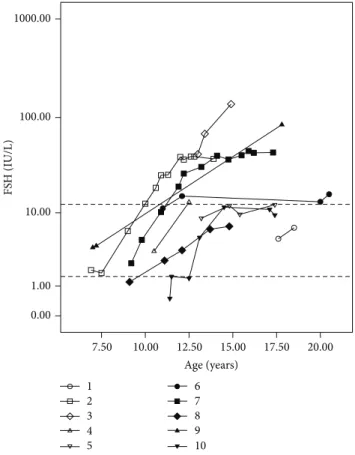
Figure 1: FSH levels measured by electrochemiluminescence at diferent ages in patients with partial gonadal dysgenesis. FSH values are presented on the�-axis on a logarithmic scale. Dotted lines on the�-axis represent the upper and lower normal limits for FSH levels in pubertal boys (1.5–12.4 IU/L).
height was calculated as (maternal height + paternal height + 12.5 cm)/2±6,5 cm [20].
he Institutional Review Board approved this study (776/2007).
3. Results
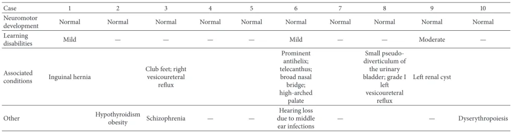
All patients had normal motor development but three pre-sented learning disabilities of unknown etiology (which were mild in two cases and moderate in one). Five had various associated conditions, including vesicoureteral relux in two cases and facial dysmorphisms in one patient. Primary hypothyroidism with negative antithyroid antibodies was diagnosed in one of the patients with a NR5A1 mutation when he was 12 years old (case 2); hearing loss due to middle ear infections, psychiatric problems, and obesity were also observed (one case each) (Table 2). here was no case of testicular neoplasia.
Results of measurements of gonadotropins (FSH and LH) and testosterone during follow-up were also analyzed. Data
Age (years)
20.00 17.50 15.00 12.50 10.00 7.50
LH (IU/L)
10.00
0.00 1.00
10 9 8 7 6
5 4 3 2 1
Figure 2: LH levels measured by electrochemiluminescence at diferent ages in patients with partial gonadal dysgenesis. FSH values are presented on the�-axis on a logarithmic scale. Dotted lines on the�-axis represent the upper and lower normal limits for LH levels in pubertal boys (1.7–8.6 IU/L).
in 3/6 patients (cases 2, 6, and 7), while LH levels were normal in all cases (Figures1and2).
At the last hormone evaluation (mean 16.5 years; range 12.6–20.5 years) FSH concentrations were high in 4/10 patients (cases 2, 3, 7, and 9), in the upper limit in four (4, 5, 6, and 10), and normal in two (1 and 8) (Figure 1). In turn, LH concentrations were high in 4/10 patients (cases 3, 6, 7, and 9), in the upper limit in one (case 1), and normal in ive (2, 4, 5, 8, and 10) (Figure 2).
Measures of testosterone could be obtained from 9/10 patients in the pubertal age range. he levels remained normal in seven cases, remained at the low limit of normality in one, and decreased in one patient (case 10), who received androgen replacement therapy (Figure 3). Nine patients had reached inal height, which ranged from−1.57 to 0.80 SDS, and in two of the eight cases for whom information on parental height was available it was lower than their genet-ically predicted range (target height ±6.5 cm). One patient (case 4) was still growing, with normal velocity and within the parental target range (Table 3).
te
rn
at
io
n
al
Jo
u
rn
al
o
f
E
n
d
o
cr
in
o
lo
g
y
5
Table 2: Neuromotor development and congenital and acquired diseases of ten patients with partial gonadal dysgenesis.
Case 1 2 3 4 5 6 7 8 9 10
Neuromotor
development Normal Normal Normal Normal Normal Normal Normal Normal Normal Normal Learning
disabilities Mild — — — — Mild — — Moderate —
Associated
conditions Inguinal hernia
Club feet; right vesicoureteral
relux
Prominent antihelix; telecanthus; broad nasal
bridge; high-arched
palate
Small pseudo-diverticulum of the urinary bladder; grade I
let vesicoureteral
relux
Let renal cyst
Other Hypothyroidism
obesity Schizophrenia — —
Hearing loss due to middle ear infections
In
te
rn
at
io
n
al
Jo
u
rn
al
o
f
E
n
d
Table 3: Data on growth, puberty, semen analysis, and surgical procedures undergone by ten patients with partial gonadal dysgenesis.
Patient 1 2 3 4 5 6 7 8 9 10
Target height
(cm) 168.3 171.5 165.5 170 182.2 169.7 — 170.5 182.5 176.7
Target height (�
score) −1.17 −0.73 −1.57 −0.94 +0.77 −0.99 — −0.87 +0.80 −0.0
Final height (cm) 170.2 175 165 ∗ 175 165.5 180 171 171 174
Final height (�
score) −0.89 −0.19 −1.51 — −0.11 −1.58 +0.57 −0.76 −0.76 +0.30
Age of pubarche 12.5 9.5 13 12.5 11.5 15 11.5 10.5 11 13.5
Tanner staging at
last visit (years) G4P5 (18.4) G5P5 (18.3) G4P4 (17.7) G4P4 (15.5) G5P5 (17.4) G5P5 (19.8) G4P4 (17.5) G4P4 (18.6) G5P5 (18.5)
G3P3#
(17.6)/G4P4§
(17.9) Testicular volume
(mL) at last visit (right/let)
NA 4/8 2/5 6/8 12/12 20/— 20/— 10/20 NA —/8
Sperm analysis (optical microscopy)
NP NP NP NP
Low viscosity, rare spz (some
motile)
High viscosity, rare spz (all
immotile)
Normal viscosity, rare spz (some
motile)
NP NP NP
Surgical procedures
Orthophalloplasty (11 mo); hypospadias correction (4 surgeries between
1 and 2 y)
Orchidopexy + hypospadias correction (2 y
8 mo)
Orchidopexy (3 y 3 mo); hypospadias correction (7 y); istula correction
(12, 13, 14, and 18 y)
Hypospadias correction (1 y
7 mo); istula correction (2 y
8 mo and 2 y 9 mo)
Hypospadias correction (1 y
4 mo); istula correction (4 y
and 5 y)
Hypospadias correction (8 y
8 mo); istula correction (9 and
12 y)
Orchidopexy (5 y)
Orchidopexy + orthophalloplasty (2 y 5 mo); istula correction (3, 4,
and 14 y)
Orchidopexy + hypospadias correction (2 y
2 m); istula correction (5 and
9 y)
Inguinal hernia correction + orchidopexy
(3 m); hypospadias correction (4 y)
Age (years)
22.00 20.00 18.00 16.00 14.00 12.00 10.00
T
est
ost
er
o
ne (n
g/mL)
10.00
8.00
6.00
4.00
2.00
0.00
10 9 8 7 6
5 4 3 2 1
Figure 3: Testosterone levels measured by electrochemilumines-cence at diferent ages in patients with partial gonadal dysgenesis. FSH values are presented on the�-axis on a linear scale. Dotted lines on the�-axis represent the upper and lower normal limits for testosterone levels in pubertal boys (2.86–8.10 ng/mL).
he seven patients who were regularly followed up in our pediatric endocrinology service had normal progression of puberty; most had low testicular volume. hree patients had a sperm count; all had severe oligozoospermia and low motility, and two had also abnormal semen viscosity.
4. Discussion
In DSD, sex assignment should be based on a precise diagnosis of the condition’s underlying etiology. Together with genital appearance and surgical options, this will allow the establishment of a prognosis on the need for lifelong replacement therapy, potential fertility, and malignancy risk [1] and also possible associated conditions. In the case of PGD, however, prognosis is not yet clearly established.
Our results showed that all patients had normal neu-romotor development, that most had normal growth, and that there was no consistent pattern of associated conditions. However, though learning disabilities are usually not a feature of DSD, in this sample it was observed in a signiicant proportion of cases (almost one-third), including a patient with moderate diiculties. Although this association may be casual, one may also consider the possibility that both conditions, testicular dysgenesis and cognitive impairment, have the same origin.
Interesting indings regarding our two patients bearing a NR5A1 mutation are hypothyroidism in one of them and schizophrenia in the other. Acquired primary hypothy-roidism has not been described as a feature of patients with PGD, with or withoutNR5A1mutations. he low prevalence of these conditions in young people aged 11–18 years (0.113%) and the fact that it is even rarer in males, with a 1 : 2.8 male to female ratio [21], make this inding noteworthy, even though expression ofNR5A1mRNA in thyroid gland is very low [22]. On the other hand, there is a recent publication of two women with mutations in this gene who had psychiatric symptoms [23].
he absence of testicular tumors in this sample demon-strates that maintenance of testes in the labioscrotal folds of patients reared as males is a relatively safe procedure, at least until the end of puberty.
Relevant indings were obtained regarding pubertal development. hough positive results from hCG stimulation tests in infancy or childhood were obtained in 4/9 patients, spontaneous pubertal development occurred in all cases. Pubertal delay was not observed, and in 9/10 cases there was normal progression of puberty, which strongly indicates that there is a good prognosis regarding spontaneous puberty in PGD patients reared as males when at least one testis may be kept in the scrotum.
In fact, in 7/9 cases testosterone levels were in the normal range during adolescence, though a progressive rise of LH in half of the cases raises the possibility that Leydig cell dysfunction may become evident in adulthood. On the other hand, high levels of FSH in most patients, sometimes observed early in adolescence, indicated that reproductive function was impaired, which could be shown in those patients whose sperm count was obtained.
Some patients with NR5A1 mutations and 46,XY PGD have been shown to have normal testosterone production in adolescence inducing spontaneous virilization [24–26], though follow-up indicated a progressive gonadal failure with elevated FSH in such cases. A similar picture was observed in our two patients withNR5A1mutations (cases 2 and 3) and also in the other eight cases without mutations in this gene. However, to the best of our knowledge, no other studies on long-term follow-up of patients with PGD reared as males are available to allow comparison with our sample.
5. Conclusions
Patients with PGD raised as males who have at least one testis in the labioscrotal region have a good prognosis for growth and spontaneous pubertal development but not for spontaneous fertility. hough additional studies are still required, our results also indicated that management of individuals with this condition should include screening for learning disabilities.
Conflict of Interests
the University Hospital and to the Cytogenetics Laboratory of the Department of Medical Genetics of State Univer-sity of Campinas (UNICAMP). his work was supported by FAPESP (2008/54776-1 and 2011/02865-3) and CNPq (301980/2009-8).
References
[1] I. A. Hughes, C. Houk, S. F. Ahmed, and P. A. Lee, “Consensus statement on management of intersex disorders,”Archives of
Disease in Childhood, vol. 91, no. 7, pp. 554–563, 2006.
[2] M. R. Scolfaro, I. A. Cardinalli, E. G. Stuchi-Perez et al., “Morphometry and histology of gonads from 13 children with dysgenetic male pseudohermaphroditism,”Archives of
Pathol-ogy & Laboratory Medicine, vol. 125, no. 5, pp. 652–656, 2001.
[3] M. Rohatgi, D. K. Gupta, P. S. N. Menon, I. C. Verma, and M. Mathur, “Mixed gonadal dysgenesis and dysgenetic male pseudohermaphroditism—a critical analysis,”he Indian
Jour-nal of Pediatrics, vol. 59, no. 4, pp. 487–500, 1992.
[4] H. E. Chemes, P. M. Muzulin, M. C. Venara et al., “Early manifestations of testicular dysgenesis in children: pathological phenotypes, karyotype correlations and precursor stages of tumour development,”APMIS, vol. 111, no. 1, pp. 12–24, 2003. [5] M. R. Scolfaro, I. A. Cardinalli, and G. Guerra Jr., “Gonadal
dysgenesis and morphometric histologic analysis,” Arquivos
Brasileiros de Endocrinologia & Metabologia, vol. 47, no. 2, pp.
128–134, 2003.
[6] S. Uehara, M. Hashiyada, K. Sato, M. Nata, T. Funato, and K. Okamura, “Complete XY gonadal dysgenesis and aspects of the SRY genotype and gonadal tumor formation,”Journal of Human
Genetics, vol. 47, no. 6, pp. 279–284, 2002.
[7] V. B. C. Rocha, G. Guerra-J´unior, A. P. Marques-de-Faria, M. P. de Mello, and A. T. Maciel-Guerra, “Complete gonadal dysgenesis in clinical practice: the 46,XY karyotype accounts for more than one third of cases,”Fertility and Sterility, vol. 96, no. 6, pp. 1431–1434, 2011.
[8] J. R. Hawkins, A. Taylor, P. N. Goodfellow, C. J. Migeon, K. D. Smith, and G. D. Berkovitz, “Evidence for increased prevalence of SRY mutations in XY females with complete rather than partial gonadal dysgenesis,”he American Journal of Human
Genetics, vol. 51, no. 5, pp. 979–984, 1992.
[9] J. S. Fuqua, J. McLaughlin, E. J. Perlman, and G. D. Berkovitz, “Analysis of the SRY gene in gonadal tissue of subjects with 46,XY gonadal dysgenesis,”Journal of Clinical Endocrinology
and Metabolism, vol. 82, no. 2, pp. 701–702, 1997.
[10] E. B. Tagliarini, J. G. Assumpc¸˜ao, M. R. Scolfaro et al., “Muta-tions inSRYandWT1genes required for gonadal development are not responsible for XY partial gonadal dysgenesis,”Brazilian
Journal of Medical and Biological Research, vol. 38, no. 1, pp. 17–
25, 2005.
[11] L. Lin and J. C. Achermann, “Steroidogenic factor-1 (SF-1,
Ad4BP, NR5A1) and disorders of testis development,”Sexual
Development, vol. 2, no. 4-5, pp. 200–209, 2008.
[12] B. K¨ohler and J. C. Achermann, “Update—steroidogenic factor 1 (SF-1, NR5A1),”Minerva Endocrinologica, vol. 35, no. 2, pp. 73–
vol. 27, no. 5, pp. 468–484, 2006.
[15] J. Słowikowska-Hilczer, M. Szarras-Czapnik, and K. Kula, “Testicular pathology in 46,XY dysgenetic male pseudo-hermaphroditism: an approach to pathogenesis of testis cancer,”
Journal of Andrology, vol. 22, no. 5, pp. 781–792, 2001.
[16] J. German, “Abnormalities of human sex chromosomes. V. A unifying concept in relation to the gonadal dysgeneses,”Clinical
Genetics, vol. 1, no. 1, pp. 15–27, 1970.
[17] E. B. Hook, “Exclusion of chromosomal mosaicism: tables of 90%, 95%, and 99% conidence limits and comments on use,”
he American Journal of Human Genetics, vol. 29, no. 1, pp. 94–
97, 1977.
[18] J. G. R. de Andrade, G. Guerra-J´unior, and A. T. Maciel-Guerra, “46,XY and 45,X/46,XY testicular dysgenesis: similar gonadal and genital phenotype, diferent prognosis,”Arquivos Brasileiros
de Endocrinologia e Metabologia, vol. 54, no. 3, pp. 331–334, 2010.
[19] World Health Organization,WHO Laboratory Manual for the
Examination and Processing of Human Semen, World Health
Organization, Geneva, Switzerland, 5th edition, 2010.
[20] J. M. Tanner, H. Goldstein, and R. H. Whitehouse, “Standards for children’s height at ages 2–9 years allowing for heights of parents,”Archives of Disease in Childhood, vol. 45, no. 244, pp. 755–762, 1970.
[21] I. Hunter, S. A. Greene, T. M. MacDonald, and A. D. Morris, “Prevalence and aetiology of hypothyroidism in the young,”
Archives of Disease in Childhood, vol. 83, no. 3, pp. 207–210,
2000.
[22] M. Nishimura, S. Naito, and T. Yokoi, “Tissue-speciic mRNA expression proiles of human nuclear receptor subfamilies,”
Drug Metabolism and Pharmacokinetics, vol. 19, no. 2, pp. 135–
149, 2004.
[23] A. S. Suwanai, T. Ishii, H. Haruna et al., “A report of two novelNR5A1mutation families: possible clinical phenotype of psychiatric symptoms of anxiety and/or depression,”Clinical
Endocrinology, vol. 78, no. 6, pp. 957–965, 2013.
[24] A. Bashamboo, B. Ferraz-De-Souza, D. Loureno et al., “Human male infertility associated with mutations in NR5A1 encoding steroidogenic factor 1,”he American Journal of Human Genet-ics, vol. 87, no. 4, pp. 505–512, 2010.
[25] S. Tantawy, L. Lin, I. Akkurt et al., “Testosterone production during puberty in two 46,XY patients with disorders of sex development and novel NR5A1 (SF-1) mutations,” European
Journal of Endocrinology, vol. 167, no. 1, pp. 125–130, 2012.
Submit your manuscripts at
http://www.hindawi.com
Stem Cells
International
Hindawi Publishing Corporation
http://www.hindawi.com Volume 2014
Hindawi Publishing Corporation
http://www.hindawi.com Volume 2014 INFLAMMATION
Hindawi Publishing Corporation
http://www.hindawi.com Volume 2014
Behavioural
Neurology
Endocrinology
International Journal of Hindawi Publishing Corporationhttp://www.hindawi.com Volume 2014
Hindawi Publishing Corporation
http://www.hindawi.com Volume 2014
Disease Markers
Hindawi Publishing Corporation
http://www.hindawi.com Volume 2014
BioMed
Research International
Oncology
Journal ofHindawi Publishing Corporation
http://www.hindawi.com Volume 2014
Hindawi Publishing Corporation
http://www.hindawi.com Volume 2014 Oxidative Medicine and Cellular Longevity Hindawi Publishing Corporation
http://www.hindawi.com Volume 2014
PPAR Research
The Scientiic
World Journal
Hindawi Publishing Corporation
http://www.hindawi.com Volume 2014
Immunology Research
Hindawi Publishing Corporation
http://www.hindawi.com Volume 2014
Journal of
Obesity
Journal ofHindawi Publishing Corporation
http://www.hindawi.com Volume 2014
Hindawi Publishing Corporation
http://www.hindawi.com Volume 2014
Computational and Mathematical Methods in Medicine
Ophthalmology
Journal ofHindawi Publishing Corporation
http://www.hindawi.com Volume 2014
Diabetes Research
Journal ofHindawi Publishing Corporation
http://www.hindawi.com Volume 2014
Hindawi Publishing Corporation
http://www.hindawi.com Volume 2014
Research and Treatment
AIDS
Hindawi Publishing Corporation
http://www.hindawi.com Volume 2014
Gastroenterology Research and Practice
Hindawi Publishing Corporation
http://www.hindawi.com Volume 2014
Parkinson’s
Disease
Evidence-Based Complementary and Alternative Medicine
Volume 2014 Hindawi Publishing Corporation