Loss of Circulating CD4 T Cells with B Cell Helper
Function during Chronic HIV Infection
Kristin L. Boswell1, Robert Paris1,2, Eli Boritz3, David Ambrozak1, Takuya Yamamoto1, Sam Darko3, Kaska Wloka1, Adam Wheatley4, Sandeep Narpala4, Adrian McDermott4, Mario Roederer5,
Richard Haubrich6, Mark Connors7, Julie Ake2, Daniel C. Douek3, Jerome Kim2, Constantinos Petrovas1*,
Richard A. Koup1
1Immunology Laboratory, Vaccine Research Center, NIAID, NIH, Bethesda, Maryland, United States of America,2United States Military HIV Research Program, Rockville, Maryland, United States of America,3Human Immunology Section, Vaccine Research Center, NIAID, NIH, Bethesda, Maryland, United States of America,4Immunology Core Section, Vaccine Research Center, NIAID, NIH, Bethesda, Maryland, United States of America,5ImmunoTechnology Section, Vaccine Research Center, National Institutes of Health, Bethesda, Maryland, United States of America,6Division of Infectious Diseases, Antiviral Research Center, University of California San Diego, San Diego, California, United States of America,7HIV-Specific Immunity Section, Laboratory of Immunoregulation, NIAID, NIH, Bethesda, Maryland, United States of America
Abstract
The interaction between follicular T helper cells (TFH) and B cells in the lymph nodes and spleen has a major impact on the
development of antigen-specific B cell responses during infection or vaccination. Recent studies described a functional equivalent of these cells among circulating CD4 T cells, referred to as peripheral TFH cells. Here, we characterize the
phenotype and in vitro B cell helper activity of peripheral TFHpopulations, as well as the effect of HIV infection on these
populations. In co-culture experiments we confirmed CXCR5+cells from HIV-uninfected donors provide help to B cells and more specifically, we identified a CCR7highCXCR5highCCR6highPD-1high CD4 T cell population that secretes IL-21 and
enhances isotype-switched immunoglobulin production. This population is significantly decreased in treatment-naı¨ve, HIV-infected individuals and can be recovered after anti-retroviral therapy. We found impaired immunoglobulin production in co-cultures from HIV-infected individuals and found no correlation between the frequency of peripheral TFHcells and
memory B cells, or with neutralization activity in untreated HIV infection in our cohort. Furthermore, we found that within the peripheral TFHpopulation, the expression level of TFH-associated genes more closely resembles a memory, non-TFH
population, as opposed to a TFHpopulation. Overall, our data identify a heterogeneous population of circulating CD4 T cells
that providesin vitrohelp to B cells, and challenges the origin of these cells as memory TFHcells.
Citation:Boswell KL, Paris R, Boritz E, Ambrozak D, Yamamoto T, et al. (2014) Loss of Circulating CD4 T Cells with B Cell Helper Function during Chronic HIV Infection. PLoS Pathog 10(1): e1003853. doi:10.1371/journal.ppat.1003853
Editor:Guido Silvestri, Emory University, United States of America
ReceivedMay 24, 2013;AcceptedNovember 12, 2013;PublishedJanuary 30, 2014
This is an open-access article, free of all copyright, and may be freely reproduced, distributed, transmitted, modified, built upon, or otherwise used by anyone for any lawful purpose. The work is made available under the Creative Commons CC0 public domain dedication.
Funding:This research was supported by the Intramural Research Program of the Vaccine Research Center, NIAID, National Institutes of Health (AI064086; RH) and CAVD grant#OPP1032325 from the Bill and Melinda Gates Foundation (RAK), the University of California San Diego Center for AIDS Research (AI36214; RH) and the San Diego AIDS Clinical Trial Group (CTU AI69432; RH). The Children’s National Medical Center (CNMC) under the auspices of the Basic Science Core of the District of Columbia Developmental Center for AIDS Research was funded by a grant from NIAID, NIH (P30AI087714). The work was also supported in part by an Interagency Agreement Y1-AI-2642-12 between the U.S. Army Medical Research and Material Command (USAMRMC) and the National Institutes of Allergy and Infectious Diseases and by a cooperative agreement (W81XWH-07-2-0067) between the Henry M. Jackson Foundation for the Advancement of Military Medicine, Inc., and the U.S. Department of Defense (DOD). The funders had no role in study design, data collection and analysis, decision to publish, or preparation of the manuscript. The opinions herein are those of the authors and should not be construed as official or representing the views of the U.S. Department of Health and Human Services, National Institute for Allergy and Infectious Diseases, the Department of Defense, or the Department of the Army.
Competing Interests:The authors have declared that no competing interests exist.
* E-mail: [email protected]
Introduction
Follicular helper CD4 T cells (TFH) are crucial for the development of antigen-specific B cells within germinal centers (GC). TFH cells interact through co-stimulatory receptors and provide essential soluble factors (i.e. IL-4, IL-21) to promote the survival, isotype switching and selection of high affinity memory B cells [1]. Phenotypic and gene signature analysis has revealed a highly conserved molecular profile of TFHcells in humans, non-human primates (NHP) and mice, which is characterized by increased expression of Bcl-6, CXCR5, PD-1, ICOS and decreased expression of CCR7 [2–4]. Human TFHcells exhibit a polarized cytokine profile characterized by compromised production of TH1 cytokines and increased secretion of IL-4, IL-10 and IL-21 [5]. Although IL-21 is characterized as a
‘‘hallmark’’ cytokine of TFHcells, other THelper subsets produce this cytokine [6].
The origin and differentiation of TFH is unclear, as previous studies found TFH cells can derive from TH1 or TH2 cells, or independently of other CD4 lineages [7–9]. However, it is well established that the transcription factor Bcl-6 regulates several molecules involved in TFH development (i.e. PD-1, IL-21R, CXCR5) [10,11]. Similarly, the fate of TFH, particularly those in the germinal center (GC-TFH), following the effector phase of the immune response is unclear. We have recently shown that NHP GC-TFHdisplay compromisedin vivocell cycling and are prone to in vitro cell death [4]. Other studies have shown that TFH can
humans, a circulating CD4 T cell population characterized by high CXCR5 expression can provide in vitro help for B cell
isotype switching and shares functional characteristics with TFH cells [14]. It was proposed that these circulating cells, termed ‘‘peripheral TFH’’ (pTFH) could represent the memory counter-parts of TFHoutside the lymphoid organs. Further investigation is needed to establish a direct relationship between TFHcells and pTFHcells.
It is becoming increasingly important to understand the interplay between CD4 T cells and B cells during HIV infection, specifically with relation to the generation of broadly neutralizing antibodies. Chronic HIV/SIV infection results in profound changes in CD4 T cell dynamics in lymph nodes characterized by TFHaccumulation and increased ability of non-TFHto egress the lymph node [4,15]. How this impacts upon the dynamics of pTFHis unknown. Elucidating the biology and dynamics of pTFH, and their ability to provide B cell help may be important for our understanding of TFHmemory formation during chronic infection, as well as the establishment of immune correlates reflecting the interactions between CD4 T cells and B cells within secondary lymphoid organs. This is of particular interest for monitoring clinical studies where the B cell arm of the immune system is under investigation [16].
Here we define, detect, quantify and characterize peripheral CD4 T cell populations that support B cell differentiation. We show that particular circulating CD4 T cell populations with distinct cytokine profiles have the capacity to help B cellsin vitro.
We further show that the frequencies of pTFH populations are significantly compromised during chronic HIV infection but can recover with antiretroviral treatment (ART), although in vitro
immunoglobulin production from HIV-infected subjects both on and off ART is reduced compared to healthy subjects. Further-more, gene expression analysis of pTFHcells and CD4 T cells in tonsil tissue suggest pTFHcells are most closely related to a non-TFH memory population within secondary lymphoid organs. Overall, our data challenge the relationship between pTFHcells and TFHmemory cells.
Results
Characterization of peripheral T follicular helper (pTFH) cells
Previous studies defined a population of circulating CD4 T cells that express CXCR5, promote the differentiation of naı¨ve B cells and induce immunoglobulin secretionin vitro[14,17]. We further
defined CXCR5high CD4 T cells from blood, analyzed their cytokine production and determined their ability to promote B cell differentiation in vitro. CXCR5high CD4 T cells were found
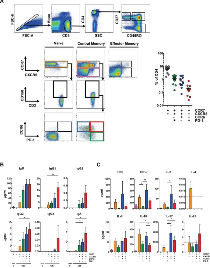
predominantly within the CD27highCD45ROhigh CD4 T cell population (hereafter referred to as central memory (CM)), in agreement with previous studies [17]. The majority of the CXCR5high CD4 T cell population also expressed CCR7 and we found the CCR7highCXCR5high population represented 6.5+/22.8% (mean+/2S.D.) of total CD4 T cells in healthy subjects (Figure 1A). The majority of CXCR5highcells expressed CD150. We further analyzed these cells based on expression of CCR6, which was previously used in combination with CXCR3 to define a pTFHsubset that promotes IgG and IgA production [14], and PD-1. CCR7highCXCR5highCCR6high cells represented 1.2+/20.9% of total CD4 T cells and a minority of these cells were PD-1high.
To analyze the ability of these populations to promote B cell differentiation, naı¨ve and CM CD4 T cells from HIV-uninfected individuals were sorted based on expression of CCR7, CXCR5, CD150, CCR6 and PD-1 (Figure 1A), and cultured with autologous naı¨ve B cells (CD19+CD272IgD+) as previously described [14,18] in the presence of staphylococcal enterotoxin B (SEB). Notably, our sorted naı¨ve B cell population did not express isotype-switched immunoglobulin (Figure S1A) and culture conditions that lacked SEB did not induce immunoglobulin production (data not shown). Naı¨ve and CM CCR7low CD4 T cells failed to promote B cell differentiation and immunoglobulin production whereas CM CCR7highCXCR5low cells induced limited production of IgM, IgG1 and IgG3 compared to the CCR7highCXCR5high populations (Figure 1B). The CCR7highCXCR5highCCR6highPD-1high population induced the greatest production of IgG1, IgG3 and IgA compared to the CXCR5lowpopulation. Prior studies defined pTFHcells based on surface expression of CXCR5, CCR6 and the lack of CXCR3 expression [14]. We found that the greatest help for immuno-globulin production was from CXCR5highCCR6high cell popula-tions and, within those, from the PD-1high cells. We did not eliminate a small population of CXCR3+cells in order to avoid removing a larger population of CXCR5highCCR6high cells that induce B cell differentiation (Figure S1B).
The cytokine profile of pTFHpopulations shared characteristics with other Thelper subsets, including TH1, TH17 and Treg cells. Supernatant from the CXCR5highCCR6highPD-1low coculture contained the greatest quantities of TNF-a, IL-2, and IL-17 compared to the CXCR5highCCR6lowPD-1high coculture (Figure 1C). Notably, the CXCR5highCCR6highPD-1high popula-tion, which promoted the greatest production of IgG1, IgG3 and IgA, showed the greatest IL-21 production, although at low levels. Overall, CXCR5high CD4 T cell populations induced B cell immunoglobulin production, although the CXCR5high CCR6highPD-1high population did so most efficiently. However, this population is not characteristic of a TFHpopulation found in secondary lymphoid organs, as coculture supernatants included a broad array of cytokines characteristic of TFHcells and multiple other Thelpersubsets.
Author Summary
Follicular T helper cells (TFH) interact with B cells within
germinal centers of lymphoid tissue to promote the survival, isotype switching and generation of high affinity memory B cells and plasma cells. Recently, a population of circulating CD4 T cells that shares phenotypic and functional characteristics with TFHcells, named peripheral
TFH cells, has been identified. The relationship between
peripheral TFHcells in the blood and TFHcells within the
lymphoid tissue remains unclear, and whether or not peripheral TFHcells can provide insight into T cell and B
cell dynamics within lymphoid tissue during infection or vaccination is not understood. Here we characterize peripheral TFH cells and show that unlike TFH cells,
peripheral TFH cells secrete a diverse array of cytokines
and decrease, rather than increase, during chronic HIV infection. Furthermore, we did not observe a relationship between peripheral TFHcells and memory B cells, or with
the production of neutralizing antibodies to HIV. Overall, our data indicate that while peripheral TFHcells share some
characteristics with TFH cells, they may not represent a
Progressive loss of pTFHcells in HIV infection
To determine the impact of HIV on pTFH populations, we compared pTFH cells from HIV-uninfected subjects and treat-ment-naı¨ve HIV-infected subjects (Table S1) as a frequency of total CD4 cells. Irrespective of how pTFHcells were defined, there was a significant decrease in the pTFH population from HIV-infected subjects compared to HIV-unHIV-infected subjects (Figure 2A). Subjects with CD4 counts greater than 200 had significantly lower pTFHpopulations, while subjects with CD4 counts less than 200 had the lowest frequency of all phenotypically defined pTFH populations. However, when we defined the CCR6highPD-1high population as a subset of the CXCR5high population, the frequency of the CCR6highPD-1high population increased in subjects with CD4 counts less than 200 (Figure S2A). The increase in PD-1high cells was likely due to immune activation in HIV infection, as we observed increases in the frequency of both PD-1high and ICOShigh cells within the CXCR5high population, with the greatest increases seen in samples with CD4 counts less than 200 (Figure S2A). We also observed a positive trend between CXCR5highPD-1high cells and serum concentrations of soluble CD14. (Figure S2A). For 10 HIV-infected individuals on whom we had longitudinal samples, we observed a loss of pTFHpopulations as a frequency of total CD4 T cells over 36 to 48 months (Figure 2B). However, the frequency of PD-1high, ICOShighand CCR6highPD-1high cells within the CXCR5high population re-mained stable (Figure S2B).
Next, we investigated the impact of ART on the frequency of pTFHwithin total CD4 T cells. Longitudinal analysis on samples from before and after 24 and 48 weeks of ART revealed a recovery of pTFHpopulations (Figure 2C). However, the frequency of PD-1high, ICOShigh and CCR6highPD-1high cells remained stable within the CXCR5high population (Figure S2C). Overall, HIV infection causes a loss of pTFH cells and ART promotes the recovery of these populations.
Impaired B cell help by pTFHcells in HIV infection
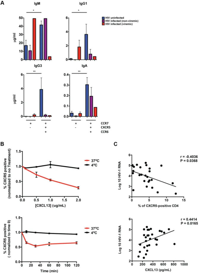
To investigate the impact of HIV on the ability of pTFHcells to support B cell differentiation, we performed co-culture experi-ments with pTFHcells from HIV-infected subjects. We focused on the CXCR5highCCR6highpopulation that included both PD-1high and PD-1low cells due to limited cell numbers in HIV-infected subjects. Similar to previous results, the CXCR5highCCR6high population from HIV-uninfected subjects supported significantly more immunoglobulin production compared to the CXCR5low population. (Figure 3A). However, for HIV-infected subjects we observed less overall immunoglobulin production when CXCR5highCCR6highCD4 T cells were co-cultivated with naı¨ve B cells. Furthermore, in viremic subjects, we observed increased IgM and IgG1 production in co-culture supernatants from the CXCR5low population, compared to HIV-uninfected subjects. Similar to HIV-uninfected subjects, we found that pTFHcells from HIV-infected subjects produced a broad spectrum of cytokines (Figure S3A).
Our data raise the possibility that some pTFHcells exhibit a CXCR5lowphenotype in HIV infection. This phenotype could be due the down regulation of CXCR5 on pTFHcells, or indicate the existence of a unique CXCR5lowpTFHpopulation in chronic HIV
infection. In order to distinguish these two possibilities, we investigated whether CXCL-13 impacts CXCR5 expression on CD4 T cells. We found that incubation of HIV-uninfected PBMC with CXCL-13 led to a decrease in frequency of CXCR5-positive CD4 T cells, presumably due to the internalization of CXCR5 (Figure 3B). Furthermore, in HIV infection we found that viral load positively correlated with CXCL-13 levels and negatively correlated with the frequency of CXCR5-positive CD4 T cells (Figure 3C). However, we did not observe a direct correlation between CXCL13 levels and the frequency of CXCR5-positive CD4 T cells. Importantly, we also found thatin vitroinfection of
CXCR5-expressing CD4 T cells did not impact CXCR5 surface expression (Figure S3B). Therefore, our data support the possibility that in untreated HIV infected individuals, increased levels of CXCL-13 could effect CXCR5 surface expression on pTFHcells.
Defective cytokine production of pTFHin HIV infection
TFH-dependent B cell differentiation requires IL-21. To characterize directly cytokine production from pTFHcells from HIV-uninfected and HIV-infected subjects, we performed intra-cellular cytokine staining afterex vivoSEB stimulation. In addition
to surface markers used to define pTFHcells, we detected CD154, IFN-c, IL-2, IL-17 and IL-21 (Figure 4A). In HIV-uninfected individuals, a minority of CD154-positive, cytokine-positive cells express a CCR7highphenotype (10.1% of IFN-cpositive cells; 28% of IL-2-positive cells; 19.4% of IL-17-positive cells and 17.9% of IL-21-positive cells), while a gradual reduction of cytokine production was found in further differentiated cells based on CXCR5 and CCR6 expression (Figure 4B). However, for all of the cytokines detected, we observed a population of cells that were CCR7highCXCR5highCCR6high, including IL-21-producing cells. Overall, we determined that a mean of 4.5% of CD154-positive IL-21-positive cells were CCR7highCXCR5highCCR6high (Figure 4B). However, this pTFHpopulation also produced IFN-c, IL-2 and IL-17 (0.8% of IFN-c positive cells; 9.0% of IL-2-positive cells and 7.1% of IL-17-IL-2-positive cells).
Next, we analyzed cytokine production from HIV-infected subjects off-treatment. Overall, we observed a loss of cytokine-producing cells from the CCR7highpopulation and a general shift towards the CXCR5lowCCR6lowpopulation (Figure 4A). Thus, we observed a loss of CCR7highCXCR5highCCR6highpTFHcells that produce IL-2, IL-17 and IL-21 (Figure 3B; IL-2: 9.0% for HIV-negative vs 2.0% for HIV-positive; IL-17: 7.1% for HIV-HIV-negative vs 2.2% for HIV-positive; IL-21: 4.5% for HIV-negative vs 1.1% for HIV-positive).
To analyze HIV-specific cells, PBMC were stimulated with Gag peptide pools and analyzed for cytokine expression. Very few IL-2-positive and IL-17-IL-2-positive cells were detected within the CM compartment (data not shown). Gag-specific IFN-c and IL-21-producing cells were detected, however, compared to SEB-stimulation fewer HIV-specific cells expressed CCR7 (4.4% vs 10.7% of IFN-c positive cells; 3.5% vs 11.9% of IL-21-positive cells for Gag and SEB stimulation, respectively). A majority of HIV-specific cells were not CCR7highCXCR5highCCR6high (Figure 4C; 0.4% of IFN-c positive cells and 0.9% of IL-21-positive cells were CCR7highCXCR5highCCR6high).
population in HIV-uninfected subjects (n= 13). Cells were not gated on CD150 for phenotypic analysis. (B) Indicated CD4 T cell populations were
cultured with autologous naı¨ve B cells (CD19highCD27lowIgD2) in the presence of SEB for 12 days and Ig concentrations were measured from
supernatants (n = 6). (C) Indicated CD4 T cell populations were cultured with autologous naı¨ve B cells in the presence of SEB for 2 days and cytokine concentrations were measured from supernatants (n = 6). Horizontal lines indicate limit of detection. Significant differences were determined using the Friedman test with Dunn’s multiple comparison post-test. *p,0.05; **p,0.01.
Overall, we observed IL-21 production from the CCR7highCXCR5highCCR6high pTFH population, although we detected the most IL-21 in non-pTFHcells, which were CCR7low and CXCR5low. In addition to IL-21, the CCR7high CXCR5highCCR6high pTFH population produced 2 and IL-17, cytokines characteristic of TH1 and TH17 cells, respectively. However, from HIV-infected individuals we observed a loss of CCR7highCXCR5highCCR6high cells making IL-2, IL-17 and IL-21.
No relationship between pTFHcells and neutralization activity
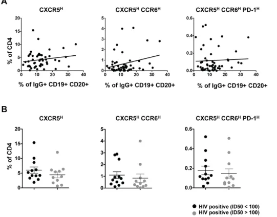
Previous studies have described a relationship between the frequency of peripheral CXCR5highcells and memory B cells and antibody titers with vaccination [16]. Therefore, we analyzed the relationship between the frequency of pTFHcells and IgG-positive memory B cells in PBMC from HIV-infected individuals. We found no significant correlation between the frequency of pTFHcells and IgG-positive B cells (Figure 5A). Similarly, we failed to detect a relationship between the frequency of pTFHand HIV-1 Env-specific antibody titers or total plasma IgG levels (data not shown).
It has also been reported that PD-1highCD4 T cells in blood are associated with cross-clade neutralizing antibody responses during HIV infection [19] and these PD-1highCD4 T cells may represent a population of pTFHcells. Thus, the relationship between pTFH cells and neutralization activity was analyzed using HIV-infected samples classified as good neutralizers (median ID50.100) or poor neutralizers (median ID50,100) [20]. Irrespective of how pTFHcells were defined, we failed to find any relationship between neutralization activity and pTFHcells (Figure 5B).
Relationship between pTFHcells and TFHcells in human tonsil
While pTFH cells induce B cell differentiation and immuno-globulin secretionin vitro, the relationship between pTFHand TFH
cells in secondary lymphoid organs remains unclear. Ourin vitro
coculture studies indicated the greatest isotype-switched immuno-globulin production was elicited from B cells cocultivated with CXCR5highCCR6high pTFH cells (Figure 1B). Therefore, we investigated the expression of CCR6 on TFH (CXCR5high PD-1high) and non-TFH(CXCR5lowPD-1low) tonsil cells to determine if the CXCR5highCCR6highpTFHpopulation is related to TFHcells within secondary lymphoid organs (Figure 6A). The lowest frequency of CCR6high cells was found within the CXCR5highPD-1highcompartment (1.5% of CXCR5highPD-1high cells) and the greatest frequency of CCR6highcells within the non-TFHcompartment (9% of CXCR5
low
PD-1lowcells; Figure 6B). Similarly, RNA sequence data from the CXCR5high CCR6highPD-1high pTFH population more closely resembles a memory, non-TFH CD4 T cell population from the tonsil (CM CD57lowPD-1dimCCR7highCCR5lowCXCR4low) as compared to the non-germinal center TFH population (CM CD57
low PD-1highCCR7lowCXCR5high) or the GC-TFHpopulation (CM PD-1highCD57high; Figure 6C). In agreement with previous reports [5,17], tonsil TFHpopulations expressed higher levels of BCL6, IL-21, and CXCL13, and lower levels of PRDM1 and S1PR1 compared to the non-TFH memory population. The pTFH population from HIV-uninfected individuals expressed
compara-ble levels of S1PR1 and PRDM1 to the non-TFH memory population in the tonsil (Figure 6). We also observed lower transcript levels of MAF, BCL6, IL-21, and CXCL-13 in the pTFH population compared to tonsillar TFHpopulations. Importantly, MAF protein expression was highest in the CCR6highPD-1high pTFH population compared to other peripheral populations, although still lower than tonsillar TFH cells. (Figure 6D). For many of the selected genes, pTFHcells from HIV-infected subjects were comparable to pTFH from HIV-uninfected individuals, however, we observed greater transcript levels of activation molecules such as ICOS and CD69. Additionally, the levels of IL-21 were decreased in pTFHcells from HIV-infected individuals, supporting earlier results (Figure 4B). Collectively, our data suggest the pTFHpopulation characterized as CXCR5highCCR6highmost closely resembles a non-TFHmemory population in the tonsil.
Discussion
The development and nature of human TFH memory cells following an effector immune response are not known. The ability to define a population of memory TFH cells in PBMC (pTFH) would help inform our understanding of CD4 T cell dynamics within lymphoid tissue during vaccination or infection. Studies of chronic infection may be helpful in this regard [21]. Whether the accumulation of TFHcells during chronic infection [4,15] impacts the TFHmemory population is of particular interest, especially if memory TFH cells migrate between lymphoid organs and peripheral tissues. Recent studies [14,16] have suggested that circulating CXCR5highCD4 T cells may represent the peripheral counterparts of TFH cells. However, the relationship between pTFHand TFHcells within secondary lymphoid organs remains unclear. Therefore, it is of great relevance to determine if pTFH cells originate from GC-TFHcells and represent a memory TFH population, reflect a precursor population that differentiates into GC-TFHupon re-exposure to antigen, or both. Our studies begin to address these issues by further defining pTFHcells, comparing pTFHcells to tonsillar TFHcells, and analyzing the effect of HIV on these cells.
In concordance with previous studies, we showed that circulating CXCR5highCD4 T cells support B cell differentiation
in vitro [14,17]. A majority of the CXCR5high cells expressed
CD150, and while CD150 was used for gating in the co-culture assays, we found it did not impact the loss of pTFHcells or effect our results with respect to loss of pTFHcells, recovery with ART or lack of association with B cell or antibody responses (data not shown). However, within the CXCR5highpopulation the expres-sion of CCR6 and PD-1 did further define pTFHpopulations with differential abilities for naı¨ve B cell help and isotype switching. Thus, pTFH cell populations support both the activation and maturation of naı¨ve B cells, and immunoglobulin isotype switching. Correspondingly, the individual pTFH populations produced cytokines associated with B cell maturation and survival, such as IL-21 [22], IL-2 [23] and IL-17 [24], in contrast to TFH cells within secondary lymphoid tissue, which display a limited cytokine profile that includes IL-4, IL-10 and IL-21, but compromised production of IL-2 and IL-17 [4]. Whether these pTFH populations represent different stages of TFH memory development or originate from separate CD4 T cell populations within lymphoid tissue [25] is still unclear.
gray circles; week 48, light gray circles). Significant differences between HIV-uninfected and HIV-infected subjects were determined using the Mann-Whitney U test. Significant differences between subjects before and after ART were determined using the Wilcoxon matched-pairs signed rank test. ***p,0.001; **p,0.01; *p,0.05.
Figure 3. Impaired B cell help by pTFHcells in HIV infection.(A) CCR7highCXCR5lowand CCR7highCXCR5highCCR6highCM CD4 T cells isolated from PBMCs were cultured with autologous naı¨ve B cells (CD19highCD27lowIgD2) in the presence of SEB for 12 days and Ig concentrations were
In order to better understand the relationship between TFHand pTFH cells, we compared gene expression levels between pTFH and tonsillar CD4 T cell populations and focused on genes important for TFH differentiation, migration, and function. We found that the pTFHpopulation with the greatest B cell helper function most closely resembled a CM, non-TFH CD4 T cell subset within the tonsil. While our studies do not directly address the relationship between GC-TFHin lymph nodes and circulating CD4 T cells from the same patients, our data challenge whether pTFH are memory TFH cells. A recent study reported that germinal center TFHcells in mice migrate throughout the follicle, but generally do not leave the follicle to enter the blood [26]. While it is conceivable that pTFH cells represent a very minor population of TFHcells that exit the follicle, it is also possible that pTFHcells are reflective of a precursor TFHpopulation that exits the lymphoid organ and enters the circulation before entering the follicle. However, while we find the CXCR5highCCR6highPD-1high pTFH population does not resemble a memory TFHpopulation, Locci and colleagues found a CXCR5+CXCR3-PD-1+ pTFH subset that functionally and transcriptionally resembles a memory TFH population [27]. A recent study in mice reported that memory TFH cells have reduced mRNA expression of TFH markers such as Bcl6, IL-21, ICOS and PD-1 compared to the effector TFHpopulation [28], indicating the expression of these molecules may change depending on the phase of infection. Therefore, further investigation of pTFH subsets and their relationship to memory and effector populations at multiple stages of infection is needed.
pTFHand naı¨ve B cell co-cultures from HIV-infected subjects produced fewer immunoglobulins compared to co-cultures from HIV-uninfected subjects. The observed defect in immunoglobulin production is likely due to impaired pTFHhelp to B cells instead of B cell dysfunction, as co-cultures included naı¨ve B cells rather than memory B cells that exhibit abnormalities in HIV infection [29]. Furthermore, while co-culture supernatants from HIV-infected subjects demonstrated a heterogeneous cytokine profile, similar to HIV-uninfected subjects, intracellular cytokine staining showed that fewer CCR7highCXCR5highCCR6high pTFH cells produced IL-2, IL-17 and IL-21 in chronic HIV infection compared to HIV-uninfected individuals. Furthermore, gene expression analysis of HIV-infected pTFH revealed fewer IL-21 and IL-4 transcripts, although the overall levels of cytokine transcripts were low.
Recent studies have shown TFHcells within secondary lymphoid organs accumulate in some donors or animals during chronic HIV/SIV infection and that TFHaccumulation is associated with GC B cell expansion and increased serum immunoglobulin concentrations [4,22,30]. In contrast to TFH cells, our studies revealed pTFHcells consistently decrease in chronic HIV infection, with disease progression resulting in a greater reduction of these compartments within the total CD4 T cell population. However, it should be noted that we were unable to analyze TFHcells within secondary lymphoid organs from these subjects and therefore we are unable to directly compare the frequency of pTFHcells and TFHcells from the same individual. The differences between the increase in TFHcells and decrease in pTFHcells may be due to differences in disease state (i.e. early vs late infection) or represent a steady state of TFH cells trafficking between the lymphoid tissue and the blood. The decreased frequency of
pTFHin the blood may indicate impaired ability of TFHto exit the lymph node in chronic HIV infection where the tissue architecture is not intact. Alternatively, the decreased frequency of pTFH in the blood may be a result of pTFH trafficking to secondary lymphoid organs. In agreement with previous studies [14,17], we found a majority of CXCR5highcells express CCR7, and it has previously been suggested that pTFHcells migrate to secondary lymphoid organs upon infection due their expression of CCR7 and CD62L [14].
A confounding factor with regard to how we interpret the decrease in pTFHcells is that we also found a reduction in the surface expression of CXCR5 on CD4 T cells in chronic HIV infection, which may result from increased sera levels of CXCL-13 [31,32]. Furthermore, our co-culture data indicate that CXCR5low CD4 T cells from viremic subjects can induce some B cell differentiation. These data support the possibility that in chronic HIV infection, a subset of functional pTFHcells may be phenotypically defined as CXCR5low. Additionally, it should be noted that analysis of cellular subsets within the CXCR5high population in chronic HIV infection revealed the frequency of CCR6highPD-1high cells increased. These results are consistent with a state of generalized immune activation, as we also observed increased surface expression of ICOS on CXCR5high and CXCR5highPD-1highcells, and a positive association between the frequency of PD-1highcells within the CXCR5highpopulation and serum concentrations of soluble CD14 [33]. Similarly, gene expression analysis indicated increased transcript levels of activa-tion markers, such as ICOS and CD69 within the pTFH population during HIV infection. Overall, these data emphasize the difficulty in defining pTFHcells in chronic HIV infection and understanding the relationship between pTFHcells and TFHcells. The uncertain definition of pTFHcells in HIV infection may provide an explanation as to why we were unable to identify correlations between pTFH populations and circulating IgG-positive memory B cells, or between pTFHcells and HIV-specific IgG (data not shown). Furthermore, we found no correlation between the frequency of pTFHand the neutralization activity of a well-characterized cohort of HIV-infected donors [20]. However, the absence of a correlation between pTFHcells and circulating HIV Env-specific IgG may also be explained by the lack of a time-dependent association (early vs. late infection) between TFHand pTFH cells, or indicate that the generation of IgG and broadly neutralizing antibodies is regulated by parameters other than pTFH, confounded by T-cell independent antibody production commonly observed in HIV infection [34] or generalized immune activation. Thus, our data challenge the application of the pTFH population as a surrogate of GC TFH-B cell interactions in chronic HIV infection. While our studies did not find a correlation between pTFH cells and neutralizing antibodies, several recent studies, each with a different definition of pTFH cells, have reported an association with antibody responses during vaccina-tion, infection or autoimmune disease [27,35–37]. Therefore, further studies are needed to establish the association between pTFH subsets and the generation of neutralizing antibodies, especially in HIV infection.
Overall, our data indicate that a range of circulating CD4 T cell populations can provide B cell help, possibly through differential secretion of soluble factors and/or cell-cell contact interactions
60 or 120 minutes at 37uC (red) or 4uC (black). The frequency of CXCR5-positve CD4 T cells was calculated and normalized to time 0. (n = 3). (C) Top: Correlative analysis showing the frequency of CM CXCR5-positive CD4 T cells versus viral load (n = 27; r =20.4036, P = 0.0368). Bottom: Correlative analysis showing the concentration of CXCL-13 in plasma or sera versus viral load (n = 27; r = 0.4414, P = 0.0165). Correlations were analyzed using the nonparametric Spearman test.
[17,35] and that HIV infection results in loss of these cells over time, but with relative increases within the CXCR5high compart-ment which may be explained by immune activation. Further-more, we did not find any association between pTFHand measures of B cell function such as HIV neutralization breadth/potency, HIV-specific IgG, or total IgG, suggesting application of this population as a surrogate of GC TFH-B cell interactions during HIV infection may be limited. A better understanding of the differentiation process and the developmental relationship be-tween pTFHsubsets and lymph node TFHcells is critical for the establishment of reliable peripheral blood CD4 T cell correlates for monitoring infection- or vaccine-associated B cell responses.
Materials and Methods
Ethics statement
Signed informed consent was obtained in accordance with the Declaration of Helsinki and approved by the appropriate Institutional Review Board. Tonsil cells were acquired from anonymized discarded pathologic specimens from Children’s National Medical Center (CNMC) under the auspices of the
Basic Science Core of the District of Columbia Developmental Center for AIDS Research. The CNMC Institutional Review Board determined that study of anonymized discarded tonsils did not constitute ‘human subjects research.’
Subjects
Fresh HIV-uninfected peripheral blood mononuclear cells (PBMC) were obtained from individuals participating in the NIH research apheresis program. Fresh HIV-infected blood was obtained from the Vaccine Research Center Clinic or Drexel University College of Medicine. Frozen HIV-infected PBMC were obtained from three study populations (Table S1). For untreated HIV infection, cells were obtained from volunteers who partici-pated in a therapeutic vaccination trial (no efficacy was observed) conducted in the 1990’s prior to the advent of combination antiretroviral therapy (cART) [38]. The second study population consisted of donors from a cohort used to identify individuals with HIV broadly neutralizing antibodies [20]. To study the effect of cART, we obtained PBMC from HIV-infected donors participat-ing in AIDS Clinical Trials Group study A5142 prior to initiation of cART and 24 and 48 weeks post-therapy [39,40]. PBMC and
Figure 4. Functional characteristics of pTFHcells and the impact of HIV.(A) Representative flow cytometry plots showing CM, CD154-positive, cytokine-positive cells after SEB stimulation. CD154-CD154-positive, cytokine-positive CD4 T cells, shown by contour plots (blue: HIV-uninfected; red: HIV-infected), are overlaid onto 2 dimensional density plots for CM CD4 T cells plotted against CCR7 and CD3, and CXCR5 and CCR6. (B) Bar graphs showing the frequency of SEB-stimulated CD154-positive, cytokine-positive cells that express CCR7, CXCR5 and CCR6 (Blue: uninfected; n = 5; Red: HIV-infected; n = 24). (C) Left: Gag-specific CD4+T cells (CD154-positive, cytokine-positive) shown as red contour plots are overlaid onto 2 dimensional density plots for CM cells CD4 T cells plotted against CCR7 and CD3, and CXCR5 and CCR6. Right: Bar graphs showing the frequency of Gag-specific CD154-positive, cytokine-positive cells that express CCR7, CXCR5 and CCR6 (n = 14). *p,0.05.
doi:10.1371/journal.ppat.1003853.g004
Figure 5. Relationship between pTFHcells and neutralization activity.(A) Correlative analysis showing the frequency (%) of CXCR5high, CXCR5highCCR6highand CXCR5highCCR6highPD-1highpopulations in total CD4 cells from HIV-infected (treatment naı¨ve) subjects (n = 50) versus the frequency of IgG+B cells in the total B population. Correlations were analyzed using the nonparametric Spearman test. (B) Pooled data showing the frequency (%) of CXCR5high, CXCR5highCCR6highand CXCR5highCCR6highPD-1highpopulations in total CD4 cells based on neutralization activity (median ID50.100 or,100). No significant differences were determined.
tonsil cells were cultured in RPMI 1640 supplemented with 10% fetal bovine serum, 2 mM L-glutamine, 100 U/mL penicillin and 100mg/mL streptomycin (Invitrogen).
Antibodies
Directly conjugated antibodies were acquired from the follow-ing: (1) BD Biosciences: CD3-H7APC, CXCR5-Alexa488 (RF8B2), CCR7-Alexa700, IgG-APC, IFN-c-Alexa700 and IL-21-Alexa647 (3A3-N2.1) (2) Beckman Coulter: CD45RO-ECD and CD27-PC5 (3) Biolegend: CCR7-BV421, CCR6-PE (TG7/ CCR6), CCR6-Alexa647 (TG7/CCR6), CD20-BV570, CD150-PE, IL-2-BV605, IL-17a-Cy5.5PerCP and CD154-Cy5PE (4) Invitrogen: CD4-Cy5.5PE, CD27-QD655, CD27-QD605 and CD19-PacBlue (5) Southern Biotech: IgD-FITC and IgD-PE (6) eBioscience: cmaf-eFluor660 (sym0F1), CXCR5-PerCP-efluor710 (MU5UBEE). Biotinylated anti-PD-1 was from R&D and streptavidin-Cy7PE (or QD655) was from Molecular Probes. The following antibodies were conjugated in our lab: CD19-QD705 and CD57-QD565. Quantum dots and Aqua amine viability dye were obtained from Invitrogen.
Polychromatic flow cytometry
Phenotypic analysis:1–26106 PBMC were incubated with Aqua-dye and surface stained with titrated amounts of anti-CD3, anti-CD4, anti-CD27, anti-CD45RO, anti-CCR7, anti-CXCR5, anti-CD150, anti-CCR6, anti-PD-1 and anti-CD19. Post-wash, cells were incubated with fluorescent-conjugated streptavidin, washed and fixed with 1% paraformaldehyde.
Intracellular cytokine staining:36106PBMC were incubat-ed in 1 mL of mincubat-edium containing brefeldin A (10 ug/mL) in the absence or presence of HIV-1 Gag-peptide pools (15mers overlapping by 11 residues; NIH AIDS Research and Reference Reagent Program) or 1 ug/mL SEB (Sigma) for 6 hours. Cells were surface stained, permeabilized (Cytofix/Cytoperm kit; BD Biosciences), and stained with anti-CD3, anti-IFN-c, anti-IL-2, anti-IL-17a, anti-IL-21 and anti-CD154. Events were collected on a modified LSRII flow cytometer (BD Immunocytometry Systems) and electronic compensation was performed with antibody capture beads (BD Biosciences). Data were analyzed using FlowJo Version 9.6 (TreeStar).
T and B cell culture
Co-culture experiments were performed with freshly isolated PBMC. 56104 CD4 T cell populations were sorted based on expression of CCR7, CXCR5, CD150, CCR6 and PD-1 and cultured with 56104 autologous naı¨ve B cells (1:1 ratio) in the presence of SEB (0.5mg/ml). Supernatants harvested on Day 2 were analyzed for cytokines using Luminex technology (Milliplex MAP Kit, HTH17MAG-14K, Millipore). The lower limit of detection (LOD) was set at the lowest concentration on the standard curve and values below the LOD were counted as zero. Supernatants collected on Day 12 were analyzed for immunoglob-ulins (Milliplex MAP Kit, HGAMMAG-301K). Some supernatants exceeded the saturation limit of the standard curves for IgM and
IgG3. These values were included in the analysis and quantified as being equivalent to the highest determined concentration.
ELISA
Soluble CD14 and CXCL-13 (R&D Systems) were measured in plasma or sera from HIV-infection patients according to the manufacturer’s instructions.
CXCL-13 treatment
Freshly isolated PBMCs were incubated with recombinant human CXCL-13 (R&D Systems) at 37uC or 4uC and analyzed for CXCR5 surface expression by FACS.
Illumina deep sequencing of messenger RNA
CD4 T cell populations were sorted from uninfected PBMC (n = 5), HIV-infected PBMC (n = 5) and uninfected human tonsils (n = 4) based on expression of CCR7, CXCR5, CD150, CCR6 and PD-1 for PBMC and CD57, PD-1, CCR7, CXCR5, CCR5 and CXCR4 for tonsils. Total RNA was purified from sorted cell populations and treated with DNAse I (Ambion) to minimize genomic DNA contamination. Polyadenylated RNA was isolated using Oligo-dT Dynabeads (Life Technologies), chemically fragmented, and used to construct barcoded Illumina Truseq libraries. Libraries were selected, quantified, pooled, size-selected and quantified again, and clustered on an Illumina Truseq Paired-End Flowcell v3. The flowcell was sequenced on an Illumina HiSeq 2000 in a 2675-base paired-end, indexed run. Adaptor sequence was trimmed from the raw sequencing reads using Trimmomatic. The trimmed sequencing reads were subsequently aligned to the human genome (hg19) using TopHat 2. Differential expression testing was done using Cufflinks 2 and visualization of differential expression was done using the R package cummerbund. Accession numbers of the selected genes are shown in Supporting Table S2.
Virus neutralization
Neutralization activity of patient sera was determined against 20 viral isolates using a TZM-bl neutralization assay as previously described [20].
In vitro infection
Freshly isolated PBMCs were stimulated with PHA (10mg/ml). After 12 hours stimulation, CXCR5highcells were sorted by FACS Aria based on surface molecule expression and infected by a multiplicity of infection (MOI) of 0.01 with either HIV NL-E or HIV NLAD8-E [41]. The infected cells were cultured in the presence of 50 U/ml recombinant human interleukin-2 (R&D) for 5 days and analyzed for CXCR5 expression by FACS.
Statistics
Experimental variables were analyzed using the nonparametric Mann-Whitney U test, the Wilcoxon matched-pairs signed rank test or the Friedman test with Dunn’s multiple comparison post-test. Correlation analysis was performed using the nonparametric
Figure 6. Relationship between pTFHcells and TFHcells in human tonsil.(A) Representative flow cytometry plots from HIV-uninfected, pediatric tonsils showing the gating scheme for determining the frequency of CCR6highcells in TFH(CXCR5highPD-1high) and non-TFHpopulations. (B) Bar graphs showing the frequency of CCR6highcells in TFHand non-TFHpopulations in human tonsils (n = 5). (C) Heatmap analysis of selected genes from RNA-seq data comparing pTFHcells (CXCR5highCCR6highPD-1high) from HIV-uninfected donors, pTFHcells from HIV-infected donors, non-TFHCD4 memory tonsil cells (CM CD57lowPD-1dimCCR7highCCR5lowCXCR4low), non-germinal center TFHtonsil cells (CM CD57lowPD-1highCCR7lowCXCR5high) and germinal center TFHtonsil cells (CM PD-1highCD57high) from HIV-uninfected donors. (D) Top: Comparison of MAF expression on CD4 T cells from blood or tonsil. Bottom: Geometric mean (MFI) of MAF expression in the indicated populations of central memory CD4 T cells normalized to MAF MFI in naı¨ve CD4 T cells.
Spearman test. Error bars depict mean+SEM in all bar graphs shown. The GraphPad Prism statistical analysis program (Graph-Pad Software, version 5.0) was used throughout.
Supporting Information
Figure S1 Characterization of peripheral TFHcells. (A)
Scatter plot depicting frequency of IgG+ and IgA+ B cells in total B (CD19+) or naı¨ve B cells (CD19+CD27low
IgDhigh) for HIV-uninfected (n = 5), HIV-infected (non-viremic; n = 7) and HIV-infected (viremic; n = 1) donors. Both surface and intracel-lular staining of IgA and IgG were used to determine frequency. (B) Left: Representative flow cytometry plots from HIV-uninfected PBMC showing CXCR3 and CCR6 expression within CXCR5high and CXCR5low CD4 T cell subsets. Right: Scatter plot comparing the frequency of CXCR3 and CCR6 subsets within the CXCR5high population from HIV-uninfected (open circles; n = 4) and HIV-infected individuals (closed circles; n = 8).
(EPS)
Figure S2 Decrease of pTFH cells in HIV infection. (A)
Pooled data showing the frequency (%) of indicated populations in CXCR5-expressing cells from PBMC from HIV-uninfected (open circles; n = 13), HIV-infected (treatment-naı¨ve), CD4 count.200 (light gray circles; n= 44), and HIV-infected (treatment-naı¨ve), CD4 count ,200 (black circles; n= 22). Significant differences
between uninfected and HIV-infected subjects were determined using the Mann-Whitney U test. ***p,0.001; **p,0.01; *p,0.05. Far Right: Correlative analysis between the frequency of CXCR5highPD-1highcells and the concentration of soluble CD14 in the sera or plasma. The trend did not reach statistical significance as determined by the Spearman test. (B) Top: Longitudinal analysis showing the frequency (%) of indicated populations in CXCR5-expressing cells from HIV-infected (treatment naı¨ve) subjects (n = 10) over 36–48 months. No significant correlations were found. Bottom: Longitudinal analysis showing CD4 counts and viral loads from HIV-infected (treatment naı¨ve) subjects (n = 10) over 36–48 months. (C) Pooled data
showing the frequency (%) of indicated populations in CXCR5-expressing cells from PBMC from HIV-uninfected subjects (open circles; n = 13) and HIV-infected subjects before (n = 14, week 0; black circles) and after ART (week 24, dark gray circles; week 48, light gray circles).
(EPS)
Figure S3 Characterization of pTFH cells in HIV
infec-tion.(A) CCR7highCXCR5lowand CCR7highCXCR5highCCR6high CD4 T cells isolated from PBMCs were cultured with autologous naı¨ve B cells (CD19highCD27lowIgD2) in the presence of SEB for 2
days and cytokine concentrations were measured from supernatants (uninfected, n = 5; infected (non-viremic), n = 4, HIV-infected (viremic), n = 0–1). Due to limited cell numbers we were unable to collect CCR7highCXCR5highCCR6highcells from viremic individuals. (B) Sorted CXCR5highcentral memory cells isolated from blood do not down-regulate surface expression of CXCR5 upon X4 or R5in vitroinfection.
(EPS)
Table S1 CD4 count, viral load and neutralization activity of subjects studied.
(EPS)
Table S2 Accession numbers of selected genes. (EPS)
Acknowledgments
We would like to thank the DC-CFAR Basic Science Core and Children’s National Medical Center (CNMC) for providing tonsils and Dr Katsikis at Drexel University School of Medicine for providing samples from HIV+ individuals.
Author Contributions
Conceived and designed the experiments: KLB RP TY CP RAK. Performed the experiments: KLB RP EB DA TY KW AW SN CP. Analyzed the data: KLB RP EB TY SD KW AW SN CP. Contributed reagents/materials/analysis tools: AM MR RH MC JA DCD JK. Wrote the paper: KLB RP TY CP RAK.
References
1. Crotty S (2011) Follicular helper CD4 T cells (TFH). Annu Rev Immunol 29: 621–663.
2. Haynes NM, Allen CD, Lesley R, Ansel KM, Killeen N, et al. (2007) Role of CXCR5 and CCR7 in follicular Th cell positioning and appearance of a programmed cell death gene-1high germinal center-associated subpopulation. J Immunol 179: 5099–5108.
3. Ma CS, Suryani S, Avery DT, Chan A, Nanan R, et al. (2009) Early commitment of naive human CD4(+) T cells to the T follicular helper (T(FH)) cell lineage is induced by IL-12. Immunol Cell Biol 87: 590–600.
4. Petrovas C, Yamamoto T, Gerner MY, Boswell KL, Wloka K, et al. (2012) CD4 T follicular helper cell dynamics during SIV infection. J Clin Invest 122: 3281– 3294.
5. Kim CH, Lim HW, Kim JR, Rott L, Hillsamer P, et al. (2004) Unique gene expression program of human germinal center T helper cells. Blood 104: 1952– 1960.
6. Yu D, Vinuesa CG (2010) The elusive identity of T follicular helper cells. Trends Immunol 31: 377–383.
7. Fahey LM, Wilson EB, Elsaesser H, Fistonich CD, McGavern DB, et al. (2011) Viral persistence redirects CD4 T cell differentiation toward T follicular helper cells. J Exp Med 208: 987–999.
8. Glatman Zaretsky A, Taylor JJ, King IL, Marshall FA, Mohrs M, et al. (2009) T follicular helper cells differentiate from Th2 cells in response to helminth antigens. J Exp Med 206: 991–999.
9. Nurieva RI, Chung Y, Hwang D, Yang XO, Kang HS, et al. (2008) Generation of T follicular helper cells is mediated by interleukin-21 but independent of T helper 1, 2, or 17 cell lineages. Immunity 29: 138–149.
10. Nurieva RI, Chung Y, Martinez GJ, Yang XO, Tanaka S, et al. (2009) Bcl6 mediates the development of T follicular helper cells. Science 325: 1001– 1005.
11. Yu D, Rao S, Tsai LM, Lee SK, He Y, et al. (2009) The transcriptional repressor Bcl-6 directs T follicular helper cell lineage commitment. Immunity 31: 457–468. 12. Luthje K, Kallies A, Shimohakamada Y, Belz GT, Light A, et al. (2012) The development and fate of follicular helper T cells defined by an IL-21 reporter mouse. Nat Immunol 13: 491–498.
13. Pepper M, Jenkins MK (2011) Origins of CD4(+) effector and central memory T cells. Nat Immunol 12: 467–471.
14. Morita R, Schmitt N, Bentebibel SE, Ranganathan R, Bourdery L, et al. (2011) Human blood CXCR5(+)CD4(+) T cells are counterparts of T follicular cells and contain specific subsets that differentially support antibody secretion. Immunity 34: 108–121.
15. Lindqvist M, van Lunzen J, Soghoian DZ, Kuhl BD, Ranasinghe S, et al. (2012) Expansion of HIV-specific T follicular helper cells in chronic HIV infection. J Clin Invest 122: 3271–3280.
16. Pallikkuth S, Parmigiani A, Silva SY, George VK, Fischl M, et al. (2012) Impaired peripheral blood T-follicular helper cell function in HIV-infected nonresponders to the 2009 H1N1/09 vaccine. Blood 120: 985–993. 17. Chevalier N, Jarrossay D, Ho E, Avery DT, Ma CS, et al. (2011) CXCR5
expressing human central memory CD4 T cells and their relevance for humoral immune responses. J Immunol 186: 5556–5568.
18. Breitfeld D, Ohl L, Kremmer E, Ellwart J, Sallusto F, et al. (2000) Follicular B helper T cells express CXC chemokine receptor 5, localize to B cell follicles, and support immunoglobulin production. J Exp Med 192: 1545–1552.
19. Mikell I, Sather DN, Kalams SA, Altfeld M, Alter G, et al. (2011) Characteristics of the earliest cross-neutralizing antibody response to HIV-1. PLoS Pathog 7: e1001251.
21. Hale JS, Youngblood B, Latner DR, Mohammed AU, Ye L, et al. (2013) Distinct Memory CD4(+) T Cells with Commitment to T Follicular Helper- and T Helper 1-Cell Lineages Are Generated after Acute Viral Infection. Immunity 38: 805–817.
22. Linterman MA, Beaton L, Yu D, Ramiscal RR, Srivastava M, et al. (2010) IL-21 acts directly on B cells to regulate Bcl-6 expression and germinal center responses. J Exp Med 207: 353–363.
23. Le Gallou S, Caron G, Delaloy C, Rossille D, Tarte K, et al. (2012) IL-2 requirement for human plasma cell generation: coupling differentiation and proliferation by enhancing MAPK-ERK signaling. J Immunol 189: 161–173. 24. Doreau A, Belot A, Bastid J, Riche B, Trescol-Biemont MC, et al. (2009)
Interleukin 17 acts in synergy with B cell-activating factor to influence B cell biology and the pathophysiology of systemic lupus erythematosus. Nat Immunol 10: 778–785.
25. Bentebibel SE, Schmitt N, Banchereau J, Ueno H (2011) Human tonsil B-cell lymphoma 6 (BCL6)-expressing CD4+T-cell subset specialized for B-cell help outside germinal centers. Proc Natl Acad Sci U S A 108: E488–497. 26. Shulman Z, Gitlin AD, Targ S, Jankovic M, Pasqual G, et al. (2013) T follicular
helper cell dynamics in germinal centers. Science 341: 673–677.
27. Locci M, Havenar-Daughton C, Landais E, Wu J, Kroenke MA, et al. (2013) Human Circulating PD-1CXCR3CXCR5 Memory Tfh Cells Are Highly Functional and Correlate with Broadly Neutralizing HIV Antibody Responses. Immunity 39: 758–769.
28. Hale JS, Youngblood B, Latner DR, Mohammed AU, Ye L, et al. (2013) Distinct memory CD4+T cells with commitment to T follicular helper- and T helper 1-cell lineages are generated after acute viral infection. Immunity 38: 805–817.
29. Malaspina A, Moir S, DiPoto AC, Ho J, Wang W, et al. (2008) CpG oligonucleotides enhance proliferative and effector responses of B Cells in HIV-infected individuals. J Immunol 181: 1199–1206.
30. Harker JA, Lewis GM, Mack L, Zuniga EI (2011) Late interleukin-6 escalates T follicular helper cell responses and controls a chronic viral infection. Science 334: 825–829.
31. Cagigi A, Mowafi F, Phuong Dang LV, Tenner-Racz K, Atlas A, et al. (2008) Altered expression of the receptor-ligand pair CXCR5/CXCL13 in B cells during chronic HIV-1 infection. Blood 112: 4401–4410.
32. Widney DP, Breen EC, Boscardin WJ, Kitchen SG, Alcantar JM, et al. (2005) Serum levels of the homeostatic B cell chemokine, CXCL13, are elevated during HIV infection. J Interferon Cytokine Res 25: 702–706.
33. Sandler NG, Wand H, Roque A, Law M, Nason MC, et al. (2011) Plasma levels of soluble CD14 independently predict mortality in HIV infection. J Infect Dis 203: 780–790.
34. He B, Qiao X, Klasse PJ, Chiu A, Chadburn A, et al. (2006) HIV-1 envelope triggers polyclonal Ig class switch recombination through a CD40-independent mechanism involving BAFF and C-type lectin receptors. J Immunol 176: 3931– 3941.
35. Bentebibel SE, Lopez S, Obermoser G, Schmitt N, Mueller C, et al. (2013) Induction of ICOS+CXCR3+CXCR5+TH Cells Correlates with Antibody Responses to Influenza Vaccination. Sci Transl Med 5: 176ra132.
36. Spensieri F, Borgogni E, Zedda L, Bardelli M, Buricchi F, et al. (2013) Human circulating influenza-CD4+ICOS1+IL-21+T cells expand after vaccination, exert helper function, and predict antibody responses. Proc Natl Acad Sci U S A 110: 14330–14335.
37. He J, Tsai LM, Leong YA, Hu X, Ma CS, et al. (2013) Circulating Precursor CCR7loPD-1hi CXCR5+ CD4+ T Cells Indicate Tfh Cell Activity and Promote Antibody Responses upon Antigen Reexposure. Immunity 39: 770– 781.
38. Birx DL, Loomis-Price LD, Aronson N, Brundage J, Davis C, et al. (2000) Efficacy testing of recombinant human immunodeficiency virus (HIV) gp160 as a therapeutic vaccine in early-stage HIV-1-infected volunteers. rgp160 Phase II Vaccine Investigators. J Infect Dis 181: 881–889.
39. Haubrich RH, Riddler SA, DiRienzo AG, Komarow L, Powderly WG, et al. (2009) Metabolic outcomes in a randomized trial of nucleoside, nonnucleoside and protease inhibitor-sparing regimens for initial HIV treatment. AIDS 23: 1109–1118.
40. Riddler SA, Haubrich R, DiRienzo AG, Peeples L, Powderly WG, et al. (2008) Class-sparing regimens for initial treatment of HIV-1 infection. N Engl J Med 358: 2095–2106.