Genetic and Molecular Epidemiological
Characterization of a Novel Adenovirus in
Antarctic Penguins Collected between 2008
and 2013
Sook-Young Lee1, Jeong-Hoon Kim2, Tae-Kun Seo2, Jin Sun No1, Hankyeom Kim3, Won-keun Kim1, Han-Gu Choi2, Sung-Ho Kang4, Jin-Won Song1*
1Department of Microbiology, College of Medicine, Korea University, Seoul, Republic of Korea,2Division of Life Sciences, Korea Polar Research Institute, Incheon, Korea,3Department of Pathology, College of Medicine, Korea University, Guro Hospital, Seoul, Korea,4Division of Polar Ocean Environment, Korea Polar Research Institute, Incheon, Korea
*jwsong@korea.ac.kr
Abstract
Antarctica is considered a relatively uncontaminated region with regard to the infectious dis-eases because of its extreme environment, and isolated geography. For the genetic charac-terization and molecular epidemiology of the newly found penguin adenovirus in Antarctica, entire genome sequencing and annual survey of penguin adenovirus were conducted. The entire genome sequences of penguin adenoviruses were completed for two Chinstrap pen-guins (Pygoscelis antarctica) and two Gentoo penguins (Pygoscelis papua). The whole
genome lengths and G+C content of penguin adenoviruses were found to be 24,630– 24,662 bp and 35.5–35.6%, respectively. Notably, the presence of putative sialidase gene was not identified in penguin adenoviruses by Rapid Amplification of cDNA Ends (RACE-PCR) as well as consensus specific PCR. The penguin adenoviruses were demonstrated to be a new species within the genusSiadenovirus, with a distance of 29.9–39.3% (amino acid, 32.1–47.9%) in DNA polymerase gene, and showed the closest relationship with tur-key adenovirus 3 (TAdV-3) in phylogenetic analysis. During the 2008–2013 study period, the penguin adenoviruses were annually detected in 22 of 78 penguins (28.2%), and the molecular epidemiological study of the penguin adenovirus indicates a predominant infec-tion in Chinstrap penguin populainfec-tion (12/30, 40%). Interestingly, the genome of penguin adenovirus could be detected in several internal samples, except the lymph node and brain. In conclusion, an analysis of the entire adenoviral genomes from Antarctic penguins was conducted, and the penguin adenoviruses, containing unique genetic character, were iden-tified as a new species within the genusSiadenovirus. Moreover, it was annually detected in
Antarctic penguins, suggesting its circulation within the penguin population.
a11111
OPEN ACCESS
Citation:Lee S-Y, Kim J-H, Seo T-K, No JS, Kim H, Kim W-k, et al. (2016) Genetic and Molecular Epidemiological Characterization of a Novel Adenovirus in Antarctic Penguins Collected between 2008 and 2013. PLoS ONE 11(6): e0157032. doi:10.1371/journal.pone.0157032
Editor:Eric J Kremer, French National Centre for Scientific Research, FRANCE
Received:February 19, 2016
Accepted:May 24, 2016
Published:June 16, 2016
Copyright:© 2016 Lee et al. This is an open access article distributed under the terms of theCreative Commons Attribution License, which permits unrestricted use, distribution, and reproduction in any medium, provided the original author and source are credited.
Data Availability Statement:All sequence files are available from the GenBank database (accession numbers KP144329, KP144330, KP279746, and KP279747).
Introduction
Adenoviruses (familyAdenoviridae) are non-enveloped, double-stranded DNA viruses with genomes ranging in size from 26 to 45 kbp. Adenoviruses infect the respiratory tract, eyes, gas-trointestinal tract, and several other organs, causing gastroenteritis and respiratory disease in many species [1]. TheAdenoviridaefamily comprises of five genera:Mastadenovirus, Aviade-novirus,Atadenovirus,Siadenovirus, andIchtadenovirus[2].Mastadenovirushas been identi-fied in mammalian species such as human, monkey, dog, cattle, swine, mouse, and bat [3–9]. Atadenovirushas been isolated from wide range of hosts, including reptiles, birds, and mam-mals [10–12].AviadenovirusandIchtadenovirushave been detected in bird species and fish, respectively [13,14]. Viruses of the genusSiadenovirushave been found in amphibian, bird, and reptile hosts [15–23]. Recently, the Chinstrap penguin adenovirus 1 (CSPAdV-1), which belongs to the genusSiadenovirus, was discovered in dead Chinstrap penguins (Pygoscelis ant-arctica) collected from Antarctica [24].
Antarctica has been isolated for long periods because of its geographical and climatic condi-tions. However, global warming, animal behavior, and human activities in Antarctica implied the potential possibilities of introduction and spread of infectious disease [25–27], and the cir-cumstantial evidences of several viral infections in Antarctic avifauna were reported, such as, adenoviruses in South Polar skuas (Catharacta maccormicki) and Chinstrap penguins and pap-illomavirus, influenza A virus, and polymavirus in Adelie penguins (Pygoscelis adeliae) [21,24,
28–30].
Here, we characterized the whole genome of the novel penguin adenoviruses as a further study of partial CSPAdV-1 [24] and examined the molecular epidemiology of these viruses in Antarctic penguins, during 2008–2013.
Materials and Methods
Samples
Seventy-eight carcasses of penguin were collected from the vicinity of the King Sejong station, Narębski Point, and Ardley Island, located on the King George Island, Antarctica, during
2008–2013, by permission in Ministry Foreign of Affairs of Republic of Korea. The penguins were composed of 30 Chinstrap penguins (CSP), 46 Gentoo penguins (Pygoscelis papua, GP), and 2 Adelie penguins (AP). No pathognomonic signs were observed in necropsy finding. Internal samples (from the lung, liver, kidney, heart, intestine, trachea, spleen, brain, lymph node, wounded-bill, and feces) of the all penguin were collected after dissection, and stored at -70°C until used.
PCR and DNA Sequencing
Total DNA was extracted from pooled internal samples using High Pure PCR Template prepa-ration kit (Roche, Indianapolis, IN, USA) according to the manufacturer’s instructions. The primer pairs specific for penguin adenovirus were used for entire genomic sequencing
(Table 1), and primers Ad_hex1514F (5’-ACATTCAGGTTCCTCAGA-3’), Ad_hex2963R
(5’-TTAT(A/G)C(C/T)GAAGCAGTTCCA-3’), Ad_hex2140F ( AGTCAGTCTAATATGAC-3’), and Ad_hex2753R (5’-GAAGAGTTCCAGTAGC-3’) were used for molecular epidemio-logical survey of adenoviral infection in penguin population. The presence of sialidase gene was tried to be verified by PCR using the primers Ad_ITR (CAATCAAAATTGATACCGCATGT), Ad_hyd112R (TCAGCAACAGCTCTGGCA), and Ad_hyd134R (AGCCATAGTACGCTTAGCA).
The final PCR volume of 50μl was composed of 10 mM dNTP, 10 pmol/ml of forward and
reverse primer, 0.25 unit of TaKaRa Ex Taq (TAKARA BIO INC. Shiga, Japan), and 50 ng of Competing Interests:The authors have declared
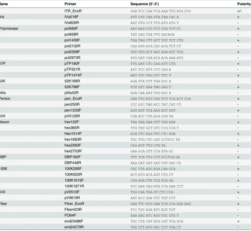
template DNA. PCR was performed under the following conditions: 1 cycle of 95°C for 5 min, followed by 14 one degree step-down cycles, each consisting of denaturation at 95°C for 40 s, with annealing from 50–37°C for 40 s, and extension at 72°C for 1–2 min. This was followed by 25 cycles consisting of denaturation at 95°C for 40 s, annealing at 42°C for 40 s, and extension at 72°C for 1–2 min, and finally, at 72°C for 5 min in a Mastercycler (Eppendorf, Germany). Extension time was altered according to the expected product size. The amplified product was purified by PCR Purification Kit (QIAGEN, Chatsworth, CA) and sequenced by Big Dye 3.1 Table 1. The list of primers used for whole genome sequencing of penguin adenoviruses.
Gene Primer Sequence (5’-3’) Polarity
ITR_EcoR GGA TCC CAA TCA AAA TTG ATA CCG
+/-IVa IVa518F ATT CAT CAA GTA CAA CAC A +
IVa626R AAT GTG CCT TTG ATG ATG T
-Polymerase pol560F AAT AAG CTG TCT GTA TCT CC +
pol658R TAT GAG TGA TTG GACAGA
-pol1439F TGA TAG CTT GCT TGT TCT CTG +
pol2132R CAA ATH ACA CAC ACA TCT CT
-pol2358F TGT GTG GCT AAT AGA ATC TCA +
pol2973R ATG GAT CAA ACA ACA AAA ATG
-pTP pTP180F TTG GAT CTC CAG ATT CTG +
pTP321R ATC TCC ATT CCT CAG A
-pTP1474F ART TTC THG GTC TTC T +
52K 52K189R AGA TTA TTT TGA GGC A
-52K798F TGT GTC ARA TWG GAG T +
pIIIa pIIIa42R AGA CAA AAT TGG AGC A
-Penton pen_EcoR GAA TTC ATG CAG TCT TCA ACT CCA +
pen250R CCC AYC TWG ACC TAT CAT CT
-pen1233F AGG AGT TCA AAG AGT GGT +
pVII pVII126R CGA ACC CTA ACA GTA TA
-Hexon hex122F TAA TAA GAA GTT TAG AGA +
hex365R TTG TAT GCT GTC CCG CCA T
-Hex1514F ACA TTC AGG TTC CTC AGA +
hex1693R TGC TTG CTC CAT CCTTCC TA
-hex2583F CAG AGT TTG CTG TA +
hex2753R GAA GCA GTT CCA GTA GC
-DBP DBP162F TTT TCA TTG CCT GCCTCA GA +
DBP448R AAA GAT GAT AAT CGT GAC CA
-100K 100K290F CAC TTA AGG AGA CAA GCA +
100K620R ACT ATG ACA AGT CTG CT
-100K1615F CGG AGA TTA TCA GCA GA +
100K1871R TCC AAA TAG ATA CCA GAA CCT
-pVII pVII510F TGG CAA TGA GT CTC CCA +
pVII619R AAT ACC GGA TTC TGT CCT
-Fiber Fiber_EcoR GAA TTC ATG GAA TCA CCA AGA AAG +
Fiber423R TCC TGC AGA ATC AGT TGT
-FO64F AAA AAC ATC AGG TGC GTG T
-end23486F TGC CTA CAT ATA CAT TCA GCA +
end24279R TGG TTT ATG TAC CCT TGA CT
terminator cycle sequencing reagents on ABI 3730 Automated DNA sequencer (Applied Bio-systems, Forest City, CA). The inverted terminal repeat (ITR) sequences of adenovirus were confirmed by modified-RACE (Rapid Amplification of cDNA Ends, TAKARA BIO INC. Shiga, Japan).
Phylogenetic analysis
The phylogenetic analysis was carried out based on the DNA polymerase and hexon sequence of penguin adenoviruses. Sequences of adenoviruses were retrieved from the GenBank. Multi-ple alignments of adenoviral sequences were generated by Clustal W method in MegAlign of DNAstar (Lasergene program version 5, DNASTAR Inc. Madison, WI). Phylogenetic trees were generated by a Bayesian inference of phylogeny throughout the MrBayes V3.1.2 software [31,32] and Maximum likelihood (ML) in methods of MEGA6.0 (Molecular Evolutionary Genetics Analysis 6.0) software [33]. WAG and GTR model contributed to approximate the posterior probabilities (pp) of trees inferred from amino acid and nucleotide alignments, respectively. The topologies of ML trees were evaluated by a bootstrap analysis of 1,000 itera-tions by using MEGA 6.0.
Results
Genetic character and genome organization
The whole genomes of 2 Chinstrap penguins (CSPAdVno3 and CSPAdVno4, GenBank acces-sion no.KP144329 and KP144330) and 2 Gentoo penguins (GPAdVno4 and GPAdVno5, KP279746 and KP279747) collected in 2010 were sequenced. The genome lengths were 24,662 bp (CSPAdVno3), 24,659 bp (CSPAdVno4), 24,630 bp (GPAdVno4), and 24,633 bp
(GPAdVno5). The G+C contents of the complete genomes were 35.5% in CSPAdV, and 35.6% in GPAdV. The G+C content of each gene ranged from 30.6–47.1%; the gene of the histone-like core protein precursor pVII was found to have the highest G+C content.
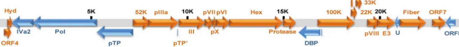
The genetic content and structure of penguin adenovirus are presented in the schematic genome map inFig 1, and contains 23 ORFs. The ORF4 reported in TAdV-3 and raptor adeno-virus 1 (RAdV-1) was also discovered in penguin adenoadeno-virus, but the sialidase gene existing between the inverted repeat (ITR) region of the 5’end and ORF4 was not detected in the pen-guin adenovirus genomes by modified RACE-PCR. The bi-directional analysis of ORF in the left-hand end of the genome between ITR of the 5’end and initial hydrophobic protein (hyd) showed only putative ORF4 (360 bp), as ORF is longer than 200 bp.
The lengths of most of the genes were identical among the various penguin adenoviruses, but a few genes, such as hexon and E3 gene, showed different lengths between CSPAdV and GPAdV (Table 2). A lack of 3 nucleotides (amino acid residue G; CAG, at nucleotide positions 722–724) in the hexon gene of CSPAdVno4 and GPAdVno4 [24], and an absence of 21 nucleo-tides (amino acid residues DGTYPFS:GATGGAACTTACCCCTTTTCT, nucleotide positions 445–465 in CSPAdV) in the E3 gene of GPAdV were identified. Moreover, there was a lack of
Fig 1. Putative genetic content of penguin adenovirus.The range of entire genome lengths of penguin adenoviruses were 24,630–24,662 bp, and the putative sialidase gene was not detected in the penguin adenovirus genomes. The genomic lengths are indicated at5,000 bp intervals.
several nucleotides in the noncoding region of CSPAdV, specifically at position 22,750–22,760 (AAATTATAGAC; on the basis of the GPAdV sequence) located between fiber and ORF7. The sequence of GPAdV was also shown to be shorter by 11nt (GCTAGTATAAA, nucleotide
position 342–356 in CSPAdV) in noncoding region, downstream of ORF4 and 13 nt (CTGTTTGGTACAA, nucleotide position 22,847–22,859 in CSPAdV) in noncoding region,
immediately before ORF7 (Table 2).
The lengths of the ITR sequences of CSPAdV and GPAdV were identical (i.e., 30 bp), and a single nucleotide difference at position 24 (C/T) was detected between CSPAdV and GPAdV.
Identification of existence of sialidase gene
The absence of sialidase gene in penguin adenovirus was verified from the 4 completely sequenced penguin adenoviruses, CSPAdVno3, CSPAdVno4, GPAdVno4, and GPAdVno5, by modified RACE-PCR. The 22 penguins that were detected with adenoviral genome were fur-ther tested for the presence of sialidase gene by PCR using the specific primer set, the primers from hyd gene and ITR region of 5’end (ITR/hyd). The size of PCR products by ITR/hyd was identified to be approximately 850 bp in the 19 penguins including the 4 penguin adenoviruses that were completely sequenced (data not shown), and the PCR results in the 3 penguins were negative.
Phylogenetic analysis
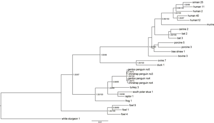
Phylogenetic analyses of entire hexon using the Bayesian and ML methods indicated that pen-guin adenoviruses clustered significantly withSiadenovirussp., as supported by the high poste-rior probabilities and bootstrap values of 100%. The analysis of the amino acid sequence of entire hexon showed the closest relationship and sharing of ancestor with TAdV-3,with high posterior probabilities (pp value 0.99) (Fig 2). The sister clades within the penguin adenovi-ruses were constructed by clustering of CSPAdVno3 and GPAdVno5, and CSPAdVno4 and GPAdVno4, respectively. Support for the clade of penguin adenoviral hexon was stronger in the phylogeny by Bayesian than by ML method. Also the phylogenetic analysis of partial DNA polymerase of 274 nt (91 aa) showed that penguin adenoviruses were clustered with the TAdV-3 (>0.92) (Fig 3). The nucleotide alignment of partial DNA polymerase showed the
clustering of Gouldian finch AdV with the Sulawesi tortoise and frog AdV (FrAdV-1) with low pp value (<50) and the first divergent of great tit AdV (Fig 3A), while the calculation
based on the amino acid alignment showed the grouping of bird-related siadenoviruses on the same branch (Fig 3B).
The entire hexon sequence of penguin adenoviruses shared 76.1–76.2% (77.2–77.4%, amino acid), 73.9–74.4% (73.4–73.5%), 73.3–73.5% (74.1–76.5%), and 68.5–68.8% (67.5–68.6%) iden-tity with the genomes of TAdV-3, South Polar skua adenovirus 1 (SPSAdV-1), raptor
Table 2. Summary of deletions of nucleotides and their position in the entire sequence of Chinstrap penguin adenovirus (CSPAdV) and Gentoo penguin adenovirus (GPAdV).
Viruses Region (position) Deficient nucleotide sequences
GPAdV Noncoding region in downstream of ORF4 (346-56nt) GCTAGTATAAA
CSPAdVno4 and GPAdVno4 Hexon (724-726nt) CAG
GPAdV E3 (455-465nt) GATGGAACTTACCCCTTTTCT
CSPAdV Noncoding region betweenfiber and ORF7 (22,750–22,760nt) CTGTTTGGTACAA
GPAdV Noncoding region, immediately before ORF7 (22,847–22,859nt) AAATTATAGAC
adenovirus 1 (RAdV-1), and frog adenovirus 1 (FrAdV-1), respectively. In addition, the entire DNA polymerase of penguin adenoviruses showed identity of 60.7–70.1% (52.1–67.9%, amino acid) with other siadenoviruses. The hexon and DNA polymerase sequences of penguin adeno-virus showed identity of 99.2–100% (99.1–99.9%) and 98.6–99.1% (97.9–99.5%), respectively. Fig 2. Phylogenetic analysis of amino acid sequences of penguin adenoviral hexon.The phylogenetic tree, based on entire hexon genome
sequences of penguin adenoviruses, was generated using the Bayesian and maximum likelihood (ML) method. The first number indicates the Bayesian posterior probability, and the second number indicates the ML bootstrap value as a percentage. Scale bars indicate the number of nucleotide substitutions per site.
doi:10.1371/journal.pone.0157032.g002
Fig 3. Phylogenetic relationships within the genusSiadenovirusbased on partial DNA polymerase sequences (a) inferred from nucleotide alignment and (b) from amino acid alignment.The phylogenetic tree was obtained with a Bayesian inference of phylogeny by the MrBayes v3.1.2 software.
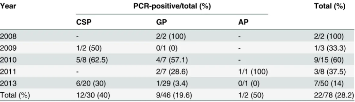
Molecular epidemiology
To investigate adenoviral infections in the penguin population from 2008 to 2013, we exam-ined 552 internal samples from 78 penguin carcasses by amplifying of a part of the hexon gene. The adenoviral genome was detected in 22 penguins (28.2%, 22/78), including 12 Chinstrap penguins (40%, 12/30), 9 Gentoo penguins (19.6%, 9/46), and an Adélie penguin (50%, 1/2). PCR positivity rate for the adenoviral genome was highest for the Chinstrap penguin popula-tion. The adenovirus detection rate was highest in 2008 (100%, 2/2), followed by 2010 (60%, 9/ 15) (Table 3). Interestingly, of the penguin adenovirus genome detected from various sample types, the PCR-positivity rate was highest in the kidney (63.6%, 14/22), followed by lung sam-ples at 36.4% (8/22), and greater than approximately 11% in the liver, heart, intestine, trachea, spleen, and fecal samples. However, the adenovirus genome was not identified in the lymph node or brain samples (Table 4). The detection rate of the penguin adenovirus genome with respect to geographic location was 20/72 (27.8%) at Narębski Point, 1/1 (100%) near the King
Sejong station, and 1/5 (20%) at Ardley Island.
Table 3. PCR positivity rate for penguin adenovirus in Chinstrap penguins (CSP), Gentoo penguins (GP), and Adelie penguins (GP), Antarctica, 2008–2013.
Year PCR-positive/total (%) Total (%)
CSP GP AP
2008 - 2/2 (100) - 2/2 (100)
2009 1/2 (50) 0/1 (0) - 1/3 (33.3)
2010 5/8 (62.5) 4/7 (57.1) - 9/15 (60)
2011 - 2/7 (28.6) 1/1 (100) 3/8 (37.5)
2013 6/20 (30) 1/29 (3.4) 0/1 (0) 7/50 (14)
Total (%) 12/30 (40) 9/46 (19.6) 1/2 (50) 22/78 (28.2)
doi:10.1371/journal.pone.0157032.t003
Table 4. Detection of the adenoviral genome in tissue and fecal samples collected from penguins infected with adenovirus by PCR.
Sample positive samples/total samples from PCR-positive penguins (%)*
Total (%)
CSP GP AP
Lung 6/12 (50) 2/9 (22.2) 0/1 (0) 8/22 (36.4)
Liver 4/12(33.3) 0/9 (0) 0/1 (0) 4/22 (18.2)
Kidney 6/12 (50) 8/9(88.9) 0/1 (0) 14/22 (63.6)
Heart 3/11 (27.3) 0/8(0) 1/1 (100) 4/20 (20)
Intestine 4/11 (36.4) 1/7 (14.3) 0/1 (0) 5/19 (26.3)
Trachea 4/11(36.4) 0/7 (0) 0/1 (0) 4/19 (21.1)
Spleen 1/7 (14.3) 0/1 (0) 0/1 (0) 1/9 (11.1)
Brain 0/7 (0) 0/3 (0) 0/1 (0) 0/11 (0)
Lymph node 0/1 (0) - - 0/1 (0)
Feces 0/6 (0) 2/3 (66.7) 0/1 (0) 2/10 (20)
*All types of tissue and fecal samples were not collected from every penguin. PCR was performed only for the collected samples. Accordingly, the total number of each sample collected from each penguin differs.
Discussion
Genetic features and phylogeny of penguin adenovirus
Our previous study suggested that based on the partial hexon gene sequence, CSPAdV merits the establishment as new species in the genusSiadenovirus[24]. In this study, the entire genome sequence and structure of GPAdV and CSPAdV were determined. The complete genomes of penguin adenoviruses (24,630–24,662 bp) were substantially shorter than those of other siadenoviruses, including SPSAdV-1 (26,340 bp), RAdV-1 (26,284 bp), TAdV-3 (26,263 bp), and FrAdV-1 (26,163 bp). The G+C content of the penguin adenoviruses (35.5–35.6%) also complied with that of four other siadenoviruses genomes (TAdV-3: 34.9%, SPSAdV-1:34.2%, RAdV-1:38.5%, FrAdV: 37.9%). The low G+C content is a character conserved across allSiadenovirusspecies, and is related with host jumping in adenoviruses [20,22]. Hence, the diverse host range of siadenoviruses can be attributed to their host switching.
Based on the phylogenetic trees of entire hexon as well as partial DNA polymerase, pen-guin adenoviruses were included within the genusSiadenovirus. In the familyAdenoviridae, a novel adenovirus species is usually defined as one detected in a new host species and having more than a 15% phylogenetic distance in DNA polymerase protein compared with previ-ously characterized adenovirus species [34,35]. The DNA polymerase gene showed the dif-ferences of29.9–39.3% (32.1–47.9%, amino acid) withSiadenovirusspecies. Furthermore, the penguin adenoviruses discovered from new host species have not been previously reported. Based on these criteria, we concluded that penguin adenoviruses were novel adenovirus in the genusSiadenovirus. The close relationship of penguin adenovirus and TAdV-3 was strongly supported by a high pp value (>0.92) in the phylogenetic analysis of entire hexon
gene and partial DNA polymerase. The phylogeny of entire hexon of penguin adenovirus showed the clustering of CSPAdVno4 and GPAdVno4 because of the deletion of an amino acid in hexon gene.
The genetic structure of the novel penguin adenovirus showed the absence of putative siali-dase gene. The lengths from 5’end to ORF4 of penguin adenoviruses are significantly shorter (758–769 bp) than that of other siadenoviruses (2,028–2,142 bp). Moreover, except the ORF4, any other ORF longer than 200 bp, between 5’end and ORF4 was not detected. The sialidase gene, named so due to its similarity to bacterial sialidase gene, is known as a putative gene that is specific to the genusSiadenovirus. Although the function of sialidase is still unknown, it may be related to entry in host cell by binding sialic acid residues [20]. The genetic structure of the novel penguin adenovirus showed the absence of putative sialidase gene. Nonetheless, their genetic characters, short genome length, low G+C contents, and phylogeny, indicated that pen-guin adenovirus belongs to the genusSiadenovirus. However, this genomic organization differ-ence, the absence of putative sialidase gene can be seen as a further species demarcation criterion. Therefore, additional studies on the function of sialidase and the presence of sialidase gene inSiadenovirussp. are necessary, since complete sequences are only available for 5species: FrAdV-1, TAdV-3, RAdV-1, SPSAdV-1, penguin AdV 1 (abbreviated as PeAdV-1).
Molecular epidemiology and infection of penguin adenovirus
The molecular epidemiological study of penguin adenovirus from 2008–2013 indicated that the infection predominantly affects the Chinstrap penguin population, and the annual detec-tion of penguin adenoviruses suggests their prevalence and circuladetec-tion in Antarctic penguin populations. However, significant divergence among the different penguin adenovirus sequences from different geographic regions was not detected.
The novel viruses in the genusSiadenovirus, Sulawesi tortoise adenovirus 1 and Gouldian finch adenovirus, cause severe systemic infections in most of the organs [22,23]. In the internal organs of penguins, the adenovirus was detected at a high rate in the kidney in addition to the lung, liver, heart, intestine, trachea, spleen, and feces. These results suggest that the penguin adenovirus causes systemic infections in penguins.
In conclusion, four penguin adenoviruses were identified from two dead Chinstrap pen-guins and two Gentoo penpen-guins, the endemic species in Antarctica [38,39]. The penguin ade-noviruses were identified as members of a new candidate species, containing unique genetic character, in the genusSiadenovirus. In addition, our molecular epidemiological data indicated that the penguin adenovirus is prevalent and circulating in Antarctic penguin populations.
Acknowledgments
We thank Jin Woo Jung, Min-Goo Lee, Kyeong Hoon Cho, and Yeong Woong Kim for assis-tance in sample collection. This work was supported by Polar Academic Program and Korea Polar Research Institute, Korea.
Author Contributions
Conceived and designed the experiments: JWS SYL. Performed the experiments: SYL. Ana-lyzed the data: SYL TKS JSN. Contributed reagents/materials/analysis tools: JHK TKS HK HGC SHK. Wrote the paper: SYL WKK.
References
1. Berk AJ.Adenoviridae: The viruses and their replication. In: Knipe DM, Howley PM, editors. Fields
virol-ogy. Vol. 2. Philadelphia: Lippincott Williams & Wilkins, 2007; p. 2355–61.
2. Davison AJ, Benko M, Harrach B. Genetic content and evolution of adenoviruses. J Gen Virol. 2003; 87:2895–908.
3. Davison AJ, Telford EA, Watson MS, McBride K, Mautner V. The DNA sequence of adenovirus type 40. J Mol Biol. 1993; 234:1308–16. PMID:8263936
4. Kovács GM, Davision AJ, Zakhartchouk AN, Harrach B. Analysis of the first complete genome sequence of an Old World monkey adenovirus reveals a lineage distinct from the six human adenovirus species. J Gen Virol. 2004; 85:2799–807. PMID:15448340
5. Morrison MD, Onions DE, Nicolson L. Complete DNA sequence of canine adenovirus type 1. J Gen Virol. 1997; 78:873–8. PMID:9129661
6. Rusvai M, Harrach B, Bánrévi A. Identification and sequence analysis of the core protein genes of
bovine adenovirus 2. Virus Res. 2000; 70:25–30. PMID:11074122
7. Aggarwal N, Mittal SK. Sequence analysis of porcine adenovirus type 3 E1 region, pIX and pIVa2 genes, and two novel open reading frames. Intervirology. 2000; 43:6–12. PMID:10773731 8. Meissner JD, Hirsch GN, LaRue EA, Fulcher RA, Spindler KR. Completion of the DNA sequence of
mouse adenovirus type 1: sequence of E2B, L1, and L2 (18–51 map units). Virus Res. 1997; 51:53–
64. PMID:9381795
9. Kohl C, Vidovszky MZ, Mühldorfer K, Dabrowski PW, RadonićA, Nitsche A, et al.Genome analysis of
bat adenovirus 2: indications of interspecies transmission. J Virol. 2012; 86:1888–92. doi:10.1128/JVI.
10. Dán A, Ruzsics Z, Russell WC, Benkö M, Harrach B. Analysis of the hexon gene sequence of bovine adenovirus type 4 provides further support for a new adenovirus genus (Atadenovirus). J Gen Virol. 1998; 79:1453–60. PMID:9634088
11. Farkas SL, Harrach B, Benkö M. Completion of the genome analysis of snake adenovirus type 1, a rep-resentative of the reptilian lineage within the novel genus Atadenovirus. Virus Res. 2008; 132:132–9.
doi:10.1016/j.virusres.2007.11.009PMID:18166240
12. Hess M, Blöcker H, Brandt P. The complete nucleotide sequence of the egg drop syndrome virus: An intermediate between mastadenoviruses and aviadenoviruses. Virology. 1997; 238:145–56. PMID:
9375018
13. Chiocca S, Kurzbauer R, Schaffner G, Baker A, Mautner V, Cotton M. The complete DNA sequence and genomic organization of the avian adenovirus CELO. J Virol. 1996; 70:2939–49. PMID:8627769 14. Kovács GM, LaPatra SE, D’Halluin JC, Benkö M. Phylogenetic analysis of the hexon and protease
genes of a fish adenovirus isolated from with sturgeon (Acipenser transmontanus) supports the pro-posal for a new adenovirus genus. Virus Res. 2003; 98:27–34. PMID:14609627
15. Davison AJ, Wright KM, Harrach B. DNA sequence of frog adenovirus. J Gen Virol. 2000; 81:2431–9. PMID:10993931
16. Pitcovski J, Mualem M, Rei-Koren Z, Krispel S, Shmueli E, Peretz Y, et al. The complete DNA
sequence and genome organization of the avian adenovirus, hemorrhagic enteritis virus. Virology. 1998; 249:307–15. PMID:9791022
17. Wellehan JF Jr, Greenacre CB, Fleming GJ, Stetter MD, Childress AL, Terrell SP. Siadenovirus
infec-tion in two psittacine bird species. Avian Pathol. 2009; 38:413–7. doi:10.1080/03079450903183660 PMID:19937528
18. Katoh H, Ohya K, Kubo M, Murata K, Yanai T, et al. A novel budgerigar-adenovirus belonging to group
II avian adenovirus of Siadenovirus. Virus Res. 2009; 144:294–7. doi:10.1016/j.virusres.2009.04.012 PMID:19394371
19. Kovács ER, Jánoska M, Dán A, Harrach B, Benko M. Recognition and partial genome characterization
by non-specific DNA amplification and PCR of a new siadenovirus species in a sample originating from Parus major, a great tit. J Virol Methods. 2010; 163:262–8. doi:10.1016/j.jviromet.2009.10.007PMID: 19854219
20. Kovács ER, Benko M. Complete sequence of raptor adenovirus 1 confirms the characteristic genome organization of siadenoviruses. Infect Genet Evol. 2011; 11:1058–65. doi:10.1016/j.meegid.2011.03. 021PMID:21463713
21. Park YM, Kim JH, Gu SH, Lee SY, Lee MG, Knag YK, et al. Full genome analysis of a novel adenovirus from the South Polar skua (Catharacta maccormicki) in Antarctica. Virology. 2012; 422:144–50. doi: 10.1016/j.virol.2011.10.008PMID:22078165
22. Joseph HM, Ballmann MZ, Garner MM, Hanley CS, Berlinski R, Erdélyi K, et al. A novel siadenovirus detected in the kidneys and liver of Gouldian finches (Erythura gouldiae). Vet Microbiol. 2014; 6: 172:35–43. doi:10.1016/j.vetmic.2014.04.006PMID:24814929
23. Rivera S, Wellehan JF, McManamon R, Innis CJ, Garner MM, Rapheal BL, et al. Systemic adenovirus infection in Sulawesi tortoises (Indotestudo forstenii) caused by a novel siadenovirus. J Vet Diagn Invest. 2009; 21:415–26. PMID:19564489
24. Lee SY, Kim JH, Park YM, Shin OS, Kim H,Kim H, et al. A novel adenovirus in Chinstrap penguin (Pygoscelis antarctica) in Antarctica. Viruses. 2014; 6:2052–61. doi:10.3390/v6052052PMID: 24811321
25. Chown SL, Lee JE, Hughes KA, Barnes J, Barrett J, Bergstrom DM, et al. Conservation. Challenges to the future conservation of the Antarctic. Science.2012; 337:158–9. doi:10.1126/science.1222821 PMID:22798586
26. Kerry K, Riddle M, Clarke J. Disease of Antarctic wildlife; 18; Australian Antarctic Division: Channel Highway, Kingston, Australia, August 1998; p 89–91.
27. Grimaldi WW, Seddon PJ, Lyver POB, Nakagawa S, Tompkins DM. Infectious disease of Antarctic
pen-guins: current status and future threats. Polar Biol. 2014; 38:591–606.
28. Hurt AC, Vijaykrishna D, Butler J, Baas C, Maurer-Stroh S, Silva-de-la-Fuente M, et al. Detection of evolutionarily distinct avian influenza A viruses in Antarctica. MBio. 2014; 5:e01098–14. doi:10.1128/
mBio.01098-14PMID:24803521
30. Varsani A, Porzig EL, Jennings S, Kraberger S, Farkas K, Julian L, et al. Identification of an avian poly-mavirus associated with Adelie penguins (Pygoscelis adeliae). J Gen Virol. 2015; 96:851–7. doi:10.
1099/vir.0.000038PMID:25537375
31. Huelsenbeck JP, Bollback JP. Empirical and hierarchical Bayesian estimation of ancestral states. Syst Biol. 2001; 50:351–66. PMID:12116580
32. Ronquist F, Huelsenbeck JP. MrBayes 3: Bayesian phylogenetic inference under mixed models.
Bioin-formatics. 2003; 19:1572–4. PMID:12912839
33. Tamura K, Stecher G, Peterson D, Filipski A, Kumar S. MEGA: Molecular Evolutionary Genetics
Analy-sis version 6.0. Mol Bio Evol. 2013; 28:2731–9.
34. Harrach B, BenkőM, Both GW, Brown M, Davision AJ, et al. Family Adenoviridae. In:King AMQ,
Lefko-wits E, Adams MJ, Carstens EB, editors. Virus Taxonomy: IXth Report of the International Committee on Taxonomy of Viruses. Vol 9. New York: Elsevier, 1995; p.125–41.
35. Benkö M, Davison AJ, Harrach B, Song JW. ICTV Official Taxonomy; Updates since the 8thReport, Vertebrate (through 2014), 2012.016aV.A.v2.Siadenovirus-Sp. Available:http://talk.ictvonline.org/files/ ictv_official_taxonomy_updates_since_the_8th_report/m/vertebrate-official/4483.aspx
36. Beach NM, Duncan RB, Larsen CT, Meng XJ, Sriranganathan N, Pierson FW. Comparison of 12 turkey hemorrhagic enteritis virus isolates allows prediction of genetic factors affecting virulence. J Gen Virol. 2009; 90:1978–85. doi:10.1099/vir.0.010090-0PMID:19386786
37. Singh AK, Berbís MÁ, Ballmann MZ, Kilcoyne M, Menéndez M, Nquyen TH, et al. Structure and sialyl-lactose binding of the carboxy-terminal head domain of the fibre from a Siadenvoirus, turkey adenovirus 3. PLoS One. 2015; 10:e0139339. doi:10.1371/journal.pone.0139339PMID:26418008
38. Trivelpiece WZ, Trivelpiece SG, Volkman NJ. Ecological segregation of Adelie, gentoo and chinstrap penguins at King George Island, Antarctica. Ecology.1987; 68:351–61.